
Table of Contents
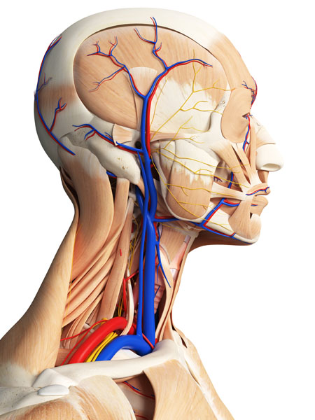
Venous Drainage of the Head

This image presents a detailed view of the venous drainage of the head, specifically of the facial region, as highlighted by the blue structures against the skeletal framework of the skull.
At the superior aspect of the image, near the forehead, we observe the supraorbital veins, which drain blood from the forehead and scalp. These veins are connected to the angular veins, seen near the bridge of the nose, which in turn receive blood from regions around the eyes and the root of the nose.
The infraorbital veins are visible just below the orbits, collecting blood from the lower eyelid, cheek, and upper lip area. They course towards the pterygoid plexus, which is a network of veins situated deep in the face, not visible on the surface here, that communicates with the cavernous sinus, a large collection of veins within the skull.
The superior labial veins are seen draining the upper lip, while the inferior labial veins drain the lower lip. These vessels merge into the facial vein, a major venous channel that runs a vertical course down the face. The facial vein is responsible for a significant portion of venous drainage from the superficial areas of the face.
Running approximately parallel to the facial vein are the veins of the lateral face, including the transverse facial vein, which drains the regions of the face over the parotid gland, an area not distinctly visible in this representation.
It’s important to note that the venous anatomy can be quite variable and anastomoses between these veins provide collateral pathways for blood to return to the heart. These veins ultimately drain into the internal jugular veins, the primary vessels that carry blood from the head and neck back to the heart, although they are not depicted in this image.
Venous Drainage of Head: Side View

In this side view, we see additional details of the venous drainage of the head and neck not visible in the previous frontal view.
Prominently, we have the external jugular vein visible as it descends over the sternocleidomastoid muscle. This vein drains the scalp and deep parts of the face, receiving blood from smaller veins such as the posterior auricular vein, which drains the region behind the ear, and the retromandibular vein, which is formed by the union of the superficial temporal vein and maxillary vein.
The superficial temporal vein is also visible here, coursing superiorly to the external ear, draining portions of the scalp. Below the external jugular vein, we can see the subclavian vein, into which the external jugular vein often drains. The subclavian vein runs under the clavicle and is a major conduit for blood returning to the heart from the upper extremities and head.
The occipital vein is shown near the back of the skull, which drains the posterior scalp and communicates with the vertebral venous plexus. This network of veins lies within the vertebral canal and is not clearly depicted here.
This lateral perspective also provides a view of the internal jugular vein, which is a larger, deeper vein that runs medial to the sternocleidomastoid muscle and carries a significant volume of venous blood from the brain, the superficial face, and the neck back toward the heart. However, the internal jugular vein is not as clearly distinguished in this representation due to the overlap of other structures.
Understanding this venous architecture is crucial in clinical contexts, as it impacts procedures ranging from venous access for injections to the management of conditions that may affect venous return, such as thrombosis or compression.
Thoracic Cavity: Anterior View
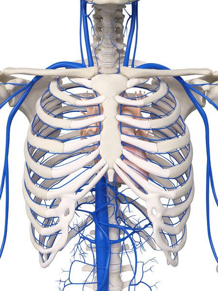
The image depicts the thoracic cavity from an anterior view, highlighting the venous system with blue-colored vessels against the bony thorax structure.
At the top of the image, we see the brachiocephalic veins, which are formed by the union of the internal jugular and subclavian veins on each side. These veins receive blood from the head, neck, and arms, and are among the major vessels that drain into the superior vena cava, the large blue vessel in the center that carries blood back to the heart.
The superior vena cava descends vertically and enters the right atrium of the heart. It is one of the two main veins by which blood is returned from the body to the heart. This vessel is not visible in the image as it is located posterior to the bony structures of the sternum and would enter the heart below the inferior border of the image.
Also visible in the image are the intercostal veins, which run between the ribs and drain the chest wall and muscles. They connect to the azygos vein system, which is a longitudinal vessel on the right side running alongside the vertebral column and is partially visible as it ascends towards the brachiocephalic veins.
The network of veins at the base of the image represents the extensive venous plexuses that are found within the thoracic cavity. They are involved in the drainage of the thoracic and abdominal walls, and the organs contained within them. These plexuses are interconnected and eventually channel the blood into the larger systemic veins like the superior vena cava.
Understanding the venous anatomy of the thoracic cavity is crucial for procedures involving central venous access, cardiac surgeries, and managing conditions affecting venous return to the heart.
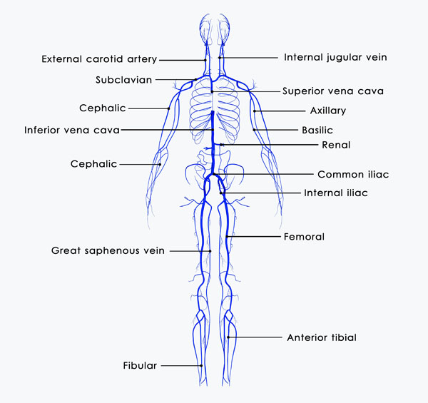
Overview of the Venous System
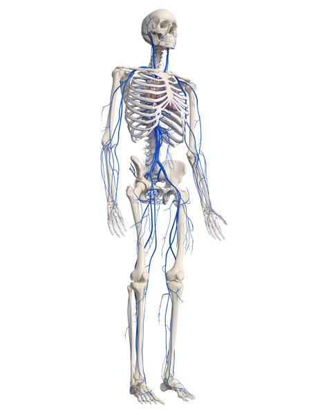
This image provides a comprehensive overview of the human venous system, depicted in blue against the skeletal system. The venous system is a network of vessels that carries deoxygenated blood from various parts of the body back to the heart.
Starting from the upper body, we see the cephalic and basilic veins in the arms, which drain blood from the radial and ulnar veins of the forearm and ascend towards the shoulder area. They merge to form the subclavian veins near the shoulders, which then join the internal jugular veins to become the brachiocephalic veins. These, in turn, feed into the superior vena cava, which carries blood from the upper half of the body into the right atrium of the heart.
The thoracic region shows the azygos vein on the right side of the vertebral column, which drains the thoracic wall and some abdominal structures into the superior vena cava. Its counterpart, the hemiazygos vein, is not visible but would be located on the left side, performing a similar function.
In the abdominal area, the image shows the inferior vena cava, the large vein that ascends along the spine and transports blood from the lower parts of the body to the heart. It receives blood from various tributaries, including the hepatic veins from the liver, the renal veins from the kidneys, and the iliac veins from the lower limbs.
The lower body highlights the great saphenous veins, which are the longest veins in the body. They run from the feet up the inside of the legs to the groin, where they drain into the femoral veins. The small saphenous veins run up the back of the calves and drain into the popliteal veins behind the knees.
The deep veins of the legs, such as the anterior and posterior tibial veins and the peroneal veins, carry blood from the feet and calves, merging into the popliteal veins and then into the femoral veins in the thigh. These join the iliac veins, which contribute to the flow into the inferior vena cava.
This extensive network is crucial for returning deoxygenated blood to the heart, where it can be reoxygenated and recirculated through the body. The veins also work against gravity, especially in the legs, and contain valves that prevent backflow, ensuring that blood travels in one direction towards the heart.
Superficial Veins of the Head and Neck
In this image, we can see the intricate network of veins interspersed among the different layers of muscle tissue in the head and neck region. The veins are depicted in blue, muscles in beige and red, and nerves in yellow.
The superficial veins are the ones closest to the skin. These include the superficial temporal vein, which drains blood from the temporal region and is seen running anteriorly to the ear. The facial vein is also visible, coursing down the face in front of the masseter muscle, the thick muscle near the jaw responsible for chewing.
Beneath these superficial veins are deeper venous structures that lie within or between the deeper muscle layers. The internal jugular vein is the predominant deep vein seen here, running deep within the neck alongside the common carotid artery and vagus nerve, forming the carotid sheath. It collects blood from the brain, the superficial parts of the face, and the neck.
Embedded within the musculature, especially around the sternocleidomastoid muscle — the prominent muscle that runs obliquely across the side of the neck — we can see the external jugular vein. It drains the scalp and deep portions of the face and neck and then descends to empty into the subclavian vein.
Further deep, the vertebral veins are visible within the transverse foramina of the cervical vertebrae. They drain the cervical spinal cord and posterior part of the skull.
These venous pathways are essential for draining deoxygenated blood from the head and neck back to the heart. They are surrounded by muscle and connective tissue, which help facilitate venous return through muscle contractions and the presence of valves within the veins to prevent backflow. The arrangement of these veins within the muscle layers also provides a protective mechanism, safeguarding these vessels from external injury.
Venous Structure of the Lower Leg

The image showcases the venous structure of the lower limb, specifically the leg, with veins highlighted in blue.
We can see the great saphenous vein, the longest vein in the body, which originates from the medial side of the dorsal venous arch in the foot. It ascends along the medial aspect of the leg, running just anterior to the medial malleolus (the bony prominence on the inner side of the ankle), and continues along the medial side of the thigh to eventually drain into the femoral vein in the groin.
Branching from the great saphenous vein are various superficial tributaries that drain the skin and subcutaneous tissues of the leg. These tributaries are visible as they traverse the surface of the muscles and subcutaneous fat.
In the lower part of the leg, we can observe the small saphenous vein, which runs along the posterior aspect of the leg. It begins at the lateral side of the dorsal venous arch, ascends behind the lateral malleolus (the bony prominence on the outer side of the ankle), and travels up the posterior aspect of the calf to drain into the popliteal vein behind the knee.
Also depicted in the image are several unnamed tributary veins that interconnect the superficial and deep venous systems. These perforating veins pass through the deep fascia of the leg, allowing blood to flow from the superficial veins into the deep veins.
The deep veins of the leg, which are typically paired with arteries and are not as clearly visible in this image, run within the muscular compartments and are crucial for returning blood from the lower extremity back to the heart. These deep veins include the anterior and posterior tibial veins, as well as the peroneal veins, which are nestled between the muscles and converge to form the popliteal vein in the knee region.
The venous network in the legs is particularly important because it must work against gravity to return blood to the heart. The veins in the lower limbs are equipped with valves that prevent the backflow of blood, and the muscle contractions in the leg help to pump the blood upwards. This is essential for maintaining proper circulation and preventing venous insufficiency.
Venous Network of the Forearm and Hand

The image displays the venous network of the forearm and hand. In this depiction, the veins are colored blue, providing a stark contrast against the muscles and tendons of the arm.
Starting proximally, near the elbow, we can identify the cephalic vein on the lateral side of the forearm. This vein is a superficial vein that arises from the dorsal venous network of the hand and ascends along the radial side of the forearm. It is frequently used for intravenous access or blood withdrawal.
On the medial side of the forearm, the basilic vein can be observed. It also originates from the dorsal venous network of the hand and ascends along the ulnar side. The basilic vein is larger and deeper than the cephalic vein as it progresses up the arm.
Connecting the cephalic and basilic veins is the median cubital vein, which is often visible just below the surface of the skin in the cubital fossa, an area on the anterior side of the elbow. This vein is commonly used for venipuncture due to its accessibility.
As we move distally towards the wrist and hand, we see the dorsal venous network of the hand. This network is a superficial system that drains blood from the back of the hand. It gives rise to the cephalic and basilic veins and is interconnected with the deeper venous system of the hand, which is not clearly visible in this image.
The deep veins of the forearm, which accompany the arteries, are not distinctly shown here but would lie deeper within the musculature. They form venae comitantes (paired veins) with the radial and ulnar arteries and are responsible for the majority of venous return from the hand and forearm.
The venous anatomy of the forearm and hand is crucial not only for venous return but also for medical procedures involving venous access, and understanding these pathways is essential for clinicians.
Venous Network of the Pelvis and Upper Legs
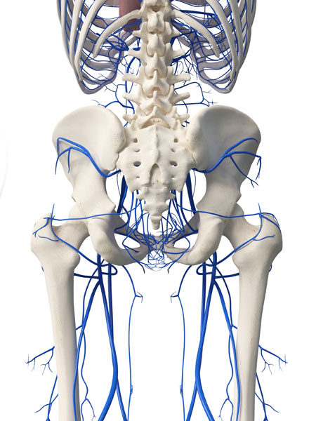
This image presents a view of the venous network surrounding the pelvis and the upper legs, with the veins depicted in blue against the backdrop of the skeletal structure.
The largest and central vein visible is the inferior vena cava, which ascends along the spine to carry deoxygenated blood from the lower body back to the heart. Emerging from the top of the pelvis and joining the inferior vena cava are the common iliac veins, which are formed by the convergence of the internal and external iliac veins on each side of the body.
The internal iliac veins drain blood from the pelvic organs, while the external iliac veins continue from the femoral veins in the thigh, which in turn receive blood from the deep veins of the leg, like the popliteal vein seen behind the knee, as well as the great saphenous vein, the longest vein in the body.
This network also shows the superficial veins that drain the lower abdomen and pelvis, such as the superficial epigastric veins, which arise near the groin area. The deep venous network, which includes the femoral veins, is responsible for the majority of venous return from the lower extremities, carrying blood from the deep tissues and muscles of the legs.
The veins in this region are essential for returning blood from the lower extremities, and they also play a role in draining the organs within the pelvis. The presence of bony landmarks, such as the pelvis and the vertebral column, provides a point of reference for medical professionals when navigating these vascular structures during interventions.
Venous Drainage System of the Brain: External
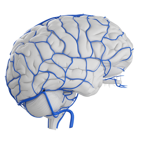
The image displays the venous drainage system of the brain superimposed on a model of the brain’s exterior. The veins are depicted in blue, contrasting against the white of the brain’s surface, or cortex.
The most prominent features are the superficial cerebral veins, which drain blood from the cortex into the larger venous sinuses. These include the superior cerebral veins, which run across the surface of the hemispheres and drain into the superior sagittal sinus, a large venous channel that runs along the top midline of the skull.
The image also shows the superficial middle cerebral vein, which courses along the lateral surface of the brain, following the Sylvian fissure. It often drains into the cavernous sinus or the superior sagittal sinus.
Inferiorly, we can see the inferior cerebral veins, which drain the undersurface of the brain and empty into the tentorial sinuses or straight sinus, another dural venous sinus.
The deep venous drainage is not visible in this image, but it would include the internal cerebral veins, the basal veins, and the great cerebral vein, all of which converge on the vein of Galen before emptying into the straight sinus.
The venous system of the brain is critical as it is responsible for draining deoxygenated blood, along with metabolic waste products, away from the brain to be reoxygenated in the lungs. The efficiency of this venous system is vital for the brain’s function and overall health. Inadequate drainage can lead to increased intracranial pressure and associated complications.
Schematic Diagram of the Venous System
The image is a schematic diagram of the human venous system, with key veins labeled to illustrate how blood is returned from various parts of the body to the heart. It is a simplified representation that highlights the major veins and their anatomical courses.
At the top of the diagram, the internal jugular vein is shown, which drains the brain and deep parts of the face and neck. It merges with the subclavian vein, coming from the arm, to form the brachiocephalic vein. The brachiocephalic veins from both sides of the body then join to form the superior vena cava, which empties into the right atrium of the heart.
The cephalic vein, a superficial vein on the lateral side of the arm, is also indicated. It eventually drains into the subclavian vein.
The diagram also shows the axillary vein, which continues from the subclavian vein and receives blood from the arm and shoulder region.
The inferior vena cava is presented as the large vertical vein running posteriorly from the lower body to the heart. It collects blood from the legs via the common iliac veins, which are formed by the confluence of the internal and external iliac veins. The renal veins, draining the kidneys, are shown connecting to the inferior vena cava as well.
The great saphenous vein is illustrated running along the length of the leg, from the foot to the groin, where it drains into the femoral vein. The femoral vein then becomes the external iliac vein, which contributes to the common iliac vein.
In the lower leg, the anterior tibial vein and fibular (peroneal) vein are labeled, indicating the deep venous drainage of the lower leg.
This diagram serves as an educational tool to understand the major pathways through which deoxygenated blood travels back to the heart. It shows the hierarchical structure of the venous system, from smaller superficial veins to larger deep veins, culminating in the superior and inferior venae cavae.
Cross-Section of a Vein
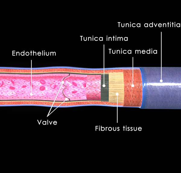
This image is a cross-sectional view of a vein, illustrating its structural layers and components.
Starting from the innermost layer, we see the endothelium, which is the thin layer of cells that lines the interior surface of the vein. This layer provides a smooth surface for blood to flow over and plays a role in regulating vascular function.
The next layer is the tunica intima, a thin layer composed of the endothelium and a subendothelial layer made up of a thin layer of connective tissue. This layer is in direct contact with the blood flowing through the vein.
Surrounding the tunica intima is the tunica media, which in veins is typically thin and composed of a few layers of smooth muscle cells and connective tissue. The tunica media’s smooth muscle allows for the regulation of the vein’s diameter under the control of the autonomic nervous system.
The outermost layer is the tunica adventitia, which is composed of connective tissue containing collagen and elastic fibers. This layer provides structural support and flexibility to the vein, allowing it to withstand changes in pressure and volume.
Also highlighted in the image is a venous valve, which is essential for the proper functioning of veins, especially in the limbs. These valves are formed from folds of the tunica intima and ensure one-way blood flow back to the heart, preventing backflow and aiding venous return against gravity.
Lastly, the image shows fibrous tissue, which provides structural integrity to the vessel and connects it with surrounding tissues, helping to anchor the vein in place.
This detailed view of the venous wall and valve is fundamental for understanding how veins maintain blood flow toward the heart, particularly in the lower extremities where blood must be transported against the force of gravity. Venous valves are key in preventing conditions such as varicose veins and venous insufficiency.
Anatomical Terms and Definitions
| Term | Definition |
|---|---|
| Angular Veins | Veins near the bridge of the nose, receiving blood from regions around the eyes and root of the nose. |
| Azygos Vein | A longitudinal vessel on the right side running alongside the vertebral column, involved in draining the thoracic wall and some abdominal structures into the superior vena cava. |
| Brachiocephalic Veins | Formed by the union of the internal jugular and subclavian veins, these veins receive blood from the head, neck, and arms. |
| Calf Muscle Pump | The mechanism by which the calf muscles assist in venous blood flow, compressing deep veins in the leg during contraction to push blood upwards. |
| Deep Vein Thrombosis (DVT) | A condition characterized by the formation of a blood clot within a deep vein, typically in the leg, which can obstruct blood flow and lead to complications like pulmonary embolism. |
| External Jugular Vein | A vein that drains the scalp and deep parts of the face, descending over the sternocleidomastoid muscle. |
| Facial Vein | A major venous channel running vertically down the face, responsible for draining venous blood from superficial areas. |
| Fenestrated Capillary | A type of capillary with pores in its endothelial lining, allowing for increased permeability and facilitating the transfer of small molecules. |
| Great Saphenous Vein | The longest vein in the body, running from the foot up the inside of the leg to the groin, where it drains into the femoral vein. |
| Inferior Vena Cava | The large vein that ascends along the spine, transporting blood from the lower parts of the body to the heart. |
| Internal Jugular Vein | A larger, deeper vein that runs medial to the sternocleidomastoid muscle, carrying venous blood from the brain, superficial face, and neck back toward the heart. |
| Popliteal Vein | A deep vein behind the knee, receiving blood from the lower leg and continuing upward to become the femoral vein in the thigh. |
| Pulmonary Embolism | A potentially life-threatening condition that occurs when a blood clot travels to the lungs and blocks one of the pulmonary arteries. |
| Subclavian Vein | Runs under the clavicle, acting as a major conduit for blood returning to the heart from the upper extremities and head. |
| Superior Vena Cava | A large vein that carries blood from the upper body into the right atrium of the heart. |
| Varicose Veins | Dilated, twisted veins that result from venous insufficiency, typically occurring in the legs. |
| Venous Insufficiency | A condition where the veins have trouble sending blood from the limbs back to the heart, often leading to varicose veins and other complications. |
| Venous Valve | A structure within veins that ensures one-way blood flow back to the heart, preventing backflow. |
