
Table of Contents
Venous Insufficiency Blood Flow
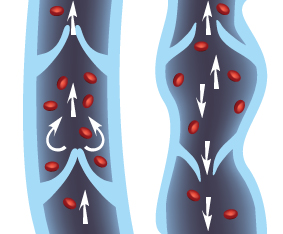
The image illustrates two segments of veins, showcasing the function of venous valves in facilitating unidirectional blood flow towards the heart.
On the left, the vein is depicted with a series of open, functioning venous valves. These valves, shown in white, are cup-shaped structures that open to allow blood to flow towards the heart (as indicated by the upward white arrows) and close to prevent backflow (as shown by the curved arrows at the valve sites), ensuring that blood continues in one direction despite the force of gravity or other factors that might otherwise cause it to pool in the lower extremities.
On the right, the vein appears to depict a condition known as venous insufficiency, where the valves are not functioning properly. This is indicated by the blood (red cells) pooling at certain points and the presence of reversed flow, as indicated by the downward arrows. The lack of effective valve closure allows blood to flow backward (reflux), which can lead to varicose veins, swelling, and other complications due to the increased pressure in the lower veins.
Overall, the image serves as a comparison between healthy venous function and a common venous disorder, emphasizing the importance of valve integrity in maintaining circulatory efficiency.
Calf Muscle Pump Flow
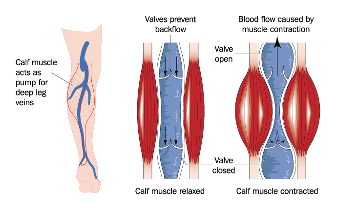
The image provides a visual explanation of the mechanism by which the calf muscles assist in venous blood flow in the legs, known as the “calf muscle pump.”
On the left, the image depicts the venous system of the lower leg with the deep veins highlighted in blue. The calf muscles are shown surrounding these veins, and the text indicates that these muscles act as a pump for the deep leg veins.
The central and right portions of the image give a closer view of how this pump mechanism works. In the central image, with the calf muscle relaxed, the vein is shown in a state of rest. The venous valves are open, and there is no blood flow indicated.
On the right, the calf muscle is contracted. This contraction compresses the deep veins in the leg, pushing blood upwards (as indicated by the black arrow). The venous valves, depicted in white, are shown in two states: the lower valve is closed to prevent backflow, and the upper valve is open, allowing blood to move upwards towards the heart.
This mechanism is essential for countering the effects of gravity, especially when standing or sitting for extended periods, which can cause blood to pool in the lower legs. The calf muscle pump is crucial for maintaining venous return from the lower extremities and for overall circulatory health.
Deep Vein Thombrosis
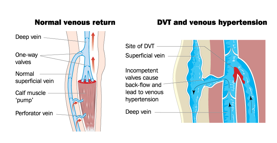
The image is a comparative illustration of two states of venous return in the leg: normal venous return versus a leg affected by deep vein thrombosis (DVT) and venous hypertension.
On the left side, labeled “Normal venous return,” the image depicts a healthy leg with efficient blood flow. The deep vein is shown in blue with one-way valves that ensure blood flows in only one direction, towards the heart (indicated by the red arrow). The normal superficial vein is also shown in blue, running parallel to the deep vein. The calf muscle, often referred to as the ‘calf pump,’ assists in propelling blood upwards through the veins when it contracts. The perforator vein, which connects the superficial to the deep vein, allows blood to pass into the deep venous system.
On the right side, labeled “DVT and venous hypertension,” the image illustrates a leg where a blockage has occurred due to DVT, indicated by the label “Site of DVT.” This blockage in the deep vein, shown in dark blue, prevents normal blood flow. The superficial vein, also in dark blue, is dilated due to the increased pressure from the obstructed deep vein. The image also indicates that there are incompetent valves, shown in red, which lead to back-flow (as shown by the blue arrows pointing downwards) and contribute to venous hypertension, a condition characterized by increased blood pressure within the venous system.
This side-by-side representation emphasizes the importance of valve function and unobstructed blood flow in maintaining venous return. It also highlights how conditions like DVT can lead to complications such as venous hypertension, which can cause pain, swelling, and varicose veins.
Varicose Vein Pathology

The image illustrates the progressive stages of varicose vein development, a condition characterized by dilated and twisted veins, typically in the legs, which results from venous insufficiency.
Stage 1: Spider veins -- These are small, fine veins that can be seen just under the skin. They are also called telangiectasias and usually do not cause symptoms.
Stage 2: Reticular varicose veins -- These are larger than spider veins and may appear raised with a bluish color. They can cause discomfort and indicate more significant venous insufficiency.
Stage 3: Venous nodes -- At this stage, the veins become even more enlarged and twisted, forming palpable nodes. This can be associated with symptoms such as aching, heaviness, or cramping in the legs.
Stage 4: Chronic venous insufficiency -- This stage is marked by persistent edema, changes in skin color, and skin texture due to long-standing venous hypertension and poor blood flow. It can lead to further complications like dermatitis.
Stage 5: Trophic ulcers or varicose eczema -- The most advanced stage of varicose vein development includes the formation of trophic ulcers, which are severe and difficult-to-heal wounds that arise from chronic venous insufficiency. Varicose eczema refers to skin changes such as thickening and inflammation that can occur with severe varicose veins.
The images depict the legs at various stages, showing the visible changes in the veins and skin that accompany the progression of this condition. It highlights the importance of early detection and treatment to prevent advancement to more severe stages.
