
Table of Contents
Basic Cardiovascular System Anatomy
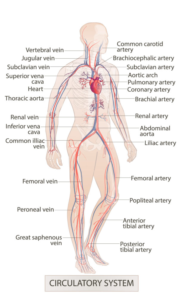
The image displayed provides a visual overview of the human circulatory system, highlighting the major arteries and veins within the body.
Starting at the top, we see the jugular vein which drains blood from the head, alongside the vertebral vein. These join the subclavian vein, which receives blood from the arm, and together they pour into the superior vena cava. This large vein carries blood into the right atrium of the heart. The heart is shown centrally in the chest cavity, with the aortic arch just above it from where the brachiocephalic artery branches off, supplying blood to the right arm and the head. Next to it, the common carotid artery ascends alongside the trachea to supply the head and neck.
To the left, we have the subclavian artery which branches from the aortic arch and follows a similar path to the subclavian vein. The pulmonary artery, which is responsible for transporting deoxygenated blood to the lungs for oxygenation, is also visible emanating from the heart. The coronary arteries, essential for supplying blood to the heart muscle, are seen branching from the base of the aorta.
Descending further, the brachial artery is seen running down the arm, whereas the thoracic aorta travels down the posterior chest wall. The renal artery and vein are responsible for blood supply to and from the kidneys. The abdominal aorta continues the descent from the thoracic aorta through the abdominal cavity, giving rise to the iliac arteries which supply the pelvic organs and lower limbs.
The common iliac vein is shown returning blood from the lower limbs, joining with its counterpart to form the inferior vena cava, which carries blood from the lower body back to the heart.
In the lower limbs, the femoral artery and vein are the principal vessels of the thigh, with the femoral artery continuing as the popliteal artery behind the knee. This artery then divides into the anterior and posterior tibial arteries to supply the lower leg and foot. The great saphenous vein, the longest vein in the body, runs medially in the leg and thigh and is commonly used for grafts in coronary artery bypass surgery. The peroneal vein runs adjacent to the fibula in the lower leg.
This intricate network of vessels is responsible for delivering oxygen and nutrients to every cell in the body and for removing carbon dioxide and metabolic wastes. The arteries, shown in red, carry oxygenated blood away from the heart, and the veins, shown in blue, carry deoxygenated blood towards the heart.
Blood Vessel Types
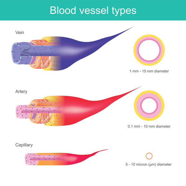
This illustration provides an educational comparison of the three primary types of blood vessels in the human circulatory system: veins, arteries, and capillaries.
At the top, we see a representation of a vein, characterized by its relatively thin wall compared to its lumen, the hollow part through which blood flows. Veins are depicted in a blue color, which traditionally indicates they carry deoxygenated blood back to the heart, except for the pulmonary veins which carry oxygenated blood. The wall layers are less distinct than in arteries and are generally less muscular and elastic. The diameter of veins can range from 1 mm to 15 mm. Valves are seen within the vein, which prevent the backflow of blood and ensure its unidirectional flow towards the heart.
Below the vein is the artery, shown in red to signify the oxygenated blood that is typically carried away from the heart to the rest of the body. Arteries have a thicker wall composed of three layers, including a thick muscular layer that allows them to withstand and regulate the high pressure of blood pumped by the heart. Their diameters can vary from 0.1 mm to 10 mm. Arteries do not have valves like veins because the pressure from the heart’s pumping action keeps blood flowing in one direction.
At the bottom, the capillary is depicted, which is the smallest type of blood vessel. Capillaries connect arteries and veins, facilitating the exchange of water, oxygen, carbon dioxide, and many other nutrients and waste substances between blood and the tissues of the body. Their walls are just one cell thick, allowing for this exchange. The diameter of capillaries is about 5 to 10 microns (micrometers), which is so small that blood cells can only pass through in a single file.
Understanding the differences in structure and function of these vessels is essential for comprehending how blood circulates throughout the body, delivering vital substances to cells and removing waste products.
Blood Circulatory System
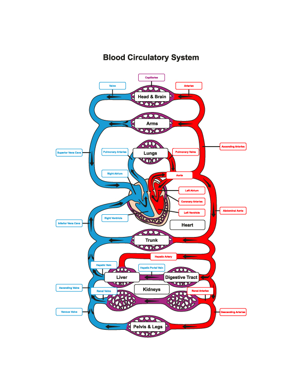
The image is a schematic representation of the human blood circulatory system, showing the major arteries, veins, and organs involved in blood circulation. It uses color-coding to differentiate between oxygenated blood (in red) and deoxygenated blood (in blue), providing a clear visual distinction between arterial and venous blood.
At the center of the diagram, the heart is depicted with its four chambers: the right atrium and ventricle, and the left atrium and ventricle. The heart acts as the pump of the circulatory system, sending deoxygenated blood to the lungs and pumping oxygenated blood to the rest of the body.
Starting from the heart, the pulmonary artery (in blue) carries deoxygenated blood to the lungs where gas exchange occurs. Oxygenated blood returns to the heart via the pulmonary veins (in red) and is then pumped out through the aorta, the largest artery in the body.
The systemic circulation is outlined, showing the major arteries that supply oxygenated blood to various parts of the body:
- The ascending aorta leads to the arterial branches that supply the head and brain, depicted at the top in red.
- The arterial branches that supply the arms are shown extending sideways from the ascending aorta.
- The descending aorta travels down the trunk, supplying oxygenated blood to the trunk and other organs such as the liver and kidneys, as well as the digestive tract.
- The descending aorta continues to supply the pelvis and legs.
The veins corresponding to these body regions are shown in blue, indicating the return of deoxygenated blood:
- The superior vena cava collects blood from the head and arms and channels it into the right atrium.
- The inferior vena cava gathers blood from the lower body regions and also empties into the right atrium.
The image also illustrates the hepatic portal vein, which carries nutrient-rich blood from the digestive tract to the liver, and the renal veins, which transport deoxygenated blood from the kidneys.
This diagram is a simplified model of the circulatory system, focusing on how blood travels from the heart to various organs and back, facilitating the distribution of oxygen, nutrients, and the removal of waste products. It encapsulates the essential pathways of both the pulmonary and systemic circulations.
Blood Microcirculation
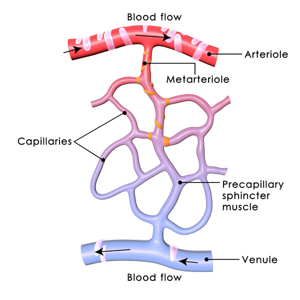
This diagram demonstrates the microcirculation within the human body, focusing on how blood passes from arterioles to venules through capillaries.
The arteriole, depicted at the top in red, is a small-diameter blood vessel in the microcirculation that extends and branches out from an artery and leads to capillaries. Arterioles are primary regulators of blood flow and pressure as they adjust to direct the flow to various parts of the body.
Emerging from the arteriole is the metarteriole, a vessel that has structural characteristics of both arterioles and capillaries. It serves as a channel through which blood can bypass the network of capillaries.
The capillaries, shown in purple, are the smallest of a body’s blood vessels and are the sites of the transfer of oxygen and other nutrients from the bloodstream to the tissues. They also collect carbon dioxide waste materials and fluids for return to the veins. They form a network that connects arterioles and venules, and their walls are only one cell thick.
Each capillary has a precapillary sphincter muscle shown as orange bands at the junction where the capillary begins. These sphincter muscles are critical in regulating blood flow into the capillary beds. They act as valves that open and close, responding to the metabolic needs of the tissues, ensuring that blood is directed to areas where it is most needed.
After the exchange of gases, nutrients, and waste products, the now deoxygenated blood enters the venules, which are depicted in blue. Venules are small blood vessels that collect blood from the capillary beds and coalesce into veins, completing the circuit back to the heart.
Arrows indicate the direction of blood flow, from the arteriole through the metarteriole and capillaries, passing through the precapillary sphincters, and eventually out through the venule. This process is essential for maintaining the physiological balance of the tissues, providing them with oxygen and nutrients, and removing metabolic waste.
Capillary Bed Anatomy
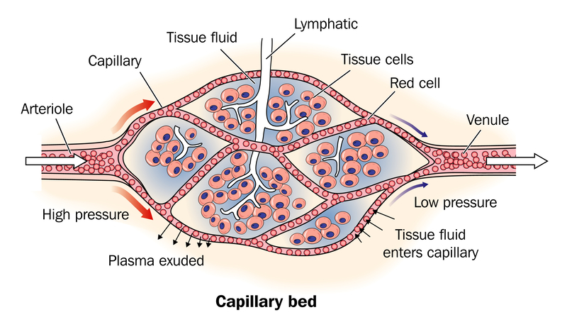
This image illustrates the capillary bed, a network of capillaries, which are the smallest blood vessels in the body’s circulatory system. Capillaries function to facilitate the exchange of materials between the blood and tissue cells.
On the left, we see an arteriole, a small branch of an artery leading into the capillary bed. The blood enters the capillary bed at high pressure, which is indicated by the red arrow and the “High pressure” label. This high pressure causes plasma, the liquid component of blood, to be exuded (or filtered out) through the thin capillary walls into the surrounding tissue. This plasma, now called tissue fluid, carries oxygen and nutrients to the tissue cells.
Within the capillary bed, the red cells, which are the erythrocytes responsible for carrying oxygen, are shown passing through the capillaries. The capillaries are surrounded by tissue fluid and tissue cells, which take up the oxygen and nutrients. The tissue cells are depicted as oval structures adjacent to the capillaries.
On the right side of the image, we have a venule, which is a small vein that the blood enters after passing through the capillary bed. By the time blood reaches the venule, it is at low pressure, as indicated by the blue arrow and “Low pressure” label. Due to the lower pressure, some of the tissue fluid is reabsorbed into the capillaries, while the excess tissue fluid is drained away by the lymphatic system, which is indicated by a thin vessel labeled “Lymphatic.”
This process of exchange between the blood and tissue cells via the capillary bed is crucial for maintaining the homeostasis of the body’s internal environment. It allows for the delivery of nutrients and oxygen to tissues and the removal of waste products. The lymphatic system also plays a vital role in returning excess tissue fluid to the circulation, helping to maintain fluid balance in the body.
Blood Flow and Capillaries: Distribution and Exchange
The circulatory system, with its intricate network of blood vessels, ensures the delivery of oxygen, nutrients, and other essential substances to every cell in the body. This section elaborates on the flow of blood and the role of capillaries in facilitating exchange processes at the cellular level.
Blood Distribution: The heart, a powerful muscular organ, pumps oxygenated blood through the arteries to supply oxygen and nutrients to the body’s tissues. Here’s an overview of the blood distribution process:
Arterial System: The arteries branch out from the heart and progressively divide into smaller vessels, forming an extensive network throughout the body. These arteries carry oxygen-rich blood away from the heart, supplying different areas, including the head, abdomen, and extremities.
Arterial Branching: As arteries extend into various regions, they undergo further branching to provide blood flow to specific organs, tissues, and cells. This branching pattern ensures that blood reaches even the tiniest capillaries, where essential exchange processes occur.
Capillaries: Capillaries are tiny, thin-walled blood vessels that connect arteries and veins. They are the site of crucial exchange processes between the bloodstream and the surrounding tissues. Let’s delve into the significance of capillaries:
Microcirculation: Capillaries form an intricate network throughout the body, allowing for microcirculation. Their small size and extensive distribution enable proximity to individual cells, ensuring efficient exchange of substances.
Oxygen and Nutrient Exchange: At the capillary level, oxygen and nutrients are exchanged between the bloodstream and the cells. Oxygen diffuses out of the capillaries into the interstitial fluid and subsequently into the cells, while waste products are carried back into the capillaries for removal.
Fluid Exchange: Capillaries also facilitate the exchange of fluid between the bloodstream and the surrounding tissues. This exchange occurs through a process called filtration, where the fluid component of blood, known as plasma, is forced out of the capillaries into the interstitial space.
Valves and Flow Regulation: Blood flow through capillaries is regulated by a series of valves that adjust the diameter of the capillaries based on the immediate needs of the cells. These valves ensure that the right amount of blood reaches the tissues, optimizing oxygen and nutrient delivery.
Red Blood Cells: While the fluid component of blood, plasma, is forced out of the capillaries, red blood cells, responsible for carrying oxygen, remain within the bloodstream. This separation allows for efficient oxygenation and exchange while preserving the structural integrity of the circulating blood.
Understanding the flow of blood and the role of capillaries in facilitating exchange processes emphasizes the intricate coordination required for optimal tissue perfusion and cellular function. The continuous circulation of blood, from the heart through the arteries, and the subsequent branching into capillaries, ensures that vital resources are delivered to cells while waste products are removed. Capillaries, with their thin walls and close proximity to cells, enable efficient exchange processes, supporting the overall health and functionality of our body’s tissues.
Conclusion: The heart’s pumping action drives the distribution of oxygenated blood through the arteries, ensuring adequate supply to various body regions. Arteries branch extensively, reaching the cellular level where capillaries take over to facilitate exchange processes. The regulated flow through capillaries allows for optimal oxygen, nutrient, and fluid exchange, while red blood cells remain within the bloodstream. Understanding the dynamic nature of blood flow and the critical role of capillaries enhances our comprehension of the intricate mechanisms that sustain cellular vitality and overall physiological processes.
Blood Vessel Types
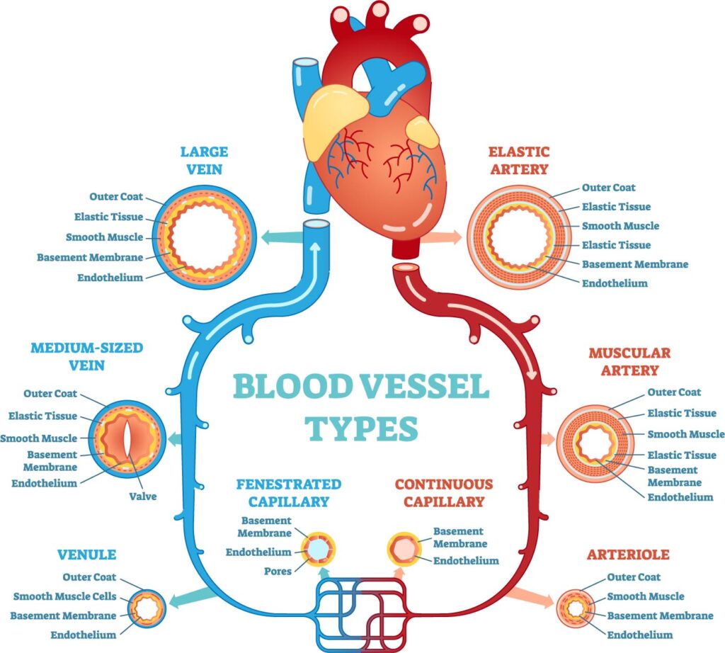
The image provides a comprehensive overview of the blood vessel types within the human circulatory system, including both the arterial and venous systems, and the microcirculation of capillaries.
Arterial versus Venous Systems: Arteries are depicted with thick walls composed of an outer coat, elastic tissue, smooth muscle, and an inner endothelial lining. The elastic arteries, like the aorta, have a prominent elastic layer allowing them to absorb the pressure created by the heart’s pumping action and help propel blood forward. Muscular arteries have a thicker layer of smooth muscle, enabling precise control of blood flow and pressure through vasoconstriction and vasodilation.
Veins, shown on the left side of the image, have thinner walls compared to arteries. They have less smooth muscle and elastic tissue, which is why they appear distensible or collapsible. Veins are equipped with valves, particularly in medium-sized veins, to prevent backflow of blood and ensure it moves toward the heart, especially against gravity.
Comparison of Venous Vessel Sizes: Large veins, such as the vena cava, have a significant outer coat and a moderate amount of elastic tissue, which allows them to transport a large volume of blood back to the heart with minimal resistance. Medium-sized veins, like the ones in the limbs, have valves that are critical for unidirectional blood flow. Venules, the smallest veins, connect capillaries to the larger veins and are primarily responsible for collecting blood from capillary beds.
Capillary Types: The image also differentiates between two types of capillaries:
- Fenestrated capillaries have pores within their endothelial lining that allow for faster exchange of water and smaller molecules between blood and tissues.
- Continuous capillaries lack pores, making them less permeable than fenestrated capillaries, and are found in most tissues such as muscle, skin, and the central nervous system.
Overall, the arterial system is characterized by thick-walled vessels that transport oxygenated blood away from the heart under high pressure, while the venous system comprises thin-walled vessels that return deoxygenated blood to the heart under lower pressure. The different sizes of the venous vessels reflect their roles: from post-capillary venules that collect blood for return, to medium-sized veins that propel it aided by muscular contractions and valves, up to large veins that carry it back to the heart. The capillary networks serve as the interface for nutrient and gas exchange between the arterial and venous systems.
Large Vein Anatomy
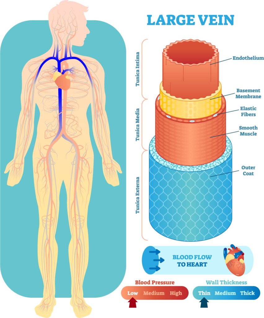
The image provides a visual representation of the circulatory system, emphasizing a large vein’s anatomy and the properties of blood flow through it.
On the left, we see an outline of a human figure with the venous system highlighted in blue, indicating the pathways by which blood returns to the heart. The veins from the lower body converge to form the inferior vena cava, while those from the head and arms form the superior vena cava, both emptying into the right atrium of the heart.
The right side of the image is a detailed cross-sectional view of a large vein, detailing its various layers:
- The innermost layer is the endothelium, a smooth lining that reduces friction and interacts with blood cells as they pass.
- Next is the basement membrane, which provides structural support for the endothelial cells.
- The elastic fibers allow the vein to stretch and accommodate varying volumes of blood.
- The smooth muscle layer enables the vein to contract and maintain blood flow towards the heart, despite the low-pressure environment.
- Lastly, the outer coat provides structural integrity and protection for the vein.
The diagram also illustrates the relationship between blood pressure and wall thickness. In large veins, blood pressure is relatively low, and the wall thickness is correspondingly thinner than in arteries, which must accommodate higher pressure. This structural difference is key to the different functions of arteries and veins: arteries must be robust to handle the surge of blood with each heartbeat, while veins require flexibility and the capacity to hold larger volumes of blood.
Additionally, the image includes an icon representing the direction of blood flow towards the heart and a comparison scale showing that the blood pressure within veins is low, and the wall thickness is relatively thin compared to arteries. This information is vital for understanding the efficiency of venous return and the mechanisms that support it, such as the venous valves not depicted in this specific illustration but crucial in preventing backflow of blood.
Medium Vein Anatomy
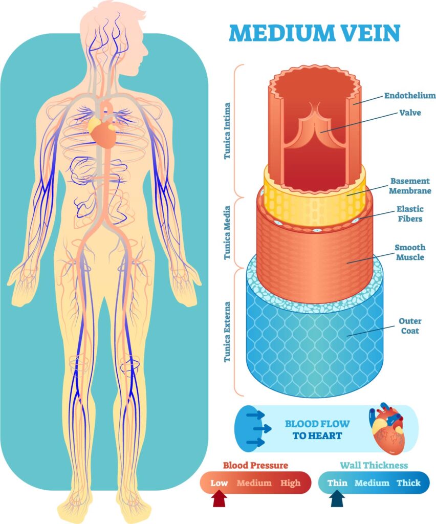
The image is divided into two parts. On the left, it depicts the human venous system with veins marked in blue, illustrating the network that returns blood to the heart. The representation shows both the superficial and deep veins throughout the body, including those in the arms, legs, and trunk.
On the right, a detailed cross-section of a medium-sized vein is highlighted. The layers from outside in are:
- The outer coat, which provides protection and structure to the vein.
- A layer of smooth muscle, which is thinner than that found in arteries, but it can contract to help move blood back towards the heart.
- Elastic fibers, which give the vein the ability to stretch and accommodate various volumes of blood.
- The basement membrane provides a supportive framework for the cellular layers.
- The endothelium is the innermost layer that lines the lumen of the vein and comes in direct contact with blood.
This cross-section also shows a valve within the vein, which is crucial in preventing backflow and ensuring unidirectional blood flow towards the heart, a feature particularly important in the extremities to counteract the effects of gravity.
The scales at the bottom indicate that the blood pressure within medium-sized veins is low to medium, and the wall thickness is moderate, thicker than capillaries but thinner than arteries. The venous system operates under lower pressure compared to the arterial system, which is reflected in the wall structure; veins have a larger lumen and thinner walls relative to their diameter.
The illustration underscores the functional design of the venous system, optimized for the low-pressure, high-volume transport of blood, and the role of venous valves in maintaining efficient circulation.
Venule Anatomy
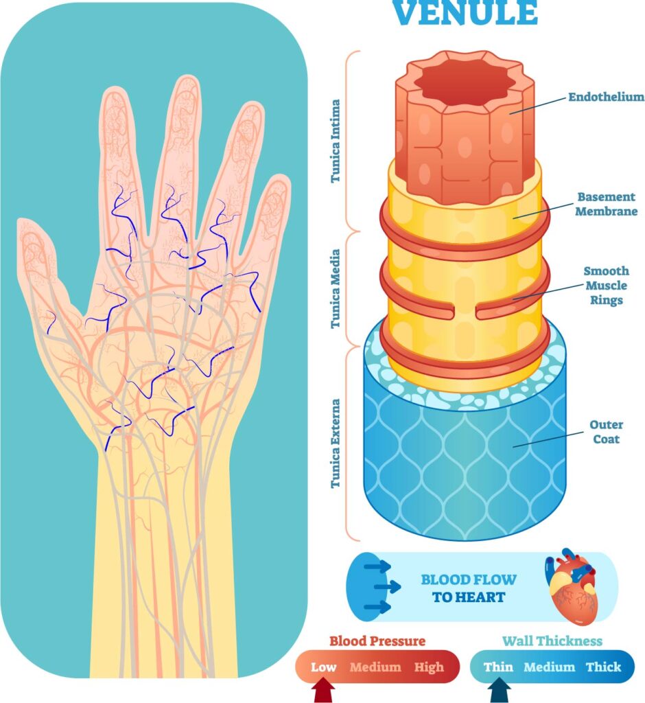
The image provides a detailed view of the venous system in the human hand and a cross-sectional diagram of a venule, which is a very small vein.
On the left, we can see a network of veins in the hand, depicted in blue, which are responsible for draining deoxygenated blood and returning it to the heart. These superficial veins are visible through the skin in many individuals.
On the right, the cross-section of a venule highlights its structure. The layers from the outside in are as follows:
- The outer coat, which is the external layer of the venule providing structural support.
- Smooth muscle rings are present but much less developed than in larger veins or arteries. These muscle cells can adjust the diameter of the venules, but their role is limited given the low pressure in these vessels.
- The basement membrane is a thin layer that supports the endothelial cells.
- The endothelium is the innermost layer of the blood vessel and comes in direct contact with the blood flowing through it.
The diagram also indicates that blood flow in venules is directed toward the heart and that the blood pressure in venules is low, as reflected by the scale indicating low blood pressure and thin wall thickness. This is consistent with the role of venules, which are to collect blood from capillary beds and begin the process of returning it to larger veins and eventually back to the heart. The structure of the venules reflects their function in the microcirculation, where the exchange of nutrients and wastes occurs, and they are not subjected to the high pressures found in the arterial system.
Fenestrated Capillary Anatomy
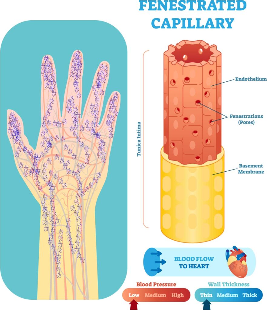
The image showcases a detailed illustration of the capillary network in the human hand and specifically highlights the structure of a fenestrated capillary.
On the left side, a dense network of capillaries is visible throughout the hand and fingers, depicted in a purplish hue. Capillaries are the smallest blood vessels in the body and are essential for the exchange of oxygen, nutrients, and waste products between the blood and the tissues.
On the right side, there’s an enlarged view of a fenestrated capillary, which is characterized by the presence of small pores, or fenestrations, within its endothelial lining. These fenestrations are represented as red dots. This type of capillary is typically found in organs that require rapid exchange of substances, such as the kidneys, intestines, and endocrine glands. The fenestrations allow for increased permeability, facilitating the transfer of small molecules.
The structure of a fenestrated capillary includes:
- The endothelium, which is the single layer of cells that line the interior surface of the capillary.
- Fenestrations, which are the pores in the endothelial cells that allow for passage of certain substances.
- The basement membrane, a thin layer of extracellular matrix that provides structural support to the endothelial cells.
The bottom of the image provides indicators for blood pressure and wall thickness. It shows that blood pressure within capillaries is low, and the wall thickness is thin, reflecting the need for capillaries to facilitate the exchange of substances without resistance. The direction of blood flow is indicated as moving towards the heart, as part of the venous return system after the exchange of gases and nutrients has occurred in the capillaries.
Anatomical Terms and Definitions
| Term | Definition |
|---|---|
| Aorta | The largest artery in the body, carrying oxygenated blood from the left ventricle to the rest of the body. |
| Arteriole | A small-diameter blood vessel in the microcirculation that extends from an artery and leads to capillaries. |
| Artery | A blood vessel that carries oxygenated blood away from the heart to the body's tissues. |
| Brachial Artery | An artery running down the arm. |
| Brachiocephalic Artery | An artery supplying blood to the right arm and head. |
| Capillary | The smallest blood vessels, connecting arteries and veins, facilitating the exchange of water, oxygen, carbon dioxide, and other nutrients and waste substances. |
| Common Carotid Artery | An artery ascending alongside the trachea to supply the head and neck. |
| Common Iliac Vein | A vein returning blood from the lower limbs, joining to form the inferior vena cava. |
| Coronary Arteries | Arteries essential for supplying blood to the heart muscle. |
| Femoral Artery | The principal artery of the thigh. |
| Femoral Vein | The principal vein of the thigh. |
| Great Saphenous Vein | The longest vein in the body, running medially in the leg and thigh. |
| Hepatic Portal Vein | A vein carrying nutrient-rich blood from the digestive tract to the liver. |
| Inferior Vena Cava | A large vein carrying blood from the lower body back to the heart. |
| Jugular Vein | A vein that drains blood from the head. |
| Metarteriole | A vessel with characteristics of both arterioles and capillaries, serving as a channel for blood to bypass capillary networks. |
| Peroneal Vein | A vein running adjacent to the fibula in the lower leg. |
| Popliteal Artery | The artery continuing from the femoral artery behind the knee. |
| Precapillary Sphincter | A muscle acting as a valve at the junction where a capillary begins, regulating blood flow into capillary beds. |
| Pulmonary Artery | An artery transporting deoxygenated blood to the lungs for oxygenation. |
| Pulmonary Veins | Veins that carry oxygenated blood from the lungs to the heart. |
| Renal Artery | An artery responsible for supplying blood to the kidneys. |
| Renal Vein | A vein responsible for carrying blood from the kidneys. |
| Subclavian Artery | An artery branching from the aortic arch, following a path similar to the subclavian vein. |
| Subclavian Vein | A vein receiving blood from the arm, joining the jugular and vertebral veins to pour into the superior vena cava. |
| Superior Vena Cava | A large vein carrying blood into the right atrium of the heart from the head, arms, and upper body. |
| Thoracic Aorta | A part of the aorta traveling down the posterior chest wall. |
| Trachea | The windpipe; a tube connecting the pharynx and larynx to the lungs, providing air flow. |
