
Table of Contents
Macular Degeneration
The image illustrates the anatomy of the eye along with the changes occurring in macular degeneration, a common eye condition that affects the macula, the central part of the retina responsible for clear vision in your direct line of sight.
In the top part of the image, a cross-sectional diagram of the eye shows the main anatomical features. The outermost layer is the sclera, the white part of the eye. Below this is the choroid, which contains blood vessels that supply the eye with nutrients. The retina is the innermost layer where light-sensitive cells convert light into electrical signals to be processed by the brain.
The macula is highlighted, indicating its importance in providing sharp, central vision. The optic disc, also known as the blind spot, is where the optic nerve exits the eye; there are no photoreceptors here, so it is insensitive to light. The optic nerve carries visual information from the retina to the brain.
Below the main diagram are three smaller images that compare the normal macula with two types of macular degeneration:
Normal: The macula appears uniform in color, indicating a healthy distribution of photoreceptor cells and absence of any abnormalities.
“Wet” Macular Degeneration: This type is characterized by the presence of abnormal blood vessels that leak fluid or blood into the region of the macula, leading to blurred vision or blind spots. The diagram shows a blob-like distortion representing bleeding or fluid leakage.
“Dry” Macular Degeneration: This more common type is depicted with yellowish spots known as drusen, which are deposits that form under the retina. Over time, these can lead to a gradual breakdown of the cells in the macula, resulting in blurred or diminished central vision.
The comparison between the normal macula and the affected ones in wet and dry macular degeneration provides a visual explanation of how this condition can impact vision. It underscores the importance of the macula in eye health and the potential severity of macular degeneration as a disease.
Glaucoma vs. Normal Eye
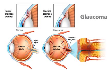
The image offers an educational comparison of a normal eye and one affected by glaucoma, highlighting the mechanisms behind this eye condition.
On the left, the top and bottom illustrations depict a normal eye. The eye has a clear fluid called aqueous humor that flows in and out of the anterior chamber, providing nutrients and maintaining intraocular pressure. The fluid drains through the trabecular meshwork, located in the angle where the iris and cornea meet. The illustration shows an unobstructed flow of aqueous humor, which is crucial for maintaining healthy eye pressure.
In the middle, the eye with glaucoma is shown. The top image indicates a blocked drainage channel, which prevents the aqueous humor from flowing out of the eye properly. As a result, as seen in the bottom image, there’s an increase in intraocular pressure, indicated by the red arrows pushing outwards. This pressure buildup can damage the optic nerve, which is responsible for transmitting visual information from the retina to the brain.
On the right, the progression of glaucoma is further illustrated by showing changes in the optic nerve due to this increased pressure. The optic nerve appears to be compressed and damaged, which can lead to vision loss. The change in coloration and the disorganized appearance of the optic nerve fibers signify the severity of damage that glaucoma can cause, potentially leading to blindness if untreated.
This visual comparison serves as a powerful tool for understanding glaucoma’s impact on eye anatomy and the importance of proper aqueous humor drainage for ocular health.
Various Conditions Affecting the Fundus
The image provides a comparative visual overview of various conditions affecting the fundus, which is the interior surface of the eye opposite the lens, including the retina, optic disc, macula, fovea, and posterior pole.
The first illustration depicts a healthy fundus with a clear, bright appearance of the retina, unobstructed and well-defined blood vessels, and a sharp optic disc where the optic nerve enters the retina.
The second illustration shows retinitis, characterized by the presence of white infiltrates and a hazy appearance, suggesting inflammation of the retina. The blood vessels appear engorged or sheathed, and there may be a slight blurring around the optic disc.
The third illustration presents retinal detachment, where the retina has pulled away from the back of the eye, indicated by the elevated sections of the retinal tissue. This detachment disrupts the normal architecture and placement of the retina.
The fourth illustration depicts an epiretinal membrane, where a thin, translucent layer has formed on the surface of the retina, causing a wrinkling or distortion of the retinal tissue that can impact vision.
The final illustration shows retinal vascular thrombosis, which is the blockage of a retinal vein, leading to hemorrhages seen as red spots and blotches, and may also cause swelling and white patches where the retina is deprived of oxygen.
Each condition illustrated can lead to vision changes or loss and requires medical attention. These depictions are valuable for educational purposes, illustrating how diseases of the fundus can alter the appearance of the eye’s interior and affect ocular health.
Diagram of the Vitreous Floaters
The image is an explanatory diagram contrasting the effects of vitreous floaters on vision compared to a normal eye.
In the top half of the diagram, a side cross-section of a normal eye is shown, indicating how light passes through the eyeball without obstruction, resulting in clear vision. This is represented by a circular field of vision that is completely unobstructed.
The bottom half of the diagram illustrates an eye affected by vitreous floaters. Here, the cross-section shows the interior of the eyeball containing various shapes labeled as “floaters.” These floaters represent debris within the vitreous humor—the clear gel that fills the space between the lens and the retina. The text explains that the passage of light through the eyeball is hindered by the presence of the vitreous floaters, which generate shadows on the retinal surface. This is visually represented by a circular field of vision with multiple shadowy shapes corresponding to the floaters, indicating disturbed vision.
Vitreous floaters are often seen as small dark spots, lines, or cobwebs in a person’s field of vision, particularly when looking at a plain, bright background like a blue sky or a white wall. They are usually harmless and are a common part of the eye’s aging process, but a sudden increase in floaters can also indicate more serious eye conditions, such as retinal detachment.
This visual tool effectively communicates how vitreous floaters can interfere with normal vision by casting shadows on the retina, leading to the perception of floating spots or threads in the visual field.
Illustration of Conjunctiva (Pink Eye)
This image is an annotated illustration of the human eye, specifically focusing on the external anatomy and the structures associated with the conjunctiva.
The key parts labeled in this diagram are:
- Ciliae: Commonly known as eyelashes, they protect the eye from debris.
- Pupilla (Pupil): The central opening of the iris that allows light to enter the eye.
- Iris: The colored part of the eye that controls the size of the pupil.
- Angulus oculi lateralis (Lateral canthus): The outer corner where the upper and lower eyelids meet.
- Tunica conjunctiva bulbi: The conjunctiva as it covers the sclera (the white of the eye).
- Limbus posterior palpebrae: The border of the eyelid near the conjunctival fornix.
- Fornix conjunctivae inferior (Inferior conjunctival fornix): The space between the palpebral conjunctiva and the bulbar conjunctiva on the lower eyelid.
- Palpebra superior (Upper eyelid): Protects the anterior part of the eye and contains the superior conjunctival fornix.
- Sulcus sclerae (Scleral sulcus): A groove or space which can refer to the area between the cornea and the sclera.
- Plica semilunaris conjunctivae: A small fold of conjunctiva that allows for movements of the eyeball.
- Caruncula lacrimalis (Lacrimal caruncle): A small, pink, globular nodule at the inner corner of the eye that contains glands producing a portion of the tear film.
- Angulus oculi medialis (Medial canthus): The inner corner where the upper and lower eyelids meet.
- Papilla lacrimalis: This term typically refers to a small elevation or bump in the conjunctiva where the lacrimal duct opens, though it is not conventionally at the inner corner.
- Limbus anterior: The junction between the cornea and the sclera.
- Tunica conjunctiva (Conjunctiva): A clear mucous membrane that lines the inside of the eyelids (palpebral conjunctiva) and covers the sclera (bulbar conjunctiva).
- Palpebra inferior (Lower eyelid): Protects the anterior part of the eye and contains the inferior conjunctival fornix.
The illustration provides a detailed view of the eye’s external and accessory structures, emphasizing the conjunctival elements which play a role in eye lubrication and protection.
Common Corneal Conditions
The image provides a comparative visualization of common corneal conditions alongside a depiction of a normal cornea.
Normal: The first illustration shows a standard, healthy cornea with a smooth, dome-like shape that is essential for proper vision. The cornea’s transparency and curvature are crucial for focusing light onto the retina.
Keratoconus: The second illustration depicts keratoconus, a condition where the cornea thins and begins to bulge into a cone-like shape, distorting vision. This irregular curvature disrupts the focus of light entering the eye, leading to visual impairment.
Bullous Keratopathy: The third illustration shows bullous keratopathy, characterized by fluid blisters (bullae) on the cornea, which result from endothelial cell damage and dysfunction. The endothelium’s primary role is to pump excess fluid out of the cornea, and its failure leads to corneal swelling and blistering, causing pain and vision distortion.
Corneal Scarring: The fourth illustration represents corneal scarring, where the transparent tissue has become opaque due to injury, infection, or inflammation. Scarring can cause the cornea to lose its clarity and smooth surface, leading to visual disturbances.
The diagram effectively educates on how various corneal conditions can alter the structure of the cornea and the potential impact on vision. Each condition depicted requires medical attention, and the treatments vary from corrective lenses in the early stages of keratoconus to corneal transplantation in severe cases of scarring or keratopathy.
Anatomy of Retinal Detachment
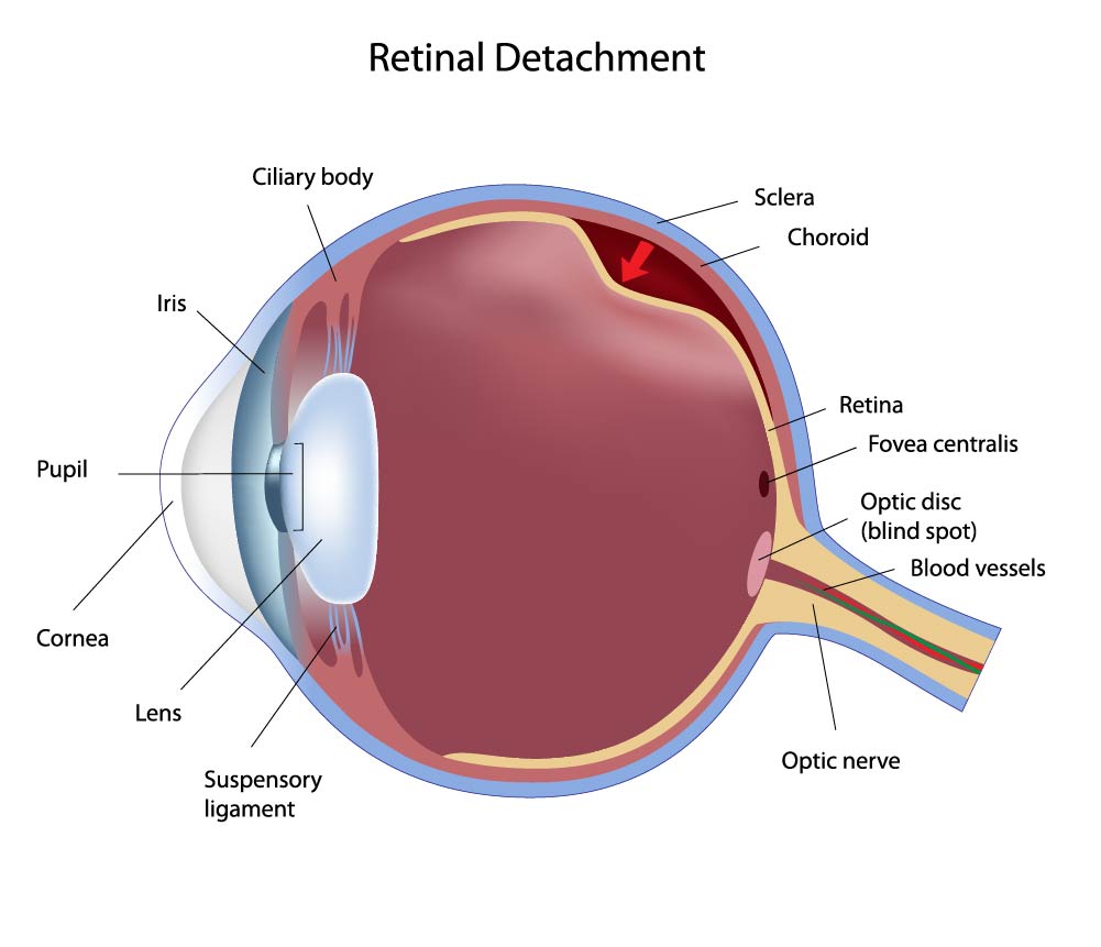
The image provides a cross-sectional illustration of the human eye, highlighting the condition known as retinal detachment.
The key structures of the eye are labeled, including:
- Ciliary Body: The part of the eye that releases the aqueous humor and contains the ciliary muscle, which controls the shape of the lens for focusing.
- Sclera: The white, outer layer of the eyeball that provides protection and maintains the shape of the eye.
- Choroid: A layer rich in blood vessels located between the retina and the sclera, supplying nutrients to the eye.
- Retina: The innermost layer of the eye, which contains light-sensitive cells that convert light into neural signals.
- Fovea Centralis: The small pit in the macula that provides the clearest vision of all.
- Optic Disc (Blind Spot): The location on the retina where the optic nerve fibers exit the eye; this area does not contain any photoreceptor cells and is thus insensitive to light.
- Blood Vessels: These provide essential nutrients and oxygen to the retinal cells.
- Optic Nerve: The nerve that transmits visual information from the retina to the brain.
- Iris: The colored part of the eye that controls the amount of light entering the eye by adjusting the size of the pupil.
- Pupil: The opening in the center of the iris that allows light to enter the eye.
- Cornea: The clear, dome-shaped surface that covers the front of the eye and contributes to the eye’s focusing power.
- Lens: The transparent structure inside the eye that focuses light rays onto the retina.
- Suspensory Ligament: The series of fibers that connect the ciliary body to the lens, helping to hold it in place.
The illustration also shows an arrow indicating the area where the retina has detached. In a normal eye, the retina is attached to the choroid layer, but in the case of retinal detachment, the retina peels away, leading to a potential loss of vision if not promptly treated. The area of detachment can be identified by the separation between the retina and the back wall of the eye, which can cause symptoms like an increase in floaters, flashes of light, or a shadow over the visual field.
Anatomy of Different Eye Conditions
The image illustrates the differences between normal vision, hyperopia (farsightedness), myopia (nearsightedness), and how these conditions can be corrected using lenses.
The top left illustration shows Normal Vision, where light rays entering the eye are focused precisely on the retina, resulting in a clear image.
The middle left illustration depicts Hyperopia, a condition where the eye is too short, causing light rays to focus behind the retina. This often leads to difficulty focusing on close objects, while distant objects may be clear.
The middle right illustration shows Hyperopia Corrected with the use of a converging lens, typically a biconvex lens. This lens bends the light rays so they converge more by the time they reach the retina, correcting the focal point to fall on the retina for clear vision.
The bottom left illustration shows Myopia, a condition where the eye is too long, causing light rays to focus in front of the retina. This results in clear vision for close objects but blurred vision for objects that are far away.
The bottom right illustration demonstrates Myopia Corrected with the use of a diverging lens, typically a biconcave lens. This lens spreads out the light rays slightly before they enter the eye, ensuring they focus directly on the retina rather than in front of it.
The term “Focal Plane” refers to the location where the light rays converge to form a clear image. In a normal eye, this is directly on the retina, but in hyperopic and myopic eyes, the focal plane is misplaced, leading to blurred vision which requires optical correction for clarity. The image serves as an educational diagram to explain common refractive errors and their corrections with lenses.
Anatomical Terms and Definitions
| Term | Definition | |||
|---|---|---|---|---|
| Arm (brachium) | The part of the upper limb located between the shoulder and the elbow. | |||
| Elbow joint | The joint connecting the arm and the forearm, comprising the articulation between the humerus and the two forearm bones, ulna, and radius. | |||
| Brachial artery | The major blood vessel of the upper arm that continues from the axillary artery to supply blood to the arm. | |||
| Radius | One of the two bones of the forearm, extending from the lateral side of the elbow to the thumb side of the wrist. | |||
| Ulna | The longer and larger of the two bones of the forearm, placed on the side opposite to the thumb. | |||
| Cornea | The transparent front part of the eye that covers the iris, pupil, and anterior chamber. | |||
| Iris | The colored part of the eye, controlling the size of the pupil and thus the amount of light reaching the retina. | |||
| Lens | The transparent structure inside the eye that focuses light rays onto the retina. | |||
| Retina | The light-sensitive layer of tissue at the back of the inner eye, which translates light into neural signals for vision. | |||
| Fovea centralis | A small central pit in the macula of the retina, composed of closely packed cones that is responsible for sharp central vision. | |||
| Optic nerve | The nerve that transmits visual information from the retina to the brain. | |||
| Macula | An area near the center of the retina that is responsible for central vision and high visual acuity. | |||
| Vitreous humor | The clear gel that fills the space between the lens and the retina in the eyeball. | |||
| Photoreceptors | Cells in the retina that respond to light; they include rods, which are responsible for vision in low light, and cones, for color vision and detail. | |||
| Sclera | The white outer layer of the eyeball that provides protection and form. | |||
| Choroid | The vascular layer of the eye between the retina and the sclera, supplying nutrients and oxygen to the eye. | |||
| Anterior chamber | The fluid-filled space inside the eye between the cornea and the iris. | |||
| Posterior chamber | The fluid-filled space directly behind the iris but in front of the lens. | |||
| Conjunctiva | A clear mucous membrane that lines the inside of the eyelids and covers the sclera. | |||
| Lacrimal gland | The gland responsible for producing tears; it is situated in the upper outer region of the orbit, above the eyeball. | |||
| Aqueous humor | The clear, watery fluid that fills the space between the cornea and the iris. | |||
| Myopia | A common vision condition also known as nearsightedness, where distant objects appear blurred. | |||
| Hyperopia | A vision condition also known as farsightedness, where close objects appear blurred. | |||
| Astigmatism | A common imperfection in the curvature of the eye's cornea or lens, causing blurred or distorted vision. | |||
| Retinal detachment | A serious condition where the retina peels away from its underlying layer of support tissue. | |||
| Macular degeneration | An eye disease that may result in blurred or no vision in the center of the visual field due to damage to the macula. | |||
| Glaucoma | A group of eye conditions that damage the optic nerve, often associated with increased pressure in the eye. | |||
| Cataracts | A condition characterized by clouding of the lens in the eye, leading to a decrease in vision. | |||
| Conjunctivitis | An eye condition diagnosed by irritation or infection of the conjunctiva, often resulting in redness and swelling. | |||
| Keratoconus | A progressive eye disease where the normally round cornea thins and begins to bulge into a cone-like shape. | |||
| Bullous Keratopathy | A condition causing swelling and blistering of the cornea due to endothelial cell dysfunction. | |||
| Corneal scarring | Opacity or scarring of the cornea often resulting from injury, infection, or inflammation. | |||
| Extraocular muscles | The muscles that control the movements of the eye and the elevation of the eyelid. | |||
| Vitreous floaters | Small flecks or threads of collagen that float in the vitreous humor and cast shadows on the retina, often seen as floaters by the individual. |
