
Table of Contents
Magnification of the Retina Anatomy
The image presents a simplified view of the eye’s anatomy alongside a magnified section of the retina, offering insight into the cellular composition and arrangement within the retinal layer.
In the larger image on the left, the entire structure of the eye is encapsulated, with the retina marked along the back inner surface. The retina is the light-sensitive layer of tissue at the back of the eye, which is essential for vision as it receives light and converts it into neural signals for the brain to process.
The smaller image on the right zooms into the retinal layer, depicting the precise organization and types of cells that compose the retina. Starting from the bottom, the layers are as follows:
Rod Cells: These are long, cylindrical cells responsible for vision at low light levels. They are highly sensitive to light and enable us to see in shades of gray in dim conditions.
Cone Cells: These are shorter, tapering cells responsible for color vision and visual acuity. They function best in bright light and enable us to see fine details and rich color variations.
Bipolar Cells: Positioned between the photoreceptors (rod and cone cells) and the ganglion cells, these cells act as direct relay cells that transmit signals from the rods and cones to the ganglion cells.
Ganglion Cells: These cells receive signals from the bipolar cells. Their axons converge to form the optic nerve, which transmits visual information to the brain.
Retinal Pigment Epithelium (RPE): This is a layer of pigmented cells adjacent to the photoreceptors. The RPE is essential for the maintenance and function of the photoreceptor cells. It helps absorb excess light to prevent light scattering, recycles visual pigments, and supplies nutrients to the retina.
The illustration provides a clear depiction of the retinal cells and their connectivity, emphasizing the complex processing that occurs within the eye before visual information is even sent to the brain. The arrangement of these cells in layers reflects the sequential processing and filtering of visual data, from the initial photodetection by rods and cones to the complex signal integration and transmission by bipolar and ganglion cells.
Schematic Representation of the Retina
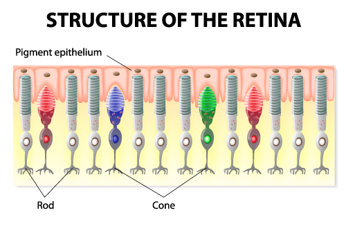
The image is a schematic representation of the structure of the retina, showcasing the two primary types of photoreceptor cells: rods and cones.
At the top layer of the diagram, the pigment epithelium is shown. This layer of the retina is vital for the health of the photoreceptor cells, as it absorbs excess light, thereby preventing light scatter that can affect visual acuity. It also plays a role in the recycling of visual pigments after phototransduction.
Below the pigment epithelium are the rod cells. Rods are illustrated here in red and are more numerous in the human retina than cones. They are highly sensitive to light and enable vision in low-light conditions but do not support color vision. Rods are responsible for peripheral vision and motion detection.
Next to the rods are the cone cells, depicted in various colors (blue, green, and red) to represent the three types of cones sensitive to different wavelengths of light: S-cones (blue light), M-cones (green light), and L-cones (red light). Cones are responsible for high-acuity central vision and color vision and are densely packed in the macula, particularly in the fovea—the region of the retina that provides the sharpest vision.
Each photoreceptor cell has an outer segment where phototransduction—conversion of light into electrical signals—takes place, an inner segment containing the cell’s organelles, a cell body with the nucleus, and a synaptic terminal that communicates with other neurons in the retina.
The arrangement of rods and cones in the image illustrates their distribution within the retina, which is a crucial factor in how we perceive light and color in various lighting conditions. This diagram is typically used for educational purposes to explain the complex process of vision and the specialized functions of different photoreceptor cells in the retina.
Photoreceptor Cells in the Retina
The image depicts two types of photoreceptor cells in the retina of the human eye: rod cells and cone cells. These cells are crucial for vision, converting light into electrical signals that the brain can interpret.
On the left, the rod cell is shown with its elongated, cylindrical shape, which is optimized for detecting low levels of light, making them crucial for night vision. The outer segment contains stacked discs, which are rich in the photopigment rhodopsin. This segment is where the initial photochemical reactions occur when light is absorbed.
Below the outer segment is the inner segment of the rod cell, which contains mitochondria. These organelles are the powerhouses of the cell, providing the energy required for the phototransduction process, which is the conversion of light into electrical signals.
The cell body contains the nucleus, which houses the cell’s genetic material. At the base of the rod cell is the synapse, where the photoreceptor cell communicates with the next neuron in the visual pathway by releasing neurotransmitters.
On the right, the cone cell is displayed with a conical outer segment. Cone cells are responsible for color vision and function best in bright light. Similar to rod cells, the outer segment of cone cells contains photopigments; however, they have different types of opsins that respond to different wavelengths of light, allowing for the perception of color.
Both rod and cone cells have a connecting cilium, which is a slender structure that links the inner and outer segments, facilitating the transport of molecules between these two compartments.
The nucleus of the cone cell is also located within the cell body, and it connects to the synapse at the bottom, similar to the rod cell, for neurotransmission.
This image succinctly illustrates the structural differences between rods and cones, correlating to their different functions in vision. Rods are more numerous and sensitive to light, contributing to vision in low-light conditions, while cones are less sensitive but essential for color vision and visual acuity.
Detailed Layers of the Retina
The image is a detailed representation of human eye anatomy, with a primary focus on the sectional view of the eye and a magnified inset detailing the layers of the retina.
The main illustration shows a cross-section of the eye, with labels for the sclera, the tough outer layer that forms the white of the eye, and the cornea, the clear front surface that covers the iris, pupil, and anterior chamber. The anterior chamber lies between the cornea and the iris, and is filled with aqueous humor, which nourishes the eye and maintains pressure.
The iris, the colored part of the eye, regulates the size of the pupil, the opening that controls the amount of light entering the eye. Behind the iris is the lens, which focuses light onto the retina. The ciliary body, attached to the lens, contains the muscle that changes the shape of the lens for focusing. The vitreous body, a clear gel filling the large space behind the lens, maintains the eye’s shape and supports the retina.
The retina is the light-sensitive layer at the back of the eye that converts light into electrical signals. The macula is the central part of the retina, responsible for detailed vision, and within the macula is the fovea, which provides the sharpest vision. The choroid beneath the retina is a layer containing blood vessels that supply the retina with nutrients and oxygen.
The optic nerve is the pathway that transmits visual signals from the retina to the brain, and the optic disc is the point on the retina where the optic nerve fibers exit the eye, also known as the blind spot because it lacks photoreceptors.
The inset magnifies the retinal layers, starting from the outermost part of the retina adjacent to the vitreous body. The layers are as follows:
- Internal Limiting Membrane: the innermost surface of the retina, facing the vitreous body.
- Nerve Fiber Layer: contains the axons of the ganglion cells that form the optic nerve.
- Ganglion Cell Layer: contains the cell bodies of ganglion cells.
- Inner Plexiform Layer: location of synapses between bipolar cell axons and ganglion cell dendrites.
- Inner Nuclear Layer: contains the nuclei and cell bodies of bipolar, horizontal, and amacrine cells.
- Outer Plexiform Layer: site of synapses between photoreceptor cells and bipolar cells.
- Outer Nuclear Layer: contains the cell bodies of the photoreceptors, rods, and cones.
- Outer Segments of Photoreceptors: the light-sensitive parts of rods and cones where phototransduction occurs.
- RPE (Retinal Pigment Epithelium): a layer of pigmented cells that nourishes the retinal visual cells and is critical for the regeneration of photoreceptor outer segments.
The illustration also denotes Bruch’s membrane, which separates the choroid from the RPE, and the choroidal vessels, which supply blood to the outer retina.
This comprehensive image serves as an excellent visual aid for understanding the complex structure of the eye and the retina’s specialized layers, crucial for the perception of visual images.
Anatomical Terms and Definitions
| Term | Definition | |||
|---|---|---|---|---|
| Arm (brachium) | The part of the upper limb located between the shoulder and the elbow. | |||
| Elbow joint | The joint connecting the arm and the forearm, comprising the articulation between the humerus and the two forearm bones, ulna, and radius. | |||
| Brachial artery | The major blood vessel of the upper arm that continues from the axillary artery to supply blood to the arm. | |||
| Radius | One of the two bones of the forearm, extending from the lateral side of the elbow to the thumb side of the wrist. | |||
| Ulna | The longer and larger of the two bones of the forearm, placed on the side opposite to the thumb. | |||
| Cornea | The transparent front part of the eye that covers the iris, pupil, and anterior chamber. | |||
| Iris | The colored part of the eye, controlling the size of the pupil and thus the amount of light reaching the retina. | |||
| Lens | The transparent structure inside the eye that focuses light rays onto the retina. | |||
| Retina | The light-sensitive layer of tissue at the back of the inner eye, which translates light into neural signals for vision. | |||
| Fovea centralis | A small central pit in the macula of the retina, composed of closely packed cones that is responsible for sharp central vision. | |||
| Optic nerve | The nerve that transmits visual information from the retina to the brain. | |||
| Macula | An area near the center of the retina that is responsible for central vision and high visual acuity. | |||
| Vitreous humor | The clear gel that fills the space between the lens and the retina in the eyeball. | |||
| Photoreceptors | Cells in the retina that respond to light; they include rods, which are responsible for vision in low light, and cones, for color vision and detail. | |||
| Sclera | The white outer layer of the eyeball that provides protection and form. | |||
| Choroid | The vascular layer of the eye between the retina and the sclera, supplying nutrients and oxygen to the eye. | |||
| Anterior chamber | The fluid-filled space inside the eye between the cornea and the iris. | |||
| Posterior chamber | The fluid-filled space directly behind the iris but in front of the lens. | |||
| Conjunctiva | A clear mucous membrane that lines the inside of the eyelids and covers the sclera. | |||
| Lacrimal gland | The gland responsible for producing tears; it is situated in the upper outer region of the orbit, above the eyeball. | |||
| Aqueous humor | The clear, watery fluid that fills the space between the cornea and the iris. | |||
| Myopia | A common vision condition also known as nearsightedness, where distant objects appear blurred. | |||
| Hyperopia | A vision condition also known as farsightedness, where close objects appear blurred. | |||
| Astigmatism | A common imperfection in the curvature of the eye's cornea or lens, causing blurred or distorted vision. | |||
| Retinal detachment | A serious condition where the retina peels away from its underlying layer of support tissue. | |||
| Macular degeneration | An eye disease that may result in blurred or no vision in the center of the visual field due to damage to the macula. | |||
| Glaucoma | A group of eye conditions that damage the optic nerve, often associated with increased pressure in the eye. | |||
| Cataracts | A condition characterized by clouding of the lens in the eye, leading to a decrease in vision. | |||
| Conjunctivitis | An eye condition diagnosed by irritation or infection of the conjunctiva, often resulting in redness and swelling. | |||
| Keratoconus | A progressive eye disease where the normally round cornea thins and begins to bulge into a cone-like shape. | |||
| Bullous Keratopathy | A condition causing swelling and blistering of the cornea due to endothelial cell dysfunction. | |||
| Corneal scarring | Opacity or scarring of the cornea often resulting from injury, infection, or inflammation. | |||
| Extraocular muscles | The muscles that control the movements of the eye and the elevation of the eyelid. | |||
| Vitreous floaters | Small flecks or threads of collagen that float in the vitreous humor and cast shadows on the retina, often seen as floaters by the individual. |
