
Table of Contents
Basic Eye Anatomy
The human body is navigated anatomically using three principal planes—imaginary lines that divide the body into distinct sections. The Sagittal Plane vertically bisects the body into right and left halves, crucial for describing movements like flexion and extension. The Coronal Plane, or frontal plane, divides the body into anterior and posterior sections, important for adduction and abduction movements. Lastly, the Transverse Plane, cutting horizontally, separates the body into superior and inferior parts, and is associated with rotational actions. These planes are indispensable in the clinical setting for precise anatomical discussions, imaging techniques, and in designing rehabilitative and athletic training programs.
The image provided is a labeled diagram of the human eye, showcasing the anatomy and various structures integral to its function.
At the front of the eye, the cornea is shown as a transparent dome-like structure. It functions as the eye’s outermost lens, controlling and focusing the entry of light. Below the cornea, the anterior chamber is a fluid-filled space that provides nutrients to the eye and maintains intraocular pressure.
The iris, the colored part of the eye, controls the diameter and size of the pupil and thus the amount of light that enters the eye. The pupil is the opening in the center of the iris through which light passes to the lens. The lens is a transparent, biconvex structure that further focuses light onto the retina. The suspensory ligaments, also known as zonules, are tiny fibers that connect the ciliary body and muscle to the lens, enabling the lens to change shape for focusing, a process known as accommodation.
Behind the lens is the posterior chamber, which, like the anterior chamber, is filled with aqueous humor that nourishes the eye and carries away waste.
The large space in the posterior part of the eye is the vitreous chamber, filled with a gel-like substance called the vitreous humor, which helps maintain the spherical shape of the eye and also acts as a shock absorber.
The innermost layer lining the back of the eye is the retina, which contains the light-sensitive cells that convert light into electrical signals. The optic nerve and retinal blood vessels are visible entering the back of the eye, indicating the flow of visual information to the brain and the blood supply to the retina, respectively.
The macula lutea is a small central area of the retina that is responsible for high-acuity vision, while the fovea centralis, located within the macula, is the point of sharpest vision.
Surrounding the retina is the choroid, a layer rich in blood vessels that supply oxygen and nutrients to the retina.
The sclera is the white, fibrous outer layer of the eye that maintains the shape of the eye and offers protection.
Lastly, the extrinsic muscles of the eye, such as the lateral and medial rectus muscles, control the movements of the eyeball, enabling it to look in different directions.
The illustration effectively summarizes the complex structure and organization of the eye, a vital organ for vision.
Detailed Cross Section of the Eye
This image displays a detailed cross-sectional view of the cornea, which is the transparent front part of the eye that covers the iris, pupil, and anterior chamber. The cornea, with its curved shape and clear structure, functions like a window that controls and focuses the entry of light into the eye.
The outermost layer, appearing as a thin, clear strip on the surface, is the epithelium. This layer serves as a barrier to protect the eye from dust, debris, and bacteria, and contains cells that have the ability to regenerate quickly, aiding in the rapid healing of superficial injuries.
Beneath the epithelium is Bowman’s layer. While it is not as distinct in this image, it is a tough layer composed of collagen fibers, which contribute to the cornea’s strength and elasticity.
The next layer, represented here by the thick, blue-colored region, is the stroma. It constitutes the bulk of the corneal thickness and is composed primarily of water and collagen fibers arranged in a precise, regular pattern. This arrangement allows the cornea to be transparent and lets light pass through without scattering.
Following the stroma is Descemet’s membrane, a thin sheet that serves as the modified basement membrane of the corneal endothelium. It’s not clearly demarcated in this image but it’s essential for maintaining the integrity and function of the endothelial cells.
The innermost layer seen here, lining the interior surface of the cornea, is the endothelium. This single layer of cells is responsible for pumping excess water out of the stroma to keep the cornea clear. If these cells are damaged or deteriorate, they do not regenerate, and the cornea may become swollen or opaque, losing its transparency and reducing vision.
The cross-sectional view also shows the uniform thickness of the corneal layers and the smooth curvature that is essential for refracting light appropriately onto the lens and subsequently onto the retina. This anatomical precision is critical for clear vision.
Illustration of the Optic Nerve
The image presents a two-part illustration focused on the optic nerve and its connection to the eye.
On the left, we see a cross-sectional view of the human eye, with the optic nerve extending from the back of the eyeball. This nerve is crucial for vision; it is composed of retinal ganglion cell axons and glial cells, and it carries visual information from the retina to the brain for processing.
The central retinal artery and vein can be seen entering and exiting the eye alongside the optic nerve, providing essential blood supply to and from the retinal tissues. These blood vessels are vital for the delivery of oxygen and nutrients to the retina and the removal of metabolic waste.
The larger image on the right provides a magnified view of the optic nerve, showing the intricate
network of blood vessels that supply it. The axons within the optic nerve are bundled together, and each is insulated by a myelin sheath, which is not explicitly shown here but is vital for the rapid conduction of electrical signals.
The optic nerve fibers are shown in yellow, illustrating how they are organized into a cohesive bundle that transmits visual information. The surrounding blood vessels ensure the nerve receives sufficient oxygen and nutrients to function properly.
The image highlights the optic nerve’s critical role in vision and the complexity of its vascular supply, emphasizing the significance of the optic nerve in maintaining healthy eyesight. It’s a clear and informative visual representation of the anatomy and importance of the optic nerve within the visual system.
Diagram of the Visual Projection Pathway
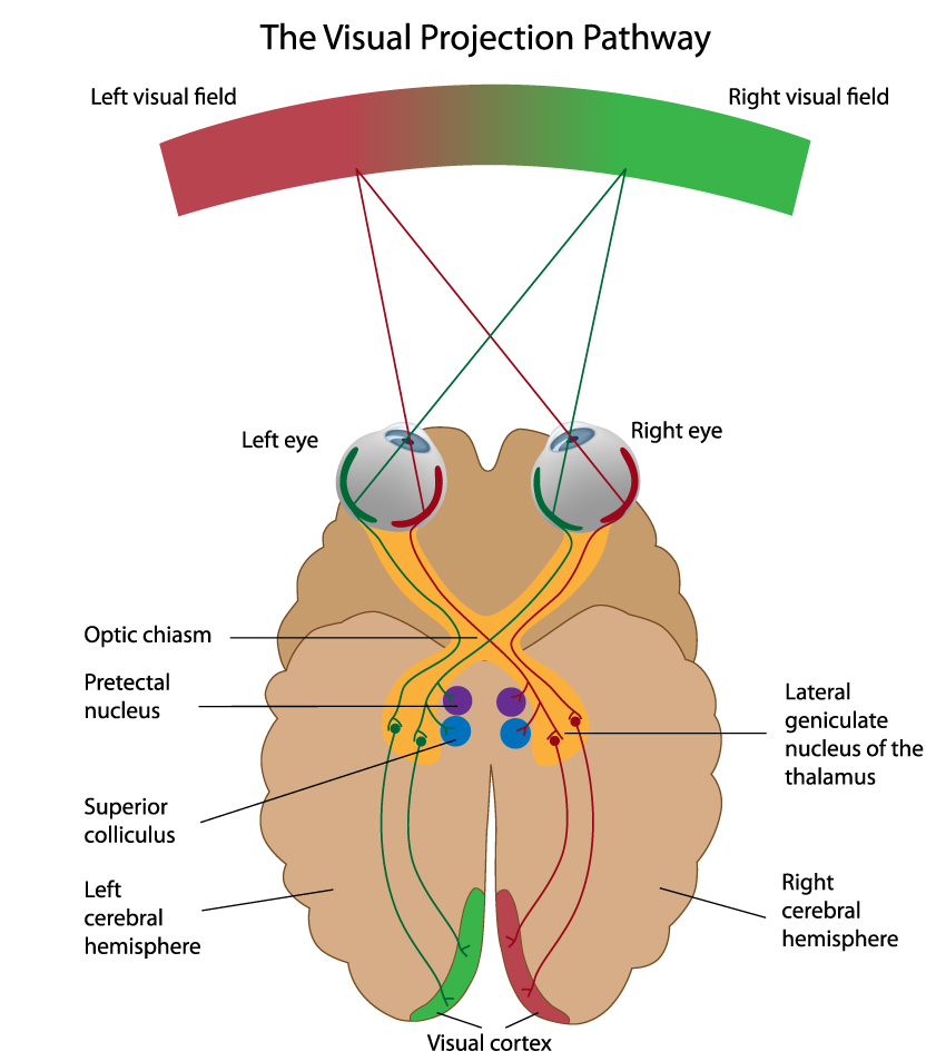
The image is a diagram illustrating the visual projection pathway, which is the route that visual information takes from the eyes to the visual cortex in the brain where it is interpreted.
At the top, two color-coded fields represent the left visual field (green) and the right visual field (red). These correspond to the areas of the external world that are visible to each eye when looking straight ahead.
The visual pathways from each eye start with the eyes themselves. The diagram shows how light from each visual field is projected onto the retina of both eyes. Notably, information from the right visual field is detected by the left side of both retinas, and vice versa for the left visual field.
The optic nerves from each eye carry the visual information towards the optic chiasm, where there is a partial crossing-over of nerve fibers. Fibers from the nasal (inner) halves of each retina cross to the opposite side of the brain, while fibers from the temporal (outer) halves remain on the same side. This crossing ensures that all information from the left visual field is processed by the right cerebral hemisphere, and all information from the right visual field is processed by the left cerebral hemisphere.
After the optic chiasm, the pathways continue as the optic tracts, which then synapse in several brain structures, including the lateral geniculate nucleus of the thalamus (shown in purple) and the superior colliculus (shown in blue). The lateral geniculate nucleus is the primary relay center for visual information on its way to the visual cortex.
From the lateral geniculate nucleus, the visual information travels through the optic radiations to the visual cortex located in the occipital lobe of the brain. This is where the visual information is interpreted, leading to visual perception.
The pretectal nuclei (shown in yellow) are involved in the reflex control of the pupil and lens.
The superior colliculus is involved in the orientation of the eyes and head towards visual stimuli.
This visual pathway is crucial for visual processing, and any damage along this pathway can result in visual field defects, which are areas of lost or reduced vision within the visual fields. The diagram serves as an educational tool, providing a simplified overview of the complex process of visual information transmission from the eyes to the brain.
External and Internal Structures of the Eye
This image is an infographic depicting the anatomy of the right eye as viewed from above, providing a comprehensive look at both the external and internal structures.
Starting with the external parts, the upper portion shows the eye lid, which protects the eye and helps distribute tear fluid across the surface of the eye. The lacrimal caruncle is a small, pink, globular nubbin of flesh located at the inner corner of the eye, which contains glands that produce a portion of the tear film. Adjacent to this is the tear duct, also known as the nasolacrimal duct, which drains tears from the eye into the nasal cavity.
Moving to the internal anatomy, the sclera is the white, fibrous outer layer of the eyeball; its main function is to provide structure, strength, and flexibility to the eye. The cornea is the transparent, dome-shaped surface that covers the front of the eye and, together with the lens, helps to refract and focus light onto the retina.
The iris is the colored part of the eye that surrounds the pupil, the central opening that regulates the amount of light entering the eye. The lens sits just behind the iris and pupil, and it is held in place by suspensory ligaments attached to the ciliary body, which contains the muscle that alters the shape of the lens for focusing, a process known as accommodation.
Between the cornea and the lens are the anterior and posterior chambers, which are filled with a clear fluid called the aqueous humor. This fluid maintains intraocular pressure and provides nutrients to the avascular structures of the eye.
The vitreous body is the clear gel that fills the space between the lens and the retina, helping to maintain the spherical shape of the eye and offering support to the retina. The retina itself is the thin layer of tissue that lines the back of the eye, containing the photoreceptor cells that convert light into electrical signals.
The fovea centralis, located in the center of the macula of the retina, is responsible for sharp central vision. The hyaloid canal is a small channel that runs through the vitreous body, which in the fetal eye allows the passage of nutrients from the optic nerve to the developing lens.
Finally, the optic nerve is depicted exiting the back of the eye. This nerve is responsible for transmitting visual information from the retina to the brain. Accompanying the optic nerve are the retinal blood vessels, which supply the retinal tissue with nutrients and oxygen.
The muscles illustrated are the lateral rectus muscle, which moves the eye outward away from the nose, and the medial rectus muscle, which moves the eye inward towards the nose.
Overall, the infographic serves as an educational tool, providing a clear and detailed visual representation of the various components of the eye and their respective roles in the complex process of vision.
Orbit and Lacrimal Anatomy of the Eye
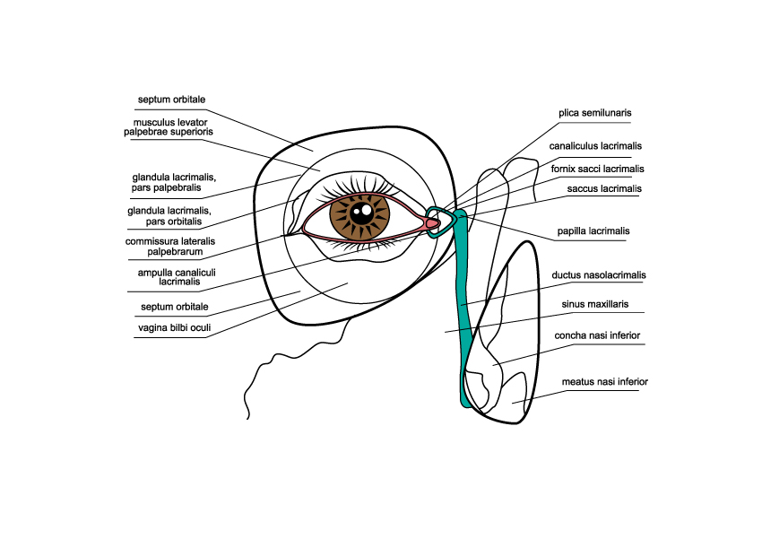
The image provides an intricate diagram of the human eye, focusing specifically on the lacrimal system and the anatomy surrounding the orbit.
Key structures labeled in the diagram are:
- Septum orbitale: A membrane that helps form the boundary between the eyelid and orbital fat.
- Musculus levator palpebrae superioris: The muscle responsible for lifting the upper eyelid.
- Glandula lacrimalis, pars palpebralis: The part of the lacrimal gland associated with the eyelid, responsible for tear production.
- Glandula lacrimalis, pars orbitalis: The portion of the lacrimal gland situated in the orbit, also involved in tear production.
- Conjunctiva: The mucous membrane that covers the front of the eye and lines the inside of the eyelids.
- Ampulla canaliculi lacrimalis: The dilated part of the lacrimal canaliculus into which tears drain.
- Septum orbitale: A thin, fibrous structure that separates the eyelid from the orbit.
- Vagina bulbi oculi (Tenon’s capsule): A membrane enveloping the eyeball, providing a socket for movement.
- Plica semilunaris: A small fold of conjunctiva that allows for movements of the eyeball.
- Canaliculus lacrimalis: Part of the lacrimal drainage system that carries tears from the lacrimal punctum towards the lacrimal sac.
- Fornix sacci lacrimalis: The upper fold or arch of the lacrimal sac where the canaliculi drain into.
- Saccus lacrimalis (Lacrimal sac): The upper dilated end of the nasolacrimal duct that collects tears from the canaliculi.
- Papilla lacrimalis: A small elevation in the conjunctiva where the lacrimal duct opens to release tears onto the eye surface.
- Ductus nasolacrimalis (Nasolacrimal duct): The duct that carries tears from the lacrimal sac into the nasal cavity.
- Sinus maxillaris (Maxillary sinus): One of the large cavities in the bones of the face, connected to the nasal cavity.
- Concha nasi inferior (Inferior nasal concha): A structure within the nasal cavity that helps to filter and humidify the air breathed through the nose.
- Meatus nasi inferior (Inferior nasal meatus): A passage within the nasal cavity below the inferior nasal concha, where the nasolacrimal duct drains.
This diagram provides a detailed overview of the lacrimal apparatus and associated orbital anatomy, showing how tears are produced and drained through the nasolacrimal system, ultimately leading to the nasal cavity.
Anatomical Illustration of the Extra-Ocular Muscles
The image is an anatomical illustration showing the lateral view of the muscles of the eye, also known as the extraocular muscles. These muscles are responsible for moving the eyeball and are crucial for binocular vision.
The muscles labeled in the diagram include:
Superior Oblique: This muscle is located at the top part of the eye and is involved in downward and outward eye movement. It passes through a fibrous loop, known as the trochlea, before attaching to the top of the eyeball.
Superior Rectus: Situated above the eye, this muscle primarily moves the eye upward. It works in coordination with the inferior oblique to control elevation of the eye.
Medial Rectus: Located on the side of the eye closest to the nose (medial side), this muscle moves the eye inward, towards the nose.
Lateral Rectus: This muscle is found on the outer side (lateral side) of the eye and is responsible for moving the eye outward, away from the nose.
Inferior Rectus: Positioned below the eye, this muscle primarily moves the eye downward. It coordinates with the superior oblique for depression of the eye.
Inferior Oblique: This muscle is located beneath the eye and helps to move the eye upward and outward. It works opposite the superior oblique muscle.
These muscles are attached to the sclera of the eye and extend back to their respective origins in the orbit. They are innervated by cranial nerves III (oculomotor nerve), IV (trochlear nerve), and VI (abducens nerve). The oculomotor nerve innervates the superior, medial, and inferior recti, as well as the inferior oblique muscle. The trochlear nerve innervates the superior oblique muscle, and the abducens nerve controls the lateral rectus muscle.
Blood Supply of the Eye
This illustration provides a detailed view of the blood supply and anatomy of the eye, particularly focusing on the vascular system and the associated structures.
At the center, the iris is shown, which is the colored part of the eye that controls the size of the pupil and, consequently, the amount of light that enters the eye. Two arterial circles can be observed in the iris: the large arterial circle and the small arterial circle. These circles are part of the intricate vascular network that provides the iris with blood.
The ciliary body is an extension of the iris. The ciliary muscle within it adjusts the shape of the lens, allowing the eye to focus on objects at different distances, a process known as accommodation. This muscle is also involved in controlling the flow of aqueous humor into the eye’s anterior chamber.
The cornea, labeled at the front of the eye, is the transparent structure that refracts light entering the eye. It is avascular, meaning it has no blood vessels, and receives nutrients from tears and aqueous humor.
The sclera, the white part of the eye, is the outer protective layer that maintains the shape of the eyeball. It extends all the way around the eye and provides an attachment for the extrinsic eye muscles, which are responsible for moving the eye.
The choroid is a layer rich in blood vessels lying between the retina and the sclera; it provides oxygen and nutrients to the outer layers of the retina.
The optic nerve is responsible for transmitting visual information from the retina to the brain. It is represented as the thick band exiting the back of the eye.
The blood vessels are highlighted, with arteries depicted in red and veins in blue. The anterior ciliary artery supplies blood to the ciliary body and the iris. The recurrent branch of the anterior ciliary artery is shown looping back towards the front of the eye.
Posteriorly, the posterior short ciliary arteries penetrate the sclera around the optic nerve to supply the choroid and the optic disc. The posterior long ciliary artery also runs forward to supply the ciliary body and the iris.
Veins are shown as return pathways for deoxygenated blood, with the return vein and the portal vein indicated.
Overall, this image effectively demonstrates the complexity of the eye’s vascular network, highlighting how blood is delivered to and from the various tissues of the eye, ensuring their proper function.
Anatomical Terms and Definitions
| Term | Definition | |||
|---|---|---|---|---|
| Arm (brachium) | The part of the upper limb located between the shoulder and the elbow. | |||
| Elbow joint | The joint connecting the arm and the forearm, comprising the articulation between the humerus and the two forearm bones, ulna, and radius. | |||
| Brachial artery | The major blood vessel of the upper arm that continues from the axillary artery to supply blood to the arm. | |||
| Radius | One of the two bones of the forearm, extending from the lateral side of the elbow to the thumb side of the wrist. | |||
| Ulna | The longer and larger of the two bones of the forearm, placed on the side opposite to the thumb. | |||
| Cornea | The transparent front part of the eye that covers the iris, pupil, and anterior chamber. | |||
| Iris | The colored part of the eye, controlling the size of the pupil and thus the amount of light reaching the retina. | |||
| Lens | The transparent structure inside the eye that focuses light rays onto the retina. | |||
| Retina | The light-sensitive layer of tissue at the back of the inner eye, which translates light into neural signals for vision. | |||
| Fovea centralis | A small central pit in the macula of the retina, composed of closely packed cones that is responsible for sharp central vision. | |||
| Optic nerve | The nerve that transmits visual information from the retina to the brain. | |||
| Macula | An area near the center of the retina that is responsible for central vision and high visual acuity. | |||
| Vitreous humor | The clear gel that fills the space between the lens and the retina in the eyeball. | |||
| Photoreceptors | Cells in the retina that respond to light; they include rods, which are responsible for vision in low light, and cones, for color vision and detail. | |||
| Sclera | The white outer layer of the eyeball that provides protection and form. | |||
| Choroid | The vascular layer of the eye between the retina and the sclera, supplying nutrients and oxygen to the eye. | |||
| Anterior chamber | The fluid-filled space inside the eye between the cornea and the iris. | |||
| Posterior chamber | The fluid-filled space directly behind the iris but in front of the lens. | |||
| Conjunctiva | A clear mucous membrane that lines the inside of the eyelids and covers the sclera. | |||
| Lacrimal gland | The gland responsible for producing tears; it is situated in the upper outer region of the orbit, above the eyeball. | |||
| Aqueous humor | The clear, watery fluid that fills the space between the cornea and the iris. | |||
| Myopia | A common vision condition also known as nearsightedness, where distant objects appear blurred. | |||
| Hyperopia | A vision condition also known as farsightedness, where close objects appear blurred. | |||
| Astigmatism | A common imperfection in the curvature of the eye's cornea or lens, causing blurred or distorted vision. | |||
| Retinal detachment | A serious condition where the retina peels away from its underlying layer of support tissue. | |||
| Macular degeneration | An eye disease that may result in blurred or no vision in the center of the visual field due to damage to the macula. | |||
| Glaucoma | A group of eye conditions that damage the optic nerve, often associated with increased pressure in the eye. | |||
| Cataracts | A condition characterized by clouding of the lens in the eye, leading to a decrease in vision. | |||
| Conjunctivitis | An eye condition diagnosed by irritation or infection of the conjunctiva, often resulting in redness and swelling. | |||
| Keratoconus | A progressive eye disease where the normally round cornea thins and begins to bulge into a cone-like shape. | |||
| Bullous Keratopathy | A condition causing swelling and blistering of the cornea due to endothelial cell dysfunction. | |||
| Corneal scarring | Opacity or scarring of the cornea often resulting from injury, infection, or inflammation. | |||
| Extraocular muscles | The muscles that control the movements of the eye and the elevation of the eyelid. | |||
| Vitreous floaters | Small flecks or threads of collagen that float in the vitreous humor and cast shadows on the retina, often seen as floaters by the individual. |
