
Table of Contents
Basic Nose Anatomy
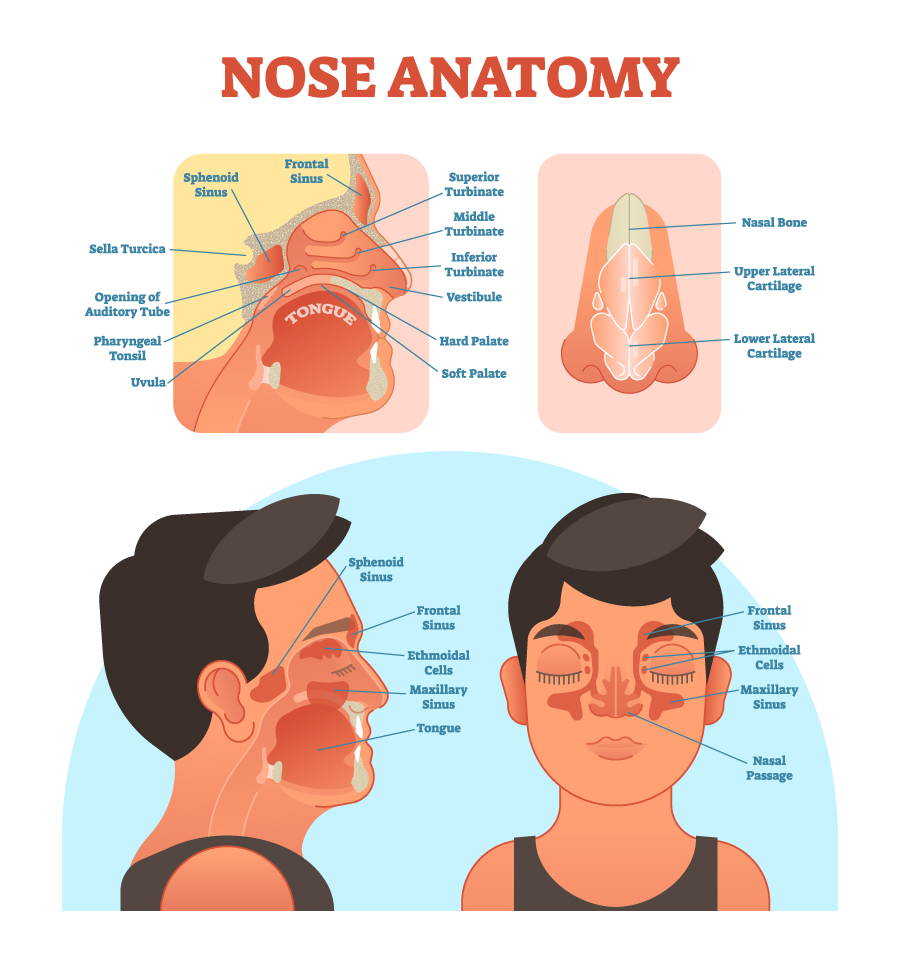
This illustration provides a comprehensive view of the nose anatomy from different angles. In the upper left section, we observe an internal side view highlighting various sinuses: the sphenoid and frontal sinuses. These are air-filled spaces within the bones of the skull that contribute to the humidification and temperature regulation of inhaled air. The sphenoid sinus is located near the sella turcica, a saddle-shaped depression in the sphenoid bone that holds the pituitary gland. The image also shows the opening of the auditory tube (Eustachian tube), which connects the nasopharynx to the middle ear, the tonsil, and parts of the mouth such as the tongue, hard palate, and soft palate.
The inset on the upper right provides a frontal view of the nasal structures, including the nasal bone, and the upper and lower lateral cartilages which contribute to the shape and structure of the nose. Below this inset, three nasal conchae or turbinates (superior, middle, and inferior) are depicted. These scroll-like bones are covered by a mucous membrane and work to filter, warm, and humidify the air entering the nasal cavity.
In the main lower part of the image, two side profiles of a human head are shown. On the left, a lateral view illustrates the frontal sinus, sphenoid sinus, ethmoidal air cells (part of the ethmoid bone), maxillary sinus, and the tongue. This view emphasizes the relationship between the nasal and oral cavities and the sinuses, which are all part of the upper respiratory tract.
On the right, we have a frontal view of a face with the skin rendered transparent over the nasal area, revealing the frontal sinuses, ethmoidal air cells, maxillary sinuses, and the nasal passage, indicating where the air flows during breathing.
Overall, the image serves as an educational tool to understand the anatomy of the nose and its adjacent structures, providing insight into how the nasal cavity is interconnected with the sinuses and the oral cavity, playing a significant role in respiration and olfaction.
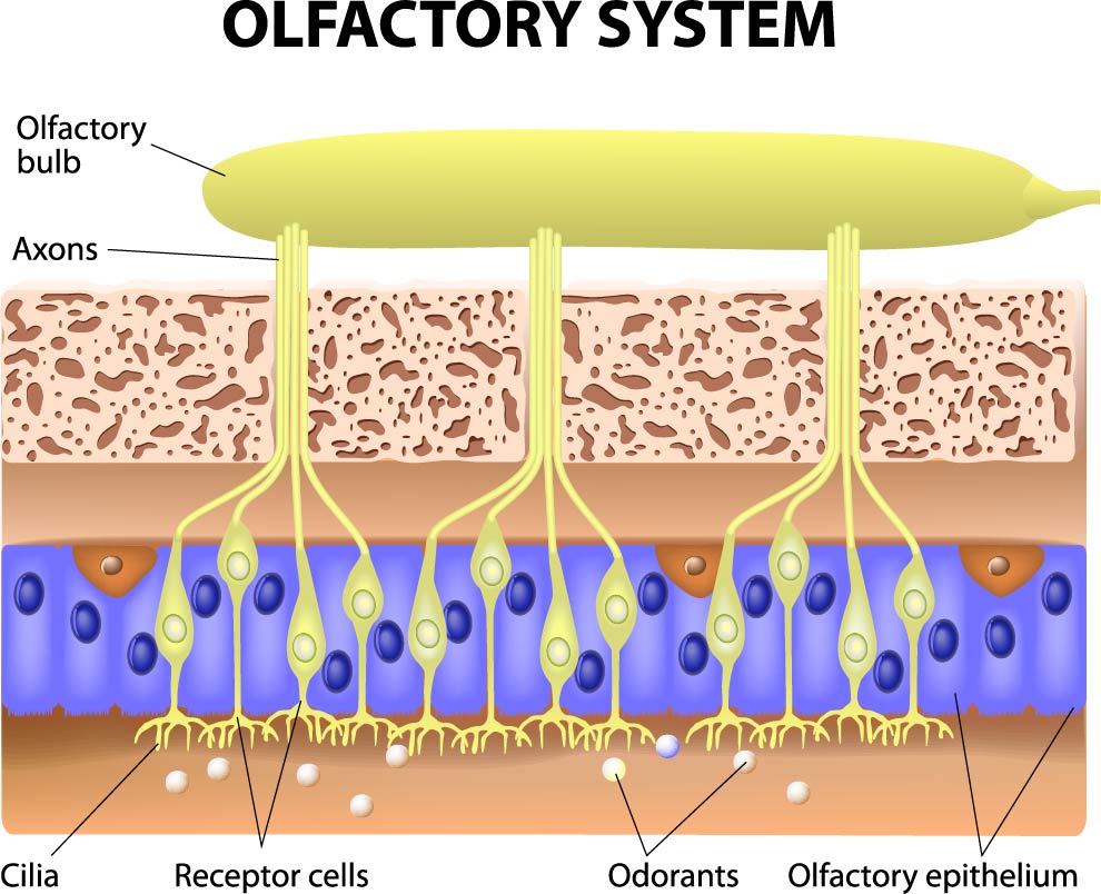
Olfactory System
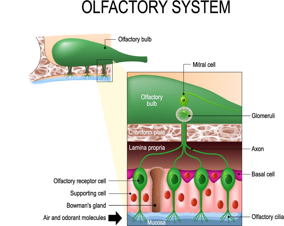
The image presents a diagram of the olfactory system, which is the sensory system responsible for the sense of smell. At the base of the diagram, we see the mucosa, which lines the nasal cavity. This mucosa contains olfactory receptor cells, which are neurons equipped with olfactory cilia. These cilia extend into the mucosal surface and are the sites where odorant molecules, carried by the air, bind and initiate the sense of smell.
Above the olfactory receptor cells are supporting cells, which provide structural and metabolic support to the olfactory receptor neurons. Among these supporting structures are Bowman’s glands, which secrete mucus onto the surface of the olfactory epithelium. This mucus is crucial as it traps and dissolves odorant molecules, allowing them to bind to the receptors on the cilia.
The olfactory receptor cells are connected to the olfactory bulb in the brain via long projections called axons, which pass through the cribriform plate of the ethmoid bone. Inside the olfactory bulb, these axons make contact with structures known as glomeruli, where they synapse with the dendrites of mitral cells. Mitral cells then process the olfactory information and send signals deeper into the brain to areas responsible for identifying, discriminating, and responding to odors.
The image’s inset shows an enlarged view of the olfactory bulb, detailing the olfactory receptor cells’ axons reaching the glomeruli and the connection to the mitral cell, which extends its processes throughout the olfactory bulb, indicating the pathway of olfactory signal transduction from the periphery to the central nervous system.
Basal cells are also indicated at the base of the olfactory epithelium; these are stem cells that give rise to new olfactory receptor cells, highlighting the regenerative capacity of the olfactory system.
The diagram’s design simplifies the complex structure and function of the olfactory system, showing the direct pathway from the detection of odorant molecules to the initial processing in the brain.
Schematic of Olfactory Epithelium
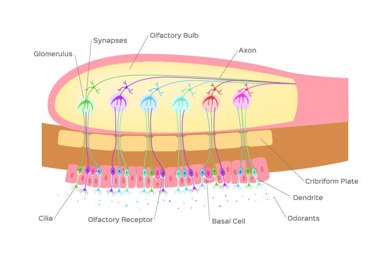
The image depicts a detailed schematic of the olfactory system’s signal transduction pathway, from the initial detection of odors to the relay of information to the brain.
At the bottom of the diagram, we see the olfactory epithelium, where the process begins. The olfactory receptors, depicted as structures with cilia, are the sensory neurons that detect odorants. These cilia increase the surface area for odorant molecules to bind to receptor proteins, initiating the olfactory response.
Above the olfactory receptors are basal cells, which are stem cells that can differentiate into new olfactory receptor neurons. This is significant because olfactory receptors have a limited lifespan and are regularly replaced.
When odorant molecules bind to receptors on the cilia, the olfactory receptor cells send electrical signals up through their axons. These axons pass through the cribriform plate—a sieve-like structure at the base of the skull—into the olfactory bulb.
Within the olfactory bulb, the axons make synaptic connections with dendrites of neurons in structures called glomeruli. Each glomerulus is a sort of processing unit. Multiple olfactory receptor neurons that detect the same type of odorant molecule converge on a single glomerulus.
The synapses within the glomeruli are points of communication where neurotransmitters are released from the axons of the olfactory receptor neurons and bind to receptors on the dendrites of other neurons in the olfactory bulb, such as mitral and tufted cells (not labeled here). This synaptic transmission is crucial for the propagation of the olfactory signal.
Finally, the mitral and tufted cells send the processed information deeper into the brain, resulting in the perception of smell. The diagram simplifies the complex neural circuitry involved in olfaction and visually conveys the essential steps of olfactory perception.
Diagram of Nasal Anatomy
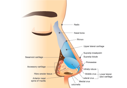
This diagram provides a detailed view of the nasal anatomy, specifically focusing on the structure of the nose and its cartilaginous components. At the top, we see the radix, which is the root or the beginning of the nose where it meets the forehead. Below the radix is the nasal bone, which forms the bridge of the nose and provides the skeletal support for the upper portion of the nasal structure.
The diagram also shows the pinion, which is not commonly referenced in basic anatomy and may refer to the outermost part of the nasal bones where they join with the cartilage.
The upper lateral cartilage is depicted beneath the nasal bone. It is one of the major cartilages of the nose and helps to shape the mid-section of the nose. These cartilages are paired, with one on each side, and they are connected to the nasal bones and the septal cartilage.
The septum is the part of the nose that separates the nasal cavity into two nostrils. It is made up of the septal cartilage, and its lower portion is called the columella, which is highlighted as the medial crus on the diagram. The columella is the tissue that links the nasal tip to the nasal base, and it defines the nasal tip’s angle and support.
The lateral crus is part of the lower lateral cartilage, often referred to as the alar cartilage, which gives shape to the nostrils. The lower lateral cartilages have a significant influence on the appearance and function of the nasal tip.
The sesamoid and accessory cartilages are smaller cartilages that provide additional structure and are often manipulated in nasal surgeries to refine the shape of the nose.
The fibro-areolar tissue is the connective tissue layer that covers the cartilages and muscles of the nose, providing a smooth contour and allowing for the flexibility of the nasal tip.
At the base of the nose, the anterior nasal spine of the maxilla is a bony projection that provides an attachment for the columella and influences the angle between the upper lip and the nose.
Overall, the illustration helps to understand the complex cartilaginous structures of the nose that contribute to its shape and function. Understanding these anatomical details is crucial for fields such as rhinoplasty and otolaryngology.
Cross-section of the Olfactory System
This illustration provides a detailed cross-section of the olfactory system, which is essential for the sense of smell. At the very top, we see the olfactory bulb, which is a neural structure of the forebrain involved in olfaction. The olfactory bulb is connected to the olfactory receptor neurons via axons, which are depicted as yellow lines extending downwards.
Below the olfactory bulb is a layer that appears speckled with dots, representing the cellular composition of the olfactory bulb, possibly the glomeruli where the initial processing of olfactory information occurs.
The main portion of the image shows the olfactory epithelium, a specialized epithelial tissue inside the nasal cavity that is responsible for detecting odors. Within this layer, we can see olfactory receptor cells, which are neurons that have receptors specifically tuned to detect different chemical compounds. These cells have cilia, which are hair-like extensions that protrude into the nasal cavity and significantly increase the surface area for capturing odorant molecules.
The blue structures represent the nuclei of the receptor cells, and the surrounding lighter blue cytoplasm extends to form the cilia. The receptor cells detect odors when odorant molecules, depicted as small white dots, bind to receptors located on the cilia.
In the background, the brownish layer represents the supporting cells of the olfactory epithelium, which provide structural support, nourishment, and insulation for the olfactory receptor neurons.
This image efficiently captures the essential elements of the olfactory system and demonstrates how odorant molecules interact with the olfactory epithelium to begin the process of olfactory perception.
Nose Structure Anatomy
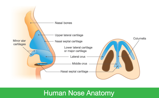
The image depicts two views of the human nose anatomy. On the left, a side profile of the nose shows the external structures, while on the right, a frontal section provides an internal perspective.
From the side view, we can see the nasal bones at the top, which form the bridge of the nose. Below these are the upper lateral cartilages, which are important for the mid-portion shape of the nose. The nasal septal cartilage is seen beneath, and it divides the nasal cavity into two nostrils internally.
Also visible in the side view are the lower lateral cartilages, sometimes referred to as the alar cartilages, which consist of the lateral and middle crura. These cartilages define the shape and support of the nasal tip and nostrils.
The minor alar cartilages are smaller cartilages that provide additional contour and structure to the nose, and they are indicated near the lower lateral cartilage.
In the frontal section, the nasal septal cartilage is seen as a vertical structure separating the nasal cavity into left and right passages. The columella, the tissue that connects the nasal tip to the base of the nose and supports the nasal tip, is indicated extending below the septal cartilage.
The image provides an informative visual aid to understand the various cartilages and bones that comprise the structure of the nose, each playing a role in the overall form and function of the nasal anatomy.
Anatomical Terms and Definitions
| Term | Definition | |||
|---|---|---|---|---|
| Olfactory Bulb | A neural structure of the forebrain involved in the sense of smell; receives neural input about odours detected by cells in the nasal cavity. | |||
| Olfactory Receptor Cells | Neurons located in the olfactory epithelium that contain receptors for odorant molecules; they send signals to the brain when these molecules are detected. | |||
| Olfactory Cilia | Hair-like extensions on the olfactory receptor cells that increase the surface area for detecting odorant molecules. | |||
| Olfactory Epithelium | A specialized epithelial tissue inside the nasal cavity responsible for detecting odors. | |||
| Axons | Long, slender projections of neurons that conduct electrical impulses away from the neuron's cell body to the olfactory bulb. | |||
| Glomeruli | Structures within the olfactory bulb where synapses form between the axons of the olfactory receptor neurons and the dendrites of other neurons. | |||
| Basal Cells | Stem cells in the olfactory epithelium that can differentiate into new olfactory receptor neurons. | |||
| Nasal Bone | The bones that form the bridge of the nose. | |||
| Upper Lateral Cartilage | Cartilage that helps to shape the mid-section of the nose. | |||
| Lower Lateral Cartilage | Also known as alar cartilage, these cartilages shape the nostrils and contribute to the form of the nasal tip. | |||
| Septal Cartilage | Cartilage forming the septum, which divides the nasal cavity into two chambers. | |||
| Columella | The tissue that links the nasal tip to the nasal base, influencing the nasal tip's angle and support. | |||
| Middle Crus | The part of the lower lateral cartilage that contributes to the shape of the nasal tip. | |||
| Lateral Crus | The part of the lower lateral cartilage that contributes to the structure and shape of the nostril walls. | |||
| Nasal Septum | The bone and cartilage wall that separates the nasal cavity into two separate chambers. | |||
| Minor Alar Cartilages | Smaller cartilages that provide additional structure and contour to the nose. | |||
| Sphenoid Sinus | An air-filled space within the sphenoid bone, part of the paranasal sinus system. | |||
| Frontal Sinus | One of the four pairs of paranasal sinuses, located in the frontal bone, just above the eyes. | |||
| Maxillary Sinus | A pair of sinuses located in the maxillary bones, the largest of the sinuses. | |||
| Ethmoidal Air Cells | Numerous small air cells within the ethmoid bone that form part of the paranasal sinus system. | |||
| Cribriform Plate | A sieve-like structure at the base of the skull that allows the passage of the olfactory nerves from the nasal cavity to the olfactory bulb. | |||
| Radix | The root or beginning of the nose where it meets the forehead. | |||
| Pinion | The outermost part of the nasal bones where they join with the cartilage. (Note: Pinion is not a standard anatomical term and may vary in use.) | |||
| Sesamoid Cartilages | Additional small cartilages found near the nasal tip, often manipulated in nasal surgery. | |||
| Fibro-Areolar Tissue | Connective tissue layer that covers the cartilages and muscles of the nose, allowing for the flexibility of the nasal tip. | |||
| Anterior Nasal Spine | A bony projection of the maxilla at the base of the nose that provides attachment for the columella. | |||
| Turbinates/Conchae | Long, narrow shelves of bone that protrude into the nasal cavity; they increase the surface area of the nasal passages, aiding in warming and humidifying inhaled air. |
