
Table of Contents
Taste Bud Anatomy
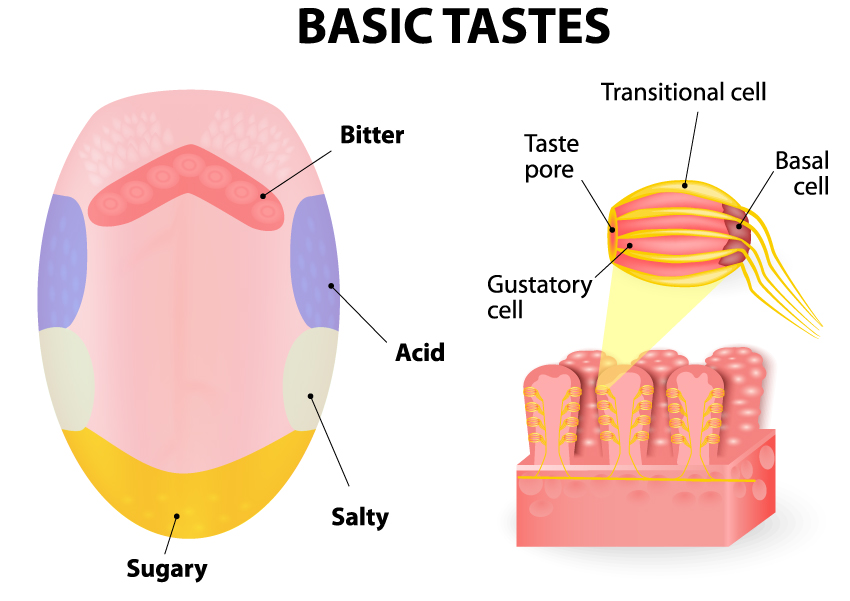
The image presents an overview of the basic tastes perceived by the human tongue and a magnified section of the taste buds where these perceptions occur. The tongue is mapped out with different regions that are more sensitive to certain tastes: the tip of the tongue is indicated as most sensitive to sugary or sweet tastes, the front sides to salty tastes, the back sides to sour or acidic tastes, and the very back of the tongue to bitter tastes.
The right side of the image illustrates a close-up view of the tongue’s surface where taste buds are located. We see a detailed structure of a taste bud, which consists of a taste pore, the opening on the tongue’s surface where tastants enter. Beneath this are gustatory cells, which are sensory cells that respond to different tastes and transmit this information to the brain. Surrounding these cells are transitional cells, which likely play a supportive role. At the bottom of the taste bud structure are basal cells, which are progenitor cells that replace old gustatory cells.
This illustration serves as an educational tool to understand the basic organization of taste regions on the tongue and the cellular composition of a taste bud, which is essential for the sensory perception of taste.
Anatomical Illustration of the Tongue
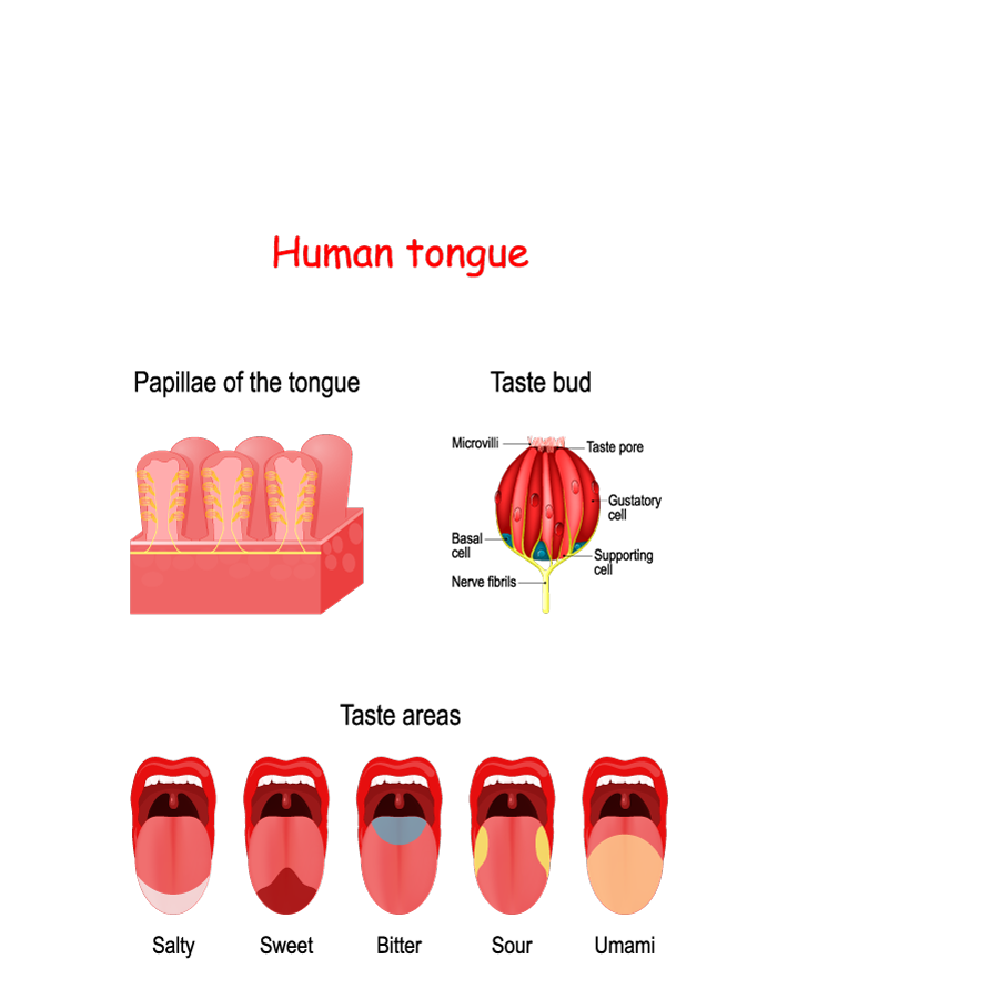
This image provides an anatomical illustration of the human tongue and its role in the perception of different taste sensations. It is divided into three main sections:
Papillae of the tongue: The top left section shows a magnified view of the papillae, which are small, nipple-like structures on the tongue’s surface. These papillae are rich with taste buds and are primarily responsible for the tongue’s texture.
Taste bud: On the top right, there is an even more magnified image of an individual taste bud, highlighting its internal structure. We can see microvilli protruding from the gustatory cells, extending into the taste pore where they can contact tastants (taste-provoking chemical substances). Below the gustatory cells are supporting cells and basal cells, which are involved in the regeneration of taste bud cells. Nerve fibers are also shown, which carry taste information to the brain.
Taste areas: The bottom section of the image depicts five different taste sensations and their corresponding areas on the tongue. Each taste sensation is represented by a different color on the tongue illustration:
- Salty: Shown at the front edges, indicating that these areas are sensitive to salt.
- Sweet: Depicted at the tip of the tongue, where sweetness is most commonly perceived.
- Bitter: Illustrated at the back of the tongue, which is most sensitive to bitter compounds.
- Sour: Located on the sides towards the back, which typically detect acidity or sourness.
- Umami: This taste, often associated with the savory flavor of amino acids like glutamate, is represented across the middle of the tongue.
This diagram serves as an educational diagram, simplifying and illustrating the locations on the tongue that are traditionally associated with the detection of different basic tastes. It’s important to note that while this representation is commonly used for educational purposes, recent research suggests that all regions of the tongue can detect all tastes to varying degrees, and the map is more of a gradient than strictly defined zones.
Cross-Section of the Taste Bud
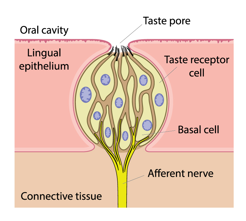
This illustration shows a detailed cross-sectional view of a taste bud, which is the sensory organ for taste located within the lingual epithelium of the tongue. The taste bud is embedded within the tongue tissue and has a narrow opening at the top known as the taste pore, which opens out into the oral cavity. This is where tastants from consumed food or drink enter the taste bud.
The main cellular components labeled within the taste bud are the taste receptor cells, which are elongated cells that extend from the base to the top of the taste bud, culminating at the taste pore. These cells contain receptors that interact with tastants and are responsible for initiating the sensory perception of taste. Each taste receptor cell has gustatory hairs that project through the taste pore to interact directly with the oral environment.
At the bottom of the taste bud are basal cells, which are precursor cells that divide and mature to replace old taste receptor cells, ensuring the taste bud maintains its sensitivity over time.
Surrounding the taste receptor cells and providing them with structural support are connective tissues, which anchor the taste bud to the surrounding tongue tissue.
Finally, the afferent nerve is depicted as a bundle of nerve fibers at the bottom of the taste bud, connecting it to the nervous system. These nerves transmit the signals generated by the taste receptor cells to the brain, where they are interpreted as specific tastes.
This diagram is used to explain the anatomical structure of a taste bud and the process by which taste signals are received and sent to the brain, contributing to our sense of taste.
Terms and Definitions
| Term | Definition | |||
|---|---|---|---|---|
| Basic Tastes | The fundamental taste sensations including sweet, salty, bitter, sour, and umami, perceived by the tongue. | |||
| Taste Bud | A sensory organ found on the tongue's surface, responsible for the perception of taste. | |||
| Taste Pore | The opening on the surface of the tongue through which tastants enter the taste bud. | |||
| Gustatory Cells | Sensory cells within a taste bud that respond to tastants and transmit taste information to the brain. | |||
| Transitional Cells | Cells surrounding gustatory cells that likely provide structural support within the taste bud. | |||
| Basal Cells | Progenitor cells at the base of the taste bud that replace old gustatory cells, maintaining the sensitivity of the taste bud. | |||
| Papillae | Nipple-like structures on the tongue’s surface that contain taste buds and contribute to the tongue’s texture. | |||
| Microvilli | Projections from gustatory cells that extend into the taste pore to interact with tastants. | |||
| Nerve Fibers | Bundles of nerve fibers that carry taste information from the taste buds to the brain. | |||
| Connective Tissue | Tissue that provides structural support to the taste buds and anchors them to the surrounding tongue tissue. | |||
| Afferent Nerve | Nerve fibers that transmit signals from taste receptor cells to the brain for taste perception. | |||
| Taste Areas | Regions of the tongue associated with sensitivity to specific tastes: salty (front edges), sweet (tip), bitter (back), sour (sides towards the back), and umami (middle). | |||
| Lingual Epithelium | The topmost layer of the tongue where the taste buds are located. | |||
| Taste Sensations | The different flavors that are detected by the taste buds: salty, sweet, bitter, sour, and umami. |
