
Table of Contents
Parasympathetic vs Sympathetic Nervous System
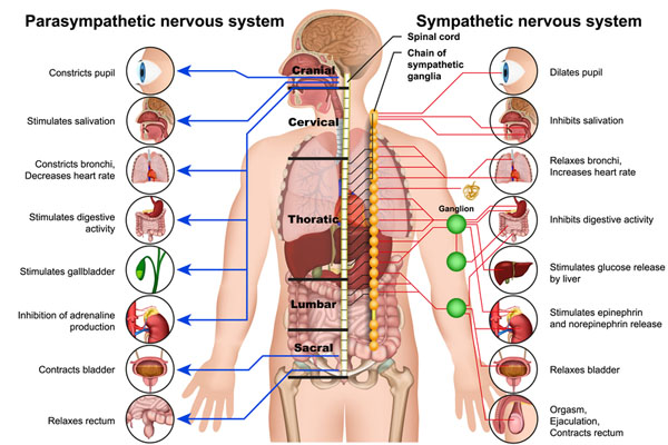
The image illustrates the differences between the parasympathetic and sympathetic nervous systems, which are two branches of the autonomic nervous system that regulate involuntary body functions.
On the left side, the parasympathetic nervous system is represented by blue lines and is responsible for “rest and digest” activities. It constricts the pupil, stimulates salivation, constricts the bronchi in the lungs and decreases heart rate, and stimulates digestive activity. It also stimulates the gallbladder, inhibits adrenaline production, contracts the bladder, and relaxes the rectum. This system is activated when the body is at rest, directing energy to body maintenance, digestion, and conserving energy.
The sympathetic nervous system, shown on the right with red lines, is involved with “fight or flight” responses. It dilates the pupil, inhibits salivation, relaxes the bronchi in the lungs and increases heart rate, and inhibits digestive activity. Furthermore, it stimulates glucose release by the liver, stimulates the release of epinephrine and norepinephrine, relaxes the bladder, and is responsible for the functions associated with orgasm, ejaculation, and contraction of the rectum. This system prepares the body for intense physical activity and is activated in response to stress or danger.
The central structure depicted is the spinal cord, with the sympathetic chain of ganglia adjacent to it. The parasympathetic nerves are shown extending from the cranial and sacral regions, while the sympathetic nerves emerge from the thoracic and lumbar regions. This anatomical distribution reflects the different origins of the parasympathetic (craniosacral) and sympathetic (thoracolumbar) fibers.
The interactions between these two systems maintain homeostasis in the body by balancing the body’s response to changing conditions, either by stimulating or by inhibiting various physiological processes.
Sympathetic Nervous System
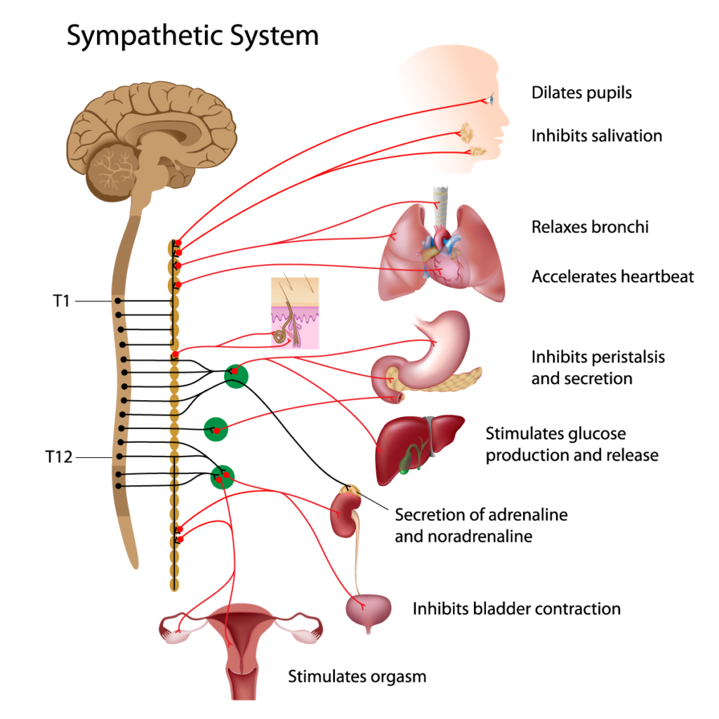
The image depicts the sympathetic nervous system, which is a part of the autonomic nervous system responsible for the body’s rapid involuntary response to dangerous or stressful situations. It shows a series of red lines that indicate the pathways from the spinal cord to various organs, representing the sympathetic nerve fibers.
At the top, we see the brain, which is the control center for the sympathetic system, though it operates largely unconsciously. The red lines connect the brain to the eyes, where the sympathetic system causes pupil dilation. This is beneficial in situations requiring heightened alertness, as it allows more light into the eyes, potentially improving vision.
Next, we see the pathways leading to the salivary glands, which are inhibited by the sympathetic nervous system, often resulting in a dry mouth during stress. The lines also extend to the lungs, where the sympathetic system relaxes the bronchi, allowing for increased air flow, which is essential during physical exertion or a fight-or-flight response.
The heart is also shown with connections from the sympathetic system, which accelerates the heartbeat to pump more blood to muscles, the brain, and other organs during emergency situations.
The sympathetic fibers then reach the stomach and intestines, where they inhibit peristalsis and secretion, effectively slowing down digestion, as the body redirects energy to more critical functions necessary for immediate survival.
Further down, we observe the connections to the liver, where the sympathetic nervous system stimulates glucose production and release, providing a quick energy source for the body.
The adrenal glands are targeted as well, where the system promotes the secretion of adrenaline and noradrenaline, hormones that collectively increase alertness, strength, and speed in response to stress.
The connections continue to the bladder, where sympathetic activity inhibits bladder contraction, preventing urination during stressful moments.
Lastly, the graphic indicates the role of the sympathetic system in stimulating orgasm, which is part of the complex interplay between the sympathetic and parasympathetic systems during sexual function.
Vertebral levels T1 to T12 are indicated, showing the origin of the sympathetic nerves from the thoracic portion of the spinal cord, highlighting the thoracolumbar outflow of the sympathetic division. The green circles represent the sympathetic ganglia where preganglionic and postganglionic neurons synapse.
Parasympathetic Nervous System
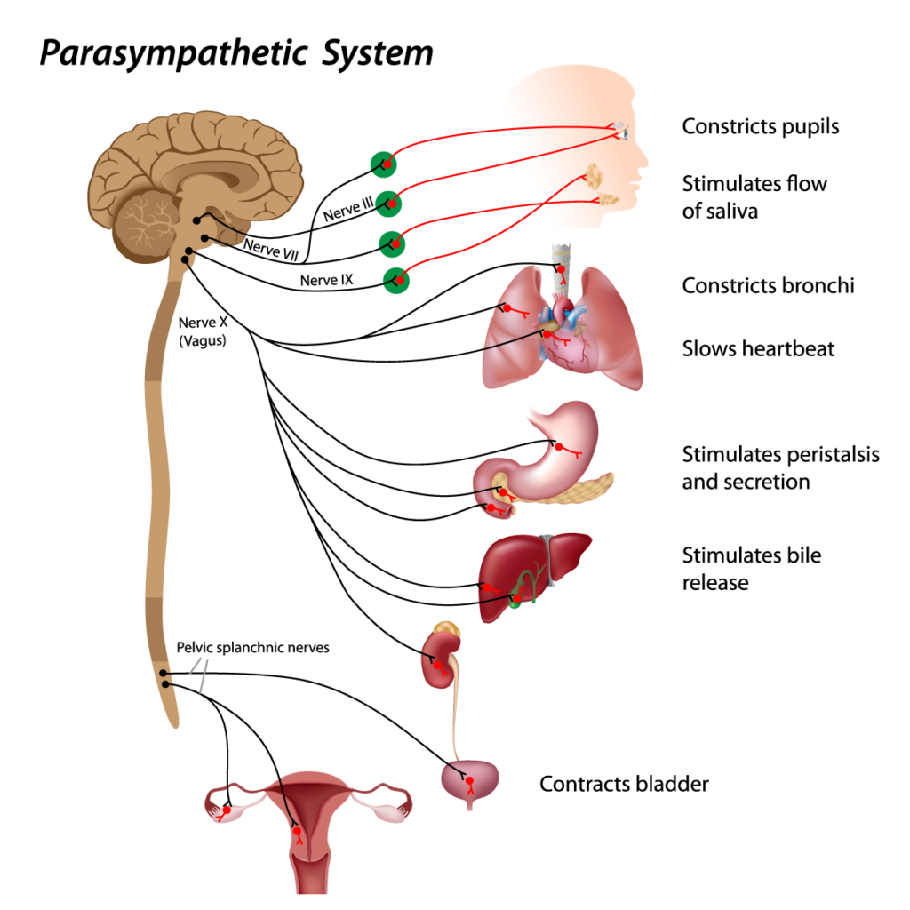
This image provides a visual representation of the parasympathetic nervous system, which is part of the autonomic nervous system often referred to as the “rest and digest” system.
Starting at the top, the parasympathetic nervous system’s impact on the eye is shown, where it constricts the pupils. This is typically in response to environments that do not require acute vision, such as in dim lighting or to protect the eyes from bright light.
The system also stimulates the flow of saliva through the salivary glands, preparing the mouth for digestion. It then travels to the lungs, where it constricts the bronchi, reducing airflow, which is suitable for a state of rest.
The heart is also under the influence of the parasympathetic nervous system, which slows the heartbeat. This conserves energy and is part of the body’s relaxation process.
Further down, the image shows how the parasympathetic nerves stimulate peristalsis and secretion in the gastrointestinal tract, facilitating digestion.
The liver is affected by the parasympathetic system to stimulate bile release, which is important for the digestion and absorption of fats.
Finally, the parasympathetic system controls the contraction of the bladder, allowing for urination, and this is part of the body’s normal excretory processes.
The nerves depicted include the cranial nerves III, VII, IX, and X (Vagus), which are some of the main conduits for parasympathetic fibers to reach the above-mentioned organs. The Vagus nerve, in particular, has a broad reach, affecting the heart, lungs, and digestive tract.
The image also shows the pelvic splanchnic nerves, which are responsible for supplying the lower half of the digestive system and the organs of the pelvis, contributing to the rest and digest functions in these areas.
The graphical representation uses red lines to trace the pathways from the origins of the parasympathetic fibers in the brainstem and sacral spinal cord to the targeted organs, emphasizing the connections and influence of the parasympathetic system on various bodily functions.
Reflex Arc
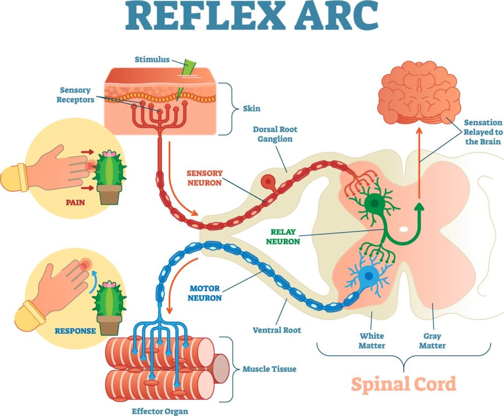
The image provides an overview of the reflex arc, which is a neural pathway that mediates an immediate response to a stimulus without conscious brain involvement.
At the top left of the image, we see a hand with sensory receptors that detect a painful stimulus—indicated by the cactus spines pressing into the skin. This sensory input starts the reflex arc.
From there, the signal travels along the sensory neuron, which is represented by the red line, towards the dorsal root ganglion. The dorsal root ganglion houses the cell bodies of the sensory neurons and is located just outside the spinal cord.
Once inside the spinal cord, the signal is relayed through a sensory neuron to a relay neuron, also known as an interneuron. This neuron, depicted in green, is located in the gray matter of the spinal cord, which is centrally located and shown in a butterfly shape. The interneuron processes the signal and then transmits it to a motor neuron.
The motor neuron, illustrated by the blue line, carries the signal away from the spinal cord through the ventral root and to the effector, which in this case is muscle tissue. The muscle then contracts in response to the painful stimulus, which is the reflex action illustrated by the hand pulling away from the cactus.
The reflex arc allows for a rapid response to harmful stimuli, protecting the body from injury. This type of response is involuntary and occurs without direct involvement from the brain. However, the sensation of pain is relayed to the brain as indicated by the curve reaching towards the brain in the image, allowing conscious recognition of the painful stimulus after the reflex action has taken place.
Anatomical Terms and Definitions
| Term | Definition |
|---|---|
| Adrenaline | Hormone increased by the sympathetic system to aid in fight or flight responses. |
| Autonomic Nervous System | System that regulates involuntary body functions, including the parasympathetic and sympathetic nervous systems. |
| Bladder | Organ whose contraction is inhibited by the sympathetic system and controlled by the parasympathetic system. |
| Bronchi | Air passages in the lungs, constricted by the parasympathetic and relaxed by the sympathetic system. |
| Craniosacral | Origin of parasympathetic nerves from the cranial and sacral regions. |
| Digestive Activity | Process stimulated by the parasympathetic and inhibited by the sympathetic system. |
| Dorsal Root Ganglion | Location housing cell bodies of sensory neurons in the reflex arc. |
| Epinephrine | Hormone, synonymous with adrenaline, released by the sympathetic nervous system. |
| Fight or Flight | Response prepared by the sympathetic system for intense physical activity. |
| Gallbladder | Organ stimulated by the parasympathetic system for bile release. |
| Heart Rate | Function decreased by the parasympathetic and increased by the sympathetic system. |
| Homeostasis | Maintenance of stable internal conditions balanced by parasympathetic and sympathetic interactions. |
| Interneuron | Neuron that relays signals in the reflex arc. |
| Liver | Organ affected by both the parasympathetic (bile release) and sympathetic (glucose release) systems. |
| Motor Neuron | Neuron that carries signals to muscles in the reflex arc. |
| Norepinephrine | Hormone released by the sympathetic system similar to adrenaline. |
| Orgasm | Function stimulated by the sympathetic system. |
| Parasympathetic Nervous System | System promoting "rest and digest" activities. |
| Pupil | Eye part constricted by the parasympathetic and dilated by the sympathetic system. |
| Reflex Arc | Pathway for rapid response to stimulus without conscious brain involvement. |
| Salivation | Process stimulated by the parasympathetic and inhibited by the sympathetic system. |
| Sensory Neuron | Neuron transmitting signals in the reflex arc. |
| Spinal Cord | Central structure in the nervous system for nerve emergence. |
| Sympathetic Nervous System | System involved in "fight or flight" responses. |
| Thoracolumbar | Origin of sympathetic nerves from the thoracic and lumbar regions. |
| Vagus Nerve | Major parasympathetic nerve affecting the heart, lungs, and digestive tract. |
