
Table of Contents
Structure of the Epidermis
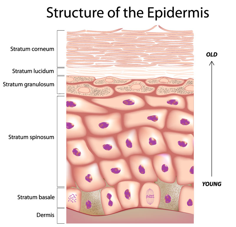
The image illustrates the structure of the epidermis, the outermost layer of the skin, in a detailed cross-section. The epidermis is depicted as a stratified squamous epithelium, composed of several distinct layers, each with a specific role in skin health and function.
From bottom to top, the labeled layers are:
Stratum Basale (Basal Layer): This is the deepest layer, resting on the dermis. It contains columnar to cuboidal basal cells, which are mitotically active, meaning they divide to form new cells. These new cells then move up through the layers of the epidermis as they differentiate.
Stratum Spinosum (Spiny Layer): Above the basal layer, this layer is named for the spiny appearance of the cells due to desmosomal connections when viewed under a microscope. It provides strength and flexibility to the skin.
Stratum Granulosum (Granular Layer): This layer consists of keratinocytes that have moved from the lower layers. These cells contain granules of keratohyalin, which play a role in water retention and give the cells a grainy appearance.
Stratum Lucidum (Clear Layer): This thin, transparent layer is found only in thick skin, like the skin of the palms and soles. It consists of dead and flattened keratinocytes that provide an additional barrier to injury and infection.
Stratum Corneum (Horny Layer): The outermost layer is made up of dead keratinocytes that have lost their nucleus and cytoplasm and are filled with keratin protein. This layer is highly durable and creates a waterproof barrier.
The progression from young cells at the bottom to old, dead cells at the top is indicated by an arrow labeled “YOUNG” at the basal layer ascending to “OLD” at the stratum corneum. Below the stratum basale is the dermis, which is not part of the epidermis but provides support and nutrition to the epidermal layers.
This cross-sectional view is essential for understanding how the skin renews itself and protects the body, with each layer playing a specialized role in skin physiology.
Cross-Sectional View of the Skin
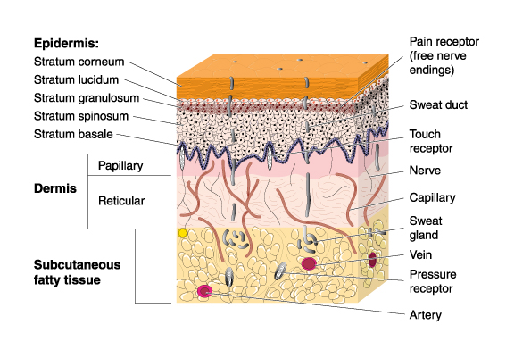
This image provides a detailed cross-sectional view of human skin, highlighting its various layers and components. The image is structured with labels pointing to specific layers and elements within those layers.
At the topmost layer, we have the Epidermis, which is divided into four distinct strata (from outermost to innermost): the Stratum corneum, Stratum lucidum, Stratum granulosum, and Stratum spinosum, ending with the Stratum basale. These layers collectively serve as the barrier of the skin, with the Stratum corneum being the outermost layer of dead cells that are continuously shed and replaced.
Below the epidermis lies the Dermis, which is further subdivided into two regions: the superficial Papillary region characterized by finger-like projections that interlock with the epidermis, increasing the surface area of contact, and the deeper Reticular region, which provides structural strength and elasticity to the skin due to its dense connective tissue.
The bottom-most layer shown is the Subcutaneous fatty tissue, which insulates the body and acts as an energy reserve, as well as providing cushioning for the skin.
Within these layers, the image also depicts various skin components and receptors:
- Pain receptors (free nerve endings) that detect painful stimuli.
- Sweat glands which are involved in thermoregulation and excretion.
- Sweat ducts leading from the sweat glands to the surface of the skin.
- Touch receptors that allow the sensation of touch.
- Nerves that transport signals to and from the brain.
- Capillaries, small blood vessels that supply the dermis with nutrients and oxygen.
- Veins which carry deoxygenated blood back to the heart.
- Pressure receptors that sense mechanical changes in the environment.
- Arteries that deliver oxygenated blood from the heart to the tissues.
The illustration clearly shows the complexity and functionality of skin, demonstrating its role as a protective barrier, a sensory interface with the environment, and an active player in the body’s thermoregulation and circulatory systems. The interplay of these elements allows the skin to perform its vital functions, including protection against pathogens, synthesis of vitamin D, sensation, and temperature regulation.
Anatomy of the Fingernail
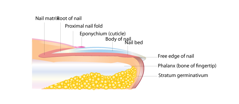
This image is a simplified representation of the anatomy of a human fingernail and its surrounding structures.
At the base of the nail, we see the Nail matrix or the Root of the nail, which is the part of the nail bed that extends beneath the skin and is responsible for producing cells that become the nail plate.
The Proximal nail fold is the skin that overlaps the nail matrix, and just below this fold is the Eponychium, commonly known as the cuticle. The cuticle is a layer of clear skin located along the bottom edge of your finger or toe. This area is particularly important for nail health and growth, as it protects the new nail from bacterial infections as it grows out from the nail root.
The Body of the nail is the hard, translucent part of the nail, composed mostly of keratin, which is the visible nail area.
The Nail bed is the skin beneath the body of the nail. The nail bed is rich in blood vessels, giving the nail its characteristic pink color, except at the base where it may look whiter.
On the tip, we have the Free edge of the nail, which is the part of the nail that has grown beyond the end of the finger or toe and is often trimmed during nail care.
Below the nail bed is the Phalanx (bone of fingertip), which is the distal portion of the fingers or toes.
Beneath the proximal nail fold and not visible from the top is the Stratum germinativum, which is the deepest layer of the epidermis and plays a key role in the generation of new skin cells, including those that form the nail plate.
Overall, the image captures the complex structure of the nail, showing how it is more than just a protective keratin plate but is a system integrated with the skin and bone of the fingertip, with specific parts like the nail matrix and eponychium playing crucial roles in nail growth and health. The nail structure supports the delicate tips of the fingers and toes, enhances tactile sensation, and aids in the manipulation of small objects.
Factors of Hair Color and Skin Texture
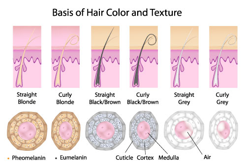
This image illustrates the factors that determine hair color and texture in humans. It’s divided into six panels, each showing a hair follicle and a cross-section of the hair shaft corresponding to different hair colors and textures.
The top row of panels depicts the hair follicles and the emerging hair shaft. The leftmost panels show a straight and curly blonde hair, followed by a straight and curly black or brown hair, and ending with straight and curly grey hair on the right. The curly hair follicles have a distinctive hook-like shape, which contributes to the hair’s curliness as it grows.
The bottom row shows cross-sections of the hair shafts corresponding to each type of hair above them. These cross-sections highlight the distribution of two types of melanin pigment: pheomelanin (which is responsible for blonde and red hair colors) and eumelanin (which determines brown and black hair colors). The presence of these pigments within the cortex of the hair shaft gives the hair its color.
In the blonde hair, we see a larger concentration of pheomelanin, which is lighter in color. In the black/brown hair, there is a higher concentration of eumelanin, giving the hair a darker appearance.
The grey hair panels show a reduction in pigment, with more air spaces within the medulla, which is the central core of the hair shaft. This lack of pigment results in the grey or white appearance of the hair.
Each hair shaft is composed of three layers:
- The Cuticle: the outer layer which is made up of overlapping scales that protect the inner layers of the hair.
- The Cortex: which contains the melanin and is responsible for the strength and color of the hair.
- The Medulla: the innermost layer which can be hollow or filled with air or keratin and is not always present in all hair types.
The structure of the hair follicle and the composition of the hair shaft play a significant role in determining the appearance of hair. For example, the shape of the follicle affects the shape of the hair shaft and thus its texture—straight or curly. The presence and proportion of different melanin types within the cortex define the hair color, and the changes in melanin production over time lead to the greying of hair.
Skin Pigmentation
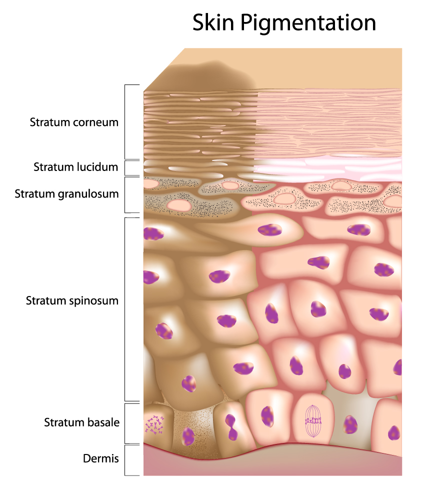
This image is a detailed representation of skin pigmentation and the layers of the skin involved in this process.
Starting from the top layer:
- The Stratum corneum is the outermost layer, consisting of dead keratinized cells that form a protective barrier.
- The Stratum lucidum is a thin, clear layer found only in certain parts of the body like the palms of the hands and the soles of the feet.
- The Stratum granulosum beneath has granules that contribute to waterproofing the skin.
In the middle section, we see the Stratum spinosum, where cells appear spiny as they begin to dry out and flatten.
The deepest epidermal layer is the Stratum basale, which is a single row of cells primarily made up of keratinocytes. This layer is significant for pigmentation because it contains melanocytes, the cells that produce melanin—the pigment responsible for the color of the skin. Melanin is visible in the image as darker spots within the cells. These melanin granules are transferred from the melanocytes to the keratinocytes and move upward through the epidermal layers, giving the skin its color.
The bottommost layer shown is the Dermis, which is not directly involved in pigmentation but provides support and nutrition to the epidermis.
This visual representation underscores the role of the Stratum basale in the pigmentation process, with melanocytes producing melanin and passing it to the surrounding keratinocytes. Skin color variation among individuals is largely due to differences in the type and amount of melanin produced by the melanocytes, which is genetically determined and can also be influenced by exposure to sunlight.
A Second Look at the Fingernail
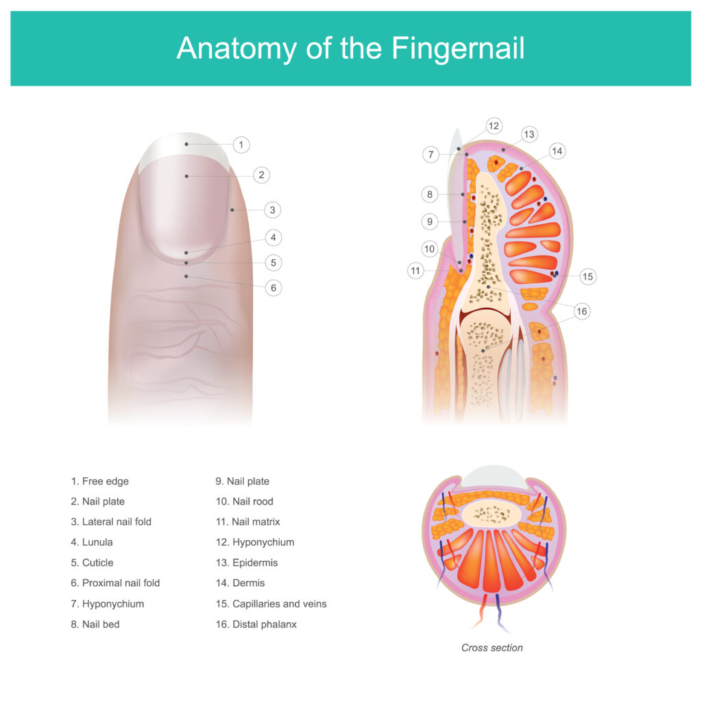
This image displays a detailed diagram of the anatomy of the fingernail, featuring both a dorsal view of the finger and cross-sectional views to illustrate the various components.
In the dorsal view on the left, we can see:
- ‘Free edge’ -- the portion of the nail that extends beyond the finger, which has no underlying skin.
- ‘Nail plate’ -- the hard, translucent part of the nail, made of keratin.
- ‘Lateral nail fold’ -- the fold of skin at the side of the nail.
- ‘Lunula’ -- the whitish half-moon shape at the base of the nail, which is part of the nail matrix.
- ‘Cuticle’ -- the thin tissue that overlaps the nail plate at the base of the nail.
- ‘Proximal nail fold’ -- the fold of skin that covers the nail matrix.
- ‘Hyponychium’ -- the area of epithelium, particularly sensitive skin, under the free edge of the nail plate.
- ‘Nail bed’ -- the skin beneath the nail plate.
In the cross-sectional view on the right, the following structures are indicated:
- ‘Nail plate’ -- as seen in cross-section.
- ‘Nail root’ -- the proximal part of the nail under the skin, where nail growth begins.
- ‘Nail matrix’ -- the tissue (often under the skin) that the nail protects, which generates the cells that become the nail plate.
- ‘Hyponychium’ -- under the free edge, as described above.
- ‘Eponychium’ -- the living tissue that emerges from the proximal nail fold and attaches to the nail plate. It is often mistaken for the cuticle.
- ‘Dermis’ -- the dense inner layer of skin beneath the epidermis, which contains nerve endings and blood vessels.
- ‘Capillaries and veins’ -- the blood vessels providing circulation to the nail bed and matrix.
- ‘Distal phalanx’ -- the bone beneath the nail bed.
The additional, smaller cross-sectional view at the bottom highlights the nail plate’s structure over the nail bed and the surrounding tissue.
Overall, the image serves as an informative tool, offering a clear visual representation of the fingernail’s structure and its different components, which are crucial for its growth and health.
Comprehensive Skin Diagram
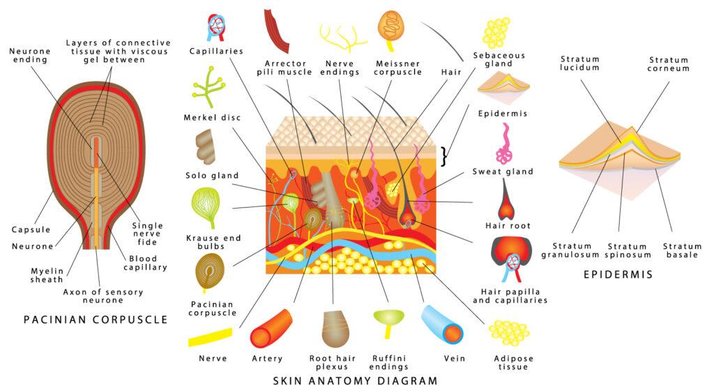
This colorful and detailed image is a comprehensive skin anatomy diagram. It combines an overview of the skin’s structure with detailed insets of various sensory receptors and skin components.
The central part of the image shows a cross-section of the skin, with the various layers labeled. The ‘Epidermis’ is the outermost layer, subdivided into the ‘Stratum basale,’ ‘Stratum spinosum,’ ‘Stratum granulosum,’ ‘Stratum lucidum’ (only present in certain parts of the body like the palms and soles), and the ‘Stratum corneum’ at the very top. The ‘Dermis’ sits below the epidermis, followed by the ‘Subcutaneous tissue’ which consists mainly of adipose tissue.
Embedded within these layers are structures such as the ‘Hair root’ and ‘Sweat gland,’ each playing vital roles in the skin’s function. The ‘Sebaceous (solo) gland’ is shown connected to the hair follicle, secreting sebum that conditions the hair and skin.
Insets around the main diagram focus on the sensory receptors and appendages. The ‘Pacinian corpuscle’ is a large, onion-like structure responsible for sensing deep pressure and vibration. Its detailed structure is shown with its many concentric layers of connective tissue that encase a nerve ending.
The ‘Meissner corpuscle,’ responsible for light touch sensitivity, is depicted as a more elongated structure within the dermal papillae. The ‘Merkel disc,’ a touch receptor, is shown with its associated nerve ending. ‘Krause end bulbs,’ thought to be involved in the sensation of cold, appear as bulbous structures. ‘Ruffini endings,’ which detect stretch and warmth, are also illustrated.
We can also see ‘Free nerve endings’ that permeate the skin layers and are involved in pain, temperature, and mechanical stress detection. ‘Arteries’ and ‘veins’ are represented, indicating the vascular supply of the skin.
The large inset on the left side is a detailed illustration of a ‘Pacinian corpuscle,’ showing its layered capsule and the nerve ending within.
In the top right corner, the diagram also depicts the ‘Layers of connective tissue with blood vessels’ and ‘Capillaries’ within the nervous structure, emphasizing the skin’s complex vascularization.
At the bottom, the labels ‘Nerve,’ ‘Artery,’ ‘Ruffini endings,’ ‘Vein,’ and ‘Adipose tissue’ are color-coded to match the structures within the main diagram.
Overall, this image illustrates the skin’s complexity as an organ, highlighting its role in protection, sensation, thermoregulation, and interaction with the environment. It underscores the diversity of specialized cells and structures that allow the skin to perform its various functions.
Terms and Definitions
| Term | Definition |
|---|---|
| Stratum Basale (Basal Layer) | The deepest layer of the epidermis, containing basal cells that are mitotically active, producing new skin cells. |
| Stratum Spinosum (Spiny Layer) | The layer above the basal layer, named for the spiny appearance of cells due to desmosomal connections, providing strength and flexibility. |
| Stratum Granulosum (Granular Layer) | The layer where keratinocytes accumulate granules of keratohyalin, contributing to water retention and barrier function. |
| Stratum Lucidum (Clear Layer) | A thin, transparent layer found only in thick skin, consisting of dead and flattened keratinocytes. |
| Stratum Corneum (Horny Layer) | The outermost layer made of dead keratinocytes that forms a durable, waterproof barrier. |
| Dermis | The supportive layer below the stratum basale, providing nutrition to the epidermis, not an epidermal layer but included for context. |
| Papillary Region | The superficial area of the dermis with projections that increase the surface area of contact with the epidermis. |
| Reticular Region | The deeper part of the dermis providing structural strength and elasticity due to dense connective tissue. |
| Subcutaneous Fatty Tissue | The bottom-most layer acting as insulation, an energy reserve, and cushioning for the skin. |
| Pain Receptors | Free nerve endings that detect painful stimuli. |
| Sweat Glands | Involved in thermoregulation and excretion. |
| Touch Receptors | Allow the sensation of touch. |
| Capillaries | Small blood vessels that supply the dermis with nutrients and oxygen. |
| Pressure Receptors | Sense mechanical changes in the environment. |
| Nail Matrix | The tissue at the base of the nail bed responsible for producing cells that become the nail plate. |
| Proximal Nail Fold | The skin overlapping the nail matrix. |
| Eponychium (Cuticle) | A layer of clear skin along the bottom edge of the finger or toe, protecting new nail from bacterial infections. |
| Nail Bed | The skin beneath the body of the nail, which is rich in blood vessels. |
| Free Edge | The part of the nail that extends beyond the finger or toe. |
| Phalanx (bone of fingertip) | The distal portion of the fingers or toes. |
| Stratum Germinativum | The deepest layer of the epidermis involved in generating new skin cells. |
| Melanocyte | A cell in the stratum basale that produces the pigment melanin, giving skin its color and protecting against UV radiation. |
| Dendritic Cells | Immune cells within the stratum spinosum that respond to pathogens. |
| Basement Membrane | A thin layer that anchors the epidermis to the dermis. |
| Lunula | The whitish half-moon shape at the base of the nail, visible part of the nail matrix. |
| Lateral Nail Fold | The fold of skin at the side of the nail. |
| Hyponychium | The area of sensitive skin under the free edge of the nail plate. |
| Nail Root | The part of the nail under the skin where nail growth begins. |
| Distal Phalanx | The bone beneath the nail bed. |
