
Table of Contents
Three Stages of Hair Loss
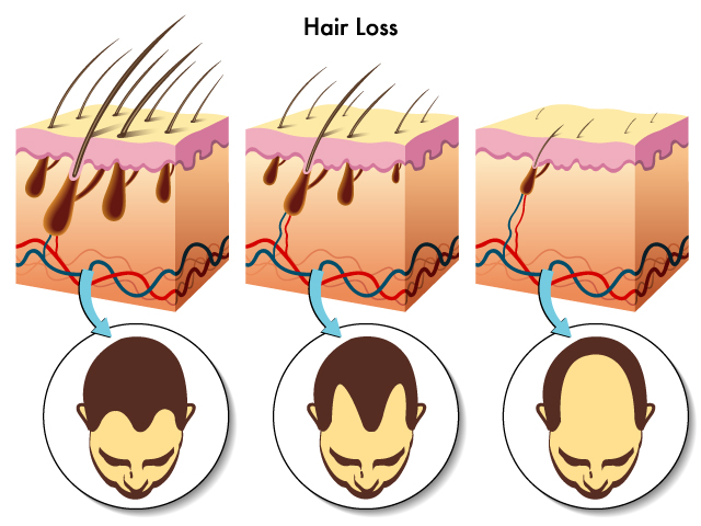
This image depicts the process of hair loss in three stages, along with a cross-sectional view of the skin and hair follicles at each stage.
In the first stage on the left, the skin section shows hair follicles with hair shafts protruding from the epidermal layer of the skin. The follicles are embedded in the dermis, and there is a rich network of blood vessels below them. The corresponding circular icon below shows a person with a full head of hair.
Moving to the middle stage, the hair follicles appear to be in a reduced state with shorter hairs not reaching the previous length, indicating that the hair growth has weakened. This is commonly associated with the miniaturization of hair follicles due to various factors such as hormonal changes, nutritional deficiencies, or genetics. The image below this stage shows a person with a noticeably receding hairline and thinner hair on top.
In the final stage on the right, the hair follicles are shown further miniaturized and the hairs have receded below the skin surface, which is now smooth, reflecting significant hair loss. This could be representative of the condition known as baldness or alopecia, where hair fails to grow back due to the follicles losing their ability to regenerate hair. The icon below shows a person with only a fringe of hair remaining around the sides and back of the head, which is a common pattern of hair loss in male-pattern baldness.
Overall, this illustration summarizes the progression of hair loss, showing how it can lead from a full head of hair to a state where the hair follicles no longer produce visible hairs above the skin. The health and function of the hair follicles are crucial for hair retention, and their gradual diminishment can lead to the common condition of baldness.
Varicella Zoster (Chickenpox)
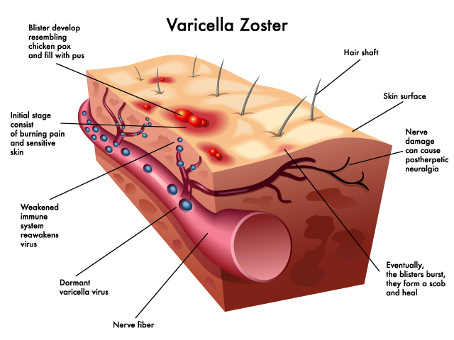
The image depicts the skin condition known as Varicella Zoster, which is the virus responsible for chickenpox in the initial infection and shingles upon reactivation.
At the skin surface, we see blisters that develop, resembling chickenpox and filling with pus, which is typical of a shingles outbreak. These blisters are a response to the viral infection in the skin cells.
Below the surface, the image illustrates the initial stage of the infection, marked by red spots representing areas of burning pain and sensitive skin. This corresponds to the prodromal symptoms of shingles, where pain often precedes the rash.
The deeper layers show the relationship between the virus and the nervous system. We see the Varicella Zoster virus reactivating within a nerve fiber. The virus, which had been dormant in the nerve ganglia after an initial chickenpox infection, can become active again, typically during periods of weakened immune system, which is also noted in the image.
The virus’s presence in the nerve fibers leads to nerve damage, which can cause postherpetic neuralgia—a condition of persistent pain in the area of the shingles rash even after the rash has cleared.
Finally, the image indicates that eventually, the blisters burst, and they form a scab as they heal. This is the body’s natural process of recovering from the blistering rash associated with shingles.
The image summarizes the progression of a shingles infection from the reactivation of the dormant virus to the development of painful skin lesions and potential nerve damage, emphasizing the connection between the virus, skin symptoms, and the nervous system.
Underlying Causes of a Wart

This image illustrates a wart and its underlying cause on human skin. A wart is an overgrowth of skin cells on the epidermis caused by the human papillomavirus (HPV).
The main part of the image shows a cross-section of the skin with a wart protruding from the surface. The wart is depicted as an irregular, rough growth on the skin, comprising clumps of epithelial cells that have proliferated excessively due to the viral infection.
Below the wart, we see the virus (HPV) within the deeper layer of the skin, indicating that it infects the cells in the basal layers of the epidermis. The virus causes these cells to grow rapidly, leading to the formation of the wart on the skin surface.
In the detailed inset, we can see the HPV particles, marked by red spots, infecting an epithelial cell. This close-up view is meant to emphasize the viral cause of the wart.
The image also shows the underlying dermis with a network of capillaries (small blood vessels), highlighted in blue and red, which supply nutrients and oxygen to the skin layers. While warts are generally considered benign, they are fed by these blood vessels, which sustain the infected and overgrown tissue.
Overall, this visualization demonstrates how a common virus can alter the behavior of skin cells, leading to a visible manifestation on the skin’s surface. Warts are typically harmless and can resolve on their own, but they are contagious and can be spread through direct contact.
Progression of a Herpes Zoster Infection
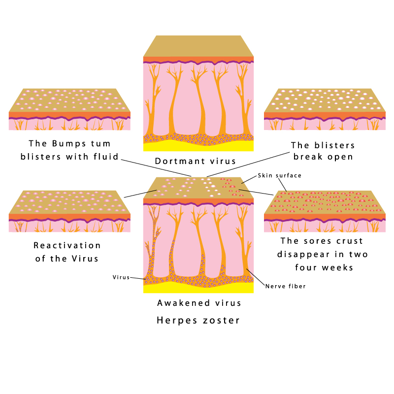
This image depicts the progression of a Herpes zoster infection, commonly known as shingles, which is a reactivation of the Varicella Zoster Virus (VZV) -- the same virus that causes chickenpox.
The first panel on the top left illustrates early symptoms, where ‘The Bumps turn blisters with fluid’. Here, we see the skin’s surface with several raised, fluid-filled blisters.
The second panel on the top middle is labeled ‘Dormant virus’. It shows a cross-section of skin with the virus lying inactive near the nerve fibers, beneath the skin. This is the stage where the virus is not causing any symptoms.
Moving to the top right panel, it says ‘The blisters break open’, depicting the stage where the blisters have ruptured, creating open sores on the skin surface.
The bottom left panel shows ‘Reactivation of the Virus’, where the VZV, which has been dormant within the nerve fibers, reactivates and travels along the nerve to the skin, causing the characteristic painful rash.
The center bottom panel is labeled ‘Awakened virus Herpes zoster’, highlighting the active phase of the virus as it causes the rash and blisters associated with shingles.
Finally, the bottom right panel indicates ‘The sores crust disappear in two four weeks’, where the blisters have dried out and formed crusts that eventually fall off, and the skin heals over a period of two to four weeks.
To summarize, this illustration captures the lifecycle of a shingles outbreak, from the initial skin bumps turning into fluid-filled blisters, to the opening and crusting of these blisters, and the eventual healing of the sores. It also shows the underlying cause, which is the reactivation of the VZV from its dormant state in the nerve fibers, leading to the painful and characteristic skin manifestations.
Four Degrees of Burns
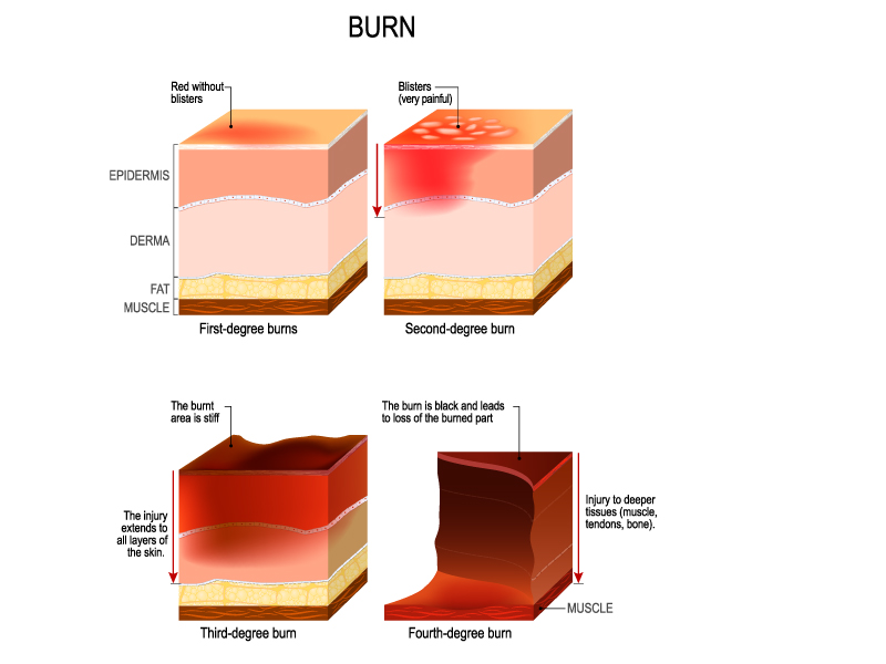
This image provides a visual explanation of the four degrees of burns that can affect the skin and underlying tissues. Each panel shows a different degree of burn severity, progressing from first to fourth degree.
The top left panel illustrates a ‘First-degree burn’, which is characterized by redness without blisters. It affects only the ‘Epidermis’, the outermost layer of the skin. First-degree burns are usually painful and may cause minor inflammation.
The top right panel depicts a ‘Second-degree burn’, identifiable by blisters and very painful. This type of burn extends through the epidermis and into the ‘Dermis’, causing more severe damage which includes swelling and the risk of infection.
The bottom left panel shows a ‘Third-degree burn’. It is described as an area that is stiff and the injury extends through all layers of the skin. In this type of burn, both the epidermis and dermis are destroyed, and there can be damage to the ‘Fat’ layer beneath. The nerve damage in third-degree burns may cause numbness.
Finally, the bottom right panel represents a ‘Fourth-degree burn’, which is the most severe. It describes an injury to deeper tissues (muscle, tendons, bones). Here, the burn extends beyond all layers of the skin and into the underlying ‘Muscle’, and potentially even deeper into tendons and bones. These burns are often life-threatening and can lead to significant scarring and loss of function.
The image effectively categorizes the burn degrees by depth of skin and tissue damage, providing a clear visual for understanding the severity and potential complications of each type.
Different Types of Scars
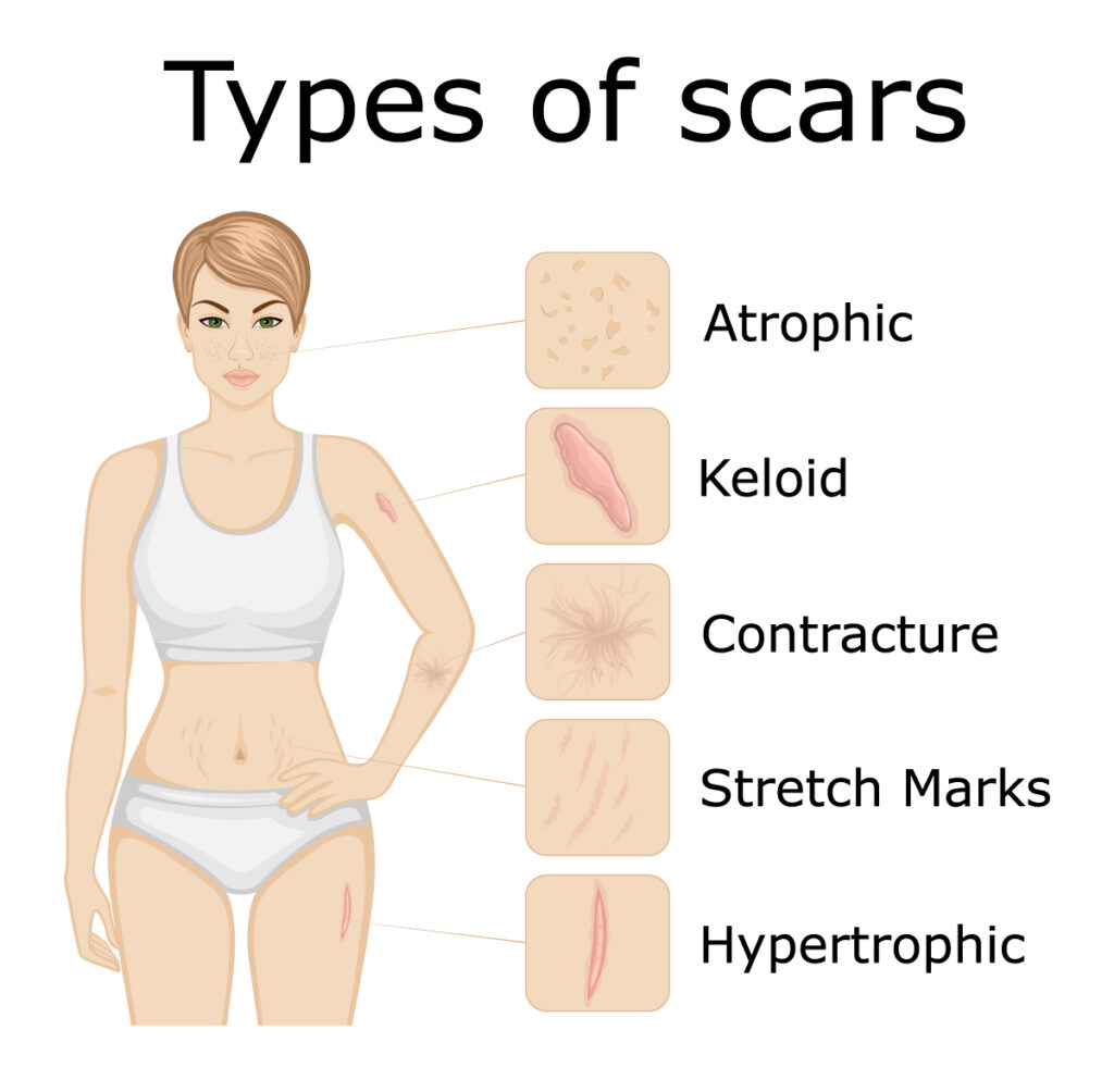
This image depicts an illustration of a woman with annotations pointing to examples of different types of scars that can form on the skin.
The ‘Atrophic’ scar is typically characterized by a sunken appearance, because the skin underlying the scar has been lost; these scars are common with conditions like acne.
A ‘Keloid’ scar is illustrated as a raised, reddish nodular scar that goes beyond the boundary of the original wound. Keloid scars are the result of an overly aggressive healing process and continue to grow, even after the wound has healed.
‘Contracture’ scars are shown as tightened skin that can limit movement. This type of scar usually forms after the skin has been burned. It may involve not just the skin but the underlying muscles and tendons, causing a contracture.
‘Stretch Marks’ are a form of scarring on the skin with an off-color hue. They are caused by tearing of the dermis, which over time may diminish but will not disappear completely.
Finally, ‘Hypertrophic’ scars are thick, raised scars that are similar to keloids but do not go beyond the boundary of the injury. They may reduce in size over time.
The image serves as an educational tool to distinguish between various scar types, showing where they might typically appear on the body and what their general characteristics are.
Common Skin Problems
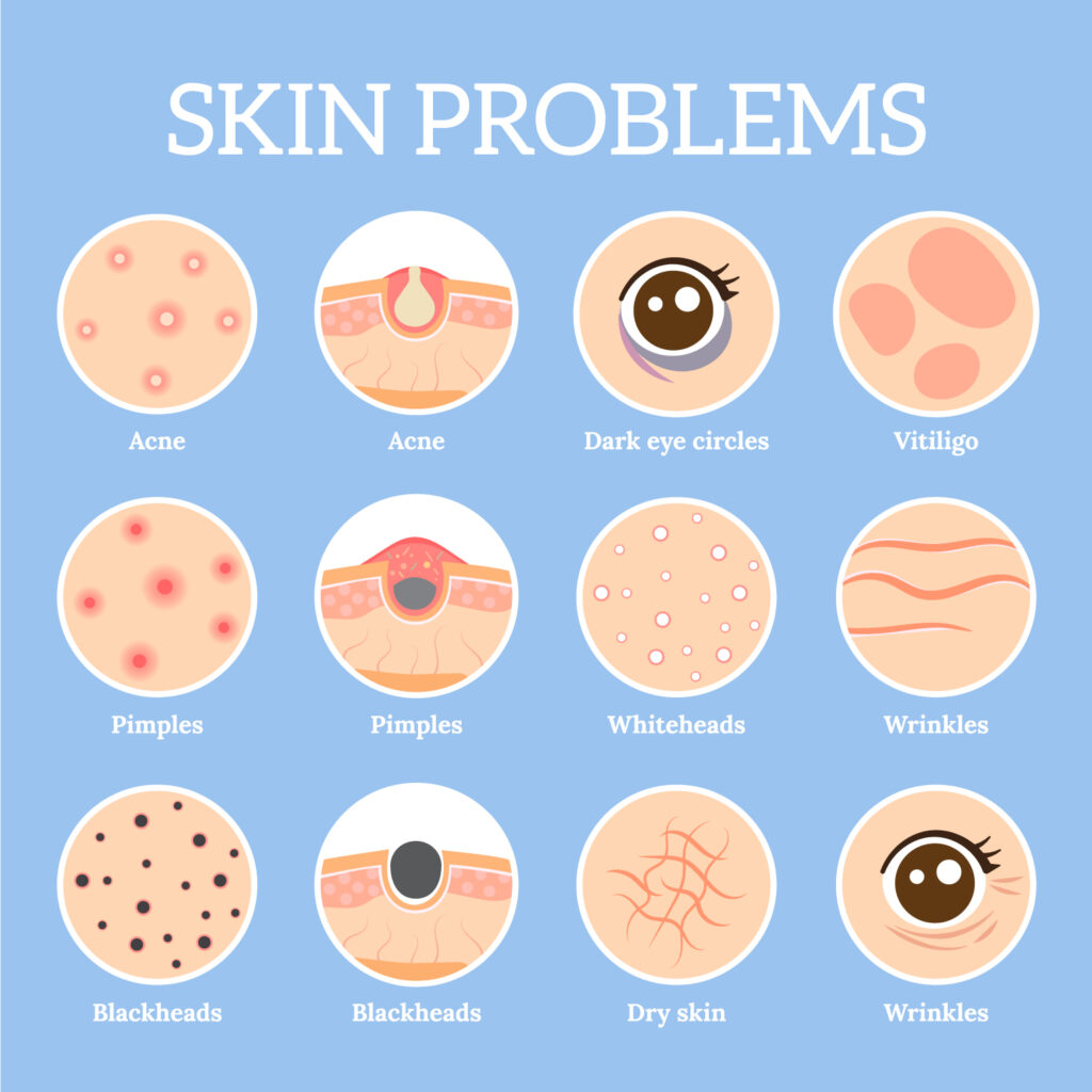
This image provides a visual overview of common skin problems, each represented by a stylized icon.
The first row shows ‘Acne’, depicted by two icons: one showing red pimples with white pus-filled tips on the skin, and the other showing a close-up cross-section of a hair follicle clogged with sebum and dead skin cells, which is typical in acne.
‘Dark eye circles’ are illustrated with an eye icon showing dark shading underneath, which can be due to various factors, including fatigue or genetics.
‘Vitiligo’ is represented by a patch of skin with a significant loss of pigmentation, which is characteristic of this condition where melanocytes (the cells that produce pigment) are destroyed.
The second row displays ‘Pimples’, similar to the acne icons, but focused on the swollen red bumps on the skin’s surface.
‘Whiteheads’ are depicted as white, raised spots on the skin, indicating a type of acne lesion that occurs when dead skin cells, oil, and bacteria become trapped within one of your pores.
‘Wrinkles’ are shown as lines and creases in the skin, commonly associated with aging.
In the third row, ‘Blackheads’ are presented as darkened pores on the skin, another type of acne that occurs when pores are clogged with oil and dead skin cells, the dark surface is due to the oxidation of the debris inside the pore.
‘Dry skin’ is depicted with a cracked texture, indicating the lack of moisture that can lead to a rough and flaky appearance.
Another ‘Wrinkles’ icon closes the visual list, this time focusing on the eye area, showing crow’s feet which are the lines that radiate from the corners of the eyes.
This image serves as an easy-to-understand guide to identifying common skin issues, each with its unique features and visual markers.
