
Table of Contents
Tennis Elbow
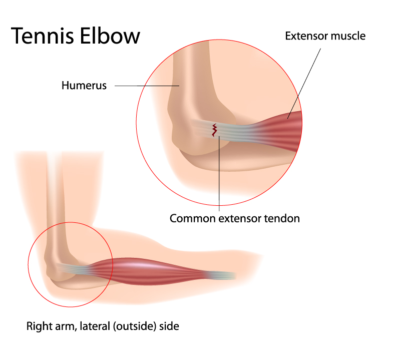
The image depicts a condition commonly known as Tennis Elbow, highlighting the lateral (outside) side of the right arm. The elbow joint, where this condition is focused, involves the distal end of the humerus, the upper arm bone, which is labeled at the top left.
Central to this condition is the common extensor tendon, depicted in the enlarged area. This tendon anchors the extensor muscles, which are responsible for extending the wrist and fingers, to the lateral epicondyle of the humerus. The image shows a zoomed-in view of the elbow where we can observe the extensor muscles transitioning into the common extensor tendon, indicating the point of convergence of these muscles before they attach to the bone.
The common extensor tendon is shown with a series of red and white striations near its attachment point, symbolizing the typical microtears and degeneration associated with Tennis Elbow. This injury is often due to repetitive motion or overuse, particularly gripping activities.
The condition’s name, Tennis Elbow, arises from its prevalence among tennis players. However, it can affect anyone who engages in activities that put a strain on the extensor muscles and tendons of the forearm.
The surrounding anatomy, including the humerus and the muscles of the forearm, are not the primary focus here but serve as a reference point to understand the location and impact of the condition on the arm’s structure and function.
When examining the condition of Tennis Elbow, it’s important to integrate our understanding of the arm’s anatomy to provide context for these considerations. The elbow joint itself is a complex structure involving the articulation of the humerus with the ulna and radius. In the case of Tennis Elbow, the extensor muscles on the lateral side of the forearm play a significant role. Overuse or repetitive motion can lead to the degeneration of the common extensor tendon, which is crucial for wrist and finger extension.
In terms of vascular supply, while not directly implicated in Tennis Elbow, the brachial artery’s continuation into the radial and ulnar arteries does supply blood to the forearm muscles. Any condition affecting the elbow could potentially influence or be influenced by the vascular supply, particularly during the healing process after injury or strain.
From a musculoskeletal perspective, the interosseous membrane and other connective tissues provide structural integrity to the forearm. While these structures are not directly involved in Tennis Elbow, their overall health and functionality can impact recovery and rehabilitation, as they support the muscles and tendons in question.
Lastly, understanding the nerve supply is also pertinent. Although Tennis Elbow primarily affects the tendons and muscles, any swelling or changes in the area could potentially affect nearby nerves. Given that the ulnar and median nerves provide the nerve supply to the forearm, careful consideration during the management of Tennis Elbow is necessary to avoid any nerve compression or injury.
In conclusion, while Tennis Elbow focuses on the tendons and muscles, a comprehensive approach to treatment and rehabilitation should consider the arm’s vascular, nervous, and musculoskeletal systems, as they are interrelated and can affect the healing process and functional recovery.
Cubital Tunnel Syndrome
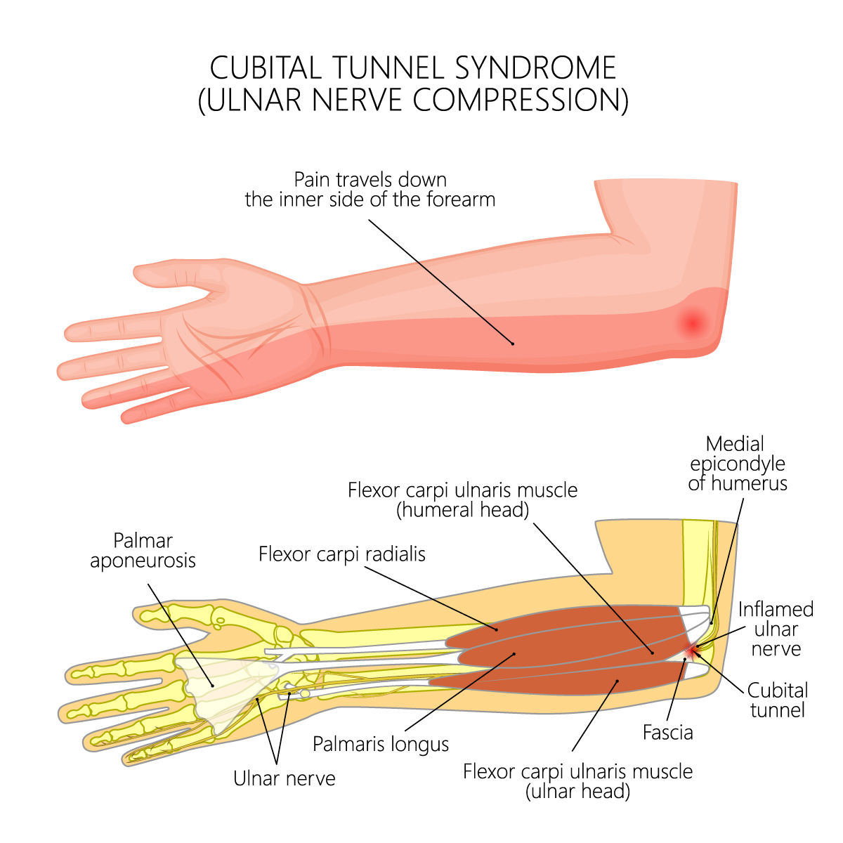
The image illustrates a condition known as Cubital Tunnel Syndrome, which is a type of ulnar nerve compression. This condition often results in pain that radiates along the inner side of the forearm. The ulnar nerve, can become inflamed within the cubital tunnel, an area near the elbow where the nerve passes under a bony bump called the medial epicondyle of the humerus.
This illustration provides a cross-sectional view of the forearm highlighting several anatomical structures. At the elbow, the flexor carpi ulnaris muscle is shown in two segments: one segment attaches to the medial epicondyle of the humerus and the other is labeled near the wrist. This muscle is responsible for flexing and adducting the wrist.
The palmaris longus, a muscle that can vary in presence among individuals and aids in wrist flexion, is also shown. Beneath it, we see the ulnar nerve running down the forearm. This nerve is responsible for the sensory and motor supply to parts of the hand. The palmar aponeurosis, which is a thickened fascial layer in the palm, is indicated at the wrist.
The flexor carpi radialis, a muscle that flexes and abducts the wrist, is adjacent to the flexor carpi ulnaris muscle. The fascia, a connective tissue that encloses muscles and other organs, is covering the area where the inflamed ulnar nerve is situated.
It is important to note that the cubital tunnel is not directly shown but is implied to be the area where the inflammation of the ulnar nerve is highlighted. The inflammation is indicated by a red area suggesting irritation or compression of the nerve within this confined space. The cubital tunnel itself is a channel that allows the ulnar nerve to travel over the elbow. When this nerve becomes compressed or irritated, it can lead to symptoms such as pain, numbness, or tingling in the forearm, hand, and fingers, which is characteristic of Cubital Tunnel Syndrome.
Golfer's Elbow Pathology
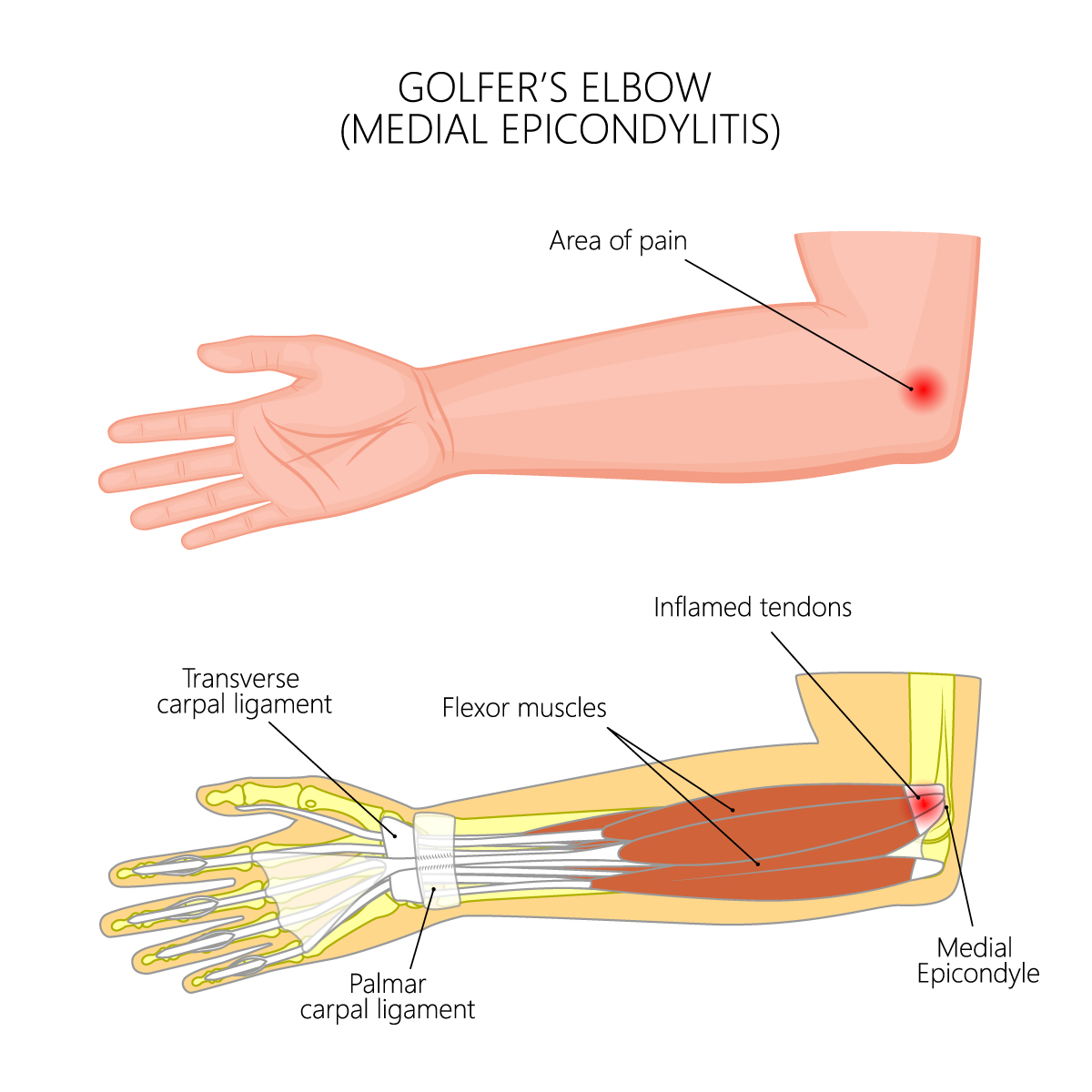
The illustration presents the condition known as Golfer’s Elbow, also medically termed Medial Epicondylitis. This condition is characterized by pain centered around the medial epicondyle of the humerus, which is on the inner side of the elbow. The pain area is highlighted on the upper part of the illustration.
This condition specifically involves inflammation of the tendons that attach the forearm flexor muscles to the medial epicondyle. The forearm flexor muscles, responsible for flexing the fingers and wrist, are shown in the lower part of the image, extending from the medial epicondyle toward the wrist and hand. The inflamed tendons are depicted in the area where the muscles converge before attaching to the medial epicondyle, noted by the presence of red shading indicating inflammation.
Also visible in the detailed view of the wrist are the transverse carpal ligament and the palmar carpal ligament. These ligaments are part of the wrist’s anatomy and play a role in the stability and movement of the wrist and hand, although they are not directly implicated in Golfer’s Elbow.
Golfer’s Elbow is a result of overuse, particularly repetitive wrist flexion or gripping activities, which can lead to the inflammation and pain shown in the image. Despite its name, the condition is not limited to golfers and can affect anyone who performs activities that put a similar strain on the forearm muscles and tendons.
In relation to the image of Golfer’s Elbow (Medial Epicondylitis), additional anatomical considerations from the knowledge source include the role of the ulnar nerve, which runs in close proximity to the medial epicondyle. While the image does not explicitly depict the nerve, it’s important to note that inflammation in the area could potentially affect the ulnar nerve, leading to symptoms such as numbness or tingling in the ring and little fingers.
From the perspective of the muscles, the forearm’s flexor muscles originate from the medial epicondyle and are responsible for various movements including flexing the wrist and fingers. Overuse or repetitive motions can strain these muscles and their tendons, contributing to the development of Golfer’s Elbow. The image shows these muscles and highlights the inflamed tendons that attach them to the medial epicondyle.
Regarding blood supply, the brachial artery, which runs down the arm and divides into the radial and ulnar arteries, supplies blood to the forearm. The ulnar artery is particularly relevant in this context as it supplies blood to the flexor muscles shown in the image. Proper vascularization is essential for delivering nutrients and oxygen to the tissues for healing and repair.
It’s also useful to mention the interosseous membrane of the forearm, which runs between the radius and ulna. While not shown in the image, this membrane is pertinent as it contributes to the overall structural integrity of the forearm and facilitates the transmission of forces across the forearm.
Finally, understanding the proximal and distal relationships of the radius and ulna is vital, as these bones form the structure of the forearm, within which the pathology of Golfer’s Elbow occurs. The forearm’s ability to rotate, pronate, and supinate is integral to many of the actions that could lead to overuse injuries such as Golfer’s Elbow.
Collectively, these additional considerations provide a more comprehensive understanding of the anatomical context in which Medial Epicondylitis occurs and its implications for the function of the arm.
