
Table of Contents
Left Hand Bones: Anterior View
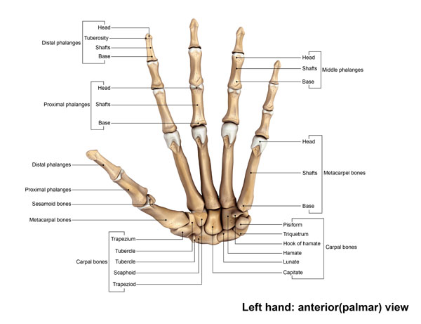
This image illustrates the skeletal structure of the left hand from an anterior or palmar view, which is what you would see if you looked directly at the palm of your hand.
At the base of the structure, we have the carpal bones, which form the wrist. These bones are arranged in two rows. The proximal row, closer to the forearm, includes the scaphoid, lunate, triquetrum, and pisiform bones. The distal row, closer to the fingers, includes the trapezium, trapezoid, capitate, and hamate bones. The hamate is notable for its hook-like projection, known as the hook of hamate.
Moving towards the fingers, the metacarpal bones form the framework of the hand itself. Each metacarpal has a base that articulates with the carpal bones, a shaft, and a head that connects to the phalanges. There are five metacarpal bones, one for each digit.
The phalanges are the bones of the fingers. Each finger has three phalanges – the proximal, middle, and distal phalanges, except for the thumb, which has only two, the proximal and distal. The distal phalanges form the tips of the fingers and have a unique structure with a tuft that allows for the attachment of the fingertip pads and nails.
Additionally, there are sesamoid bones, typically found within tendons, which vary in number and placement among individuals. In the hand, they are commonly found at the base of the thumb.
Overall, this intricate arrangement of bones allows for the complex movements of the wrist and fingers, enabling a wide range of activities from fine motor skills to grasping and manipulation of objects.
Skeletal Anatomy: Articulation Points
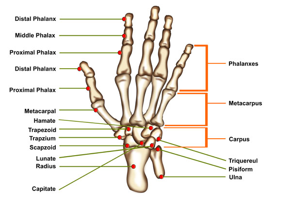
In this image, we see the skeletal anatomy of a hand, with a focus on the articulation points, which are highlighted by red dots. These points of articulation are where the bones meet and are connected by joints, allowing for movement.
Starting at the fingertips, there are three sets of phalanges for each finger: the distal, middle, and proximal phalanges. The distal phalanges are at the tips of the fingers, the middle phalanges are in the center, and the proximal phalanges connect to the metacarpal bones. The thumb is an exception, having only a distal and a proximal phalanx, with no middle phalanx.
The metacarpal bones run from the wrist to connect to the proximal phalanges of the fingers. These are long bones that form the palm of the hand. Each metacarpal bone articulates with a carpal bone at its base and a proximal phalanx at its head.
The carpus, or wrist area, is made up of eight small carpal bones. The capitate bone is centrally located and articulates with the base of the third metacarpal. The hamate is next to the capitate and features a hook-like projection. The trapezoid and trapezium articulate with the second and first metacarpal bones, respectively. The scaphoid bone is located near the thumb and is one of the most commonly fractured carpal bones. The lunate bone articulates with the radius, one of the two bones in the forearm.
The triquetral and pisiform are on the ulnar side of the wrist, which is the side opposite the thumb. The pisiform is a sesamoid bone within the tendon of the flexor carpi ulnaris muscle. The radius and ulna are the two bones of the forearm, with the radius being on the thumb side and the ulna on the little finger side.
This complex arrangement of bones and their joints allows the hand to perform a wide range of movements, from precise motions to powerful grips.
Color-coded Illustration of the Hand Bones
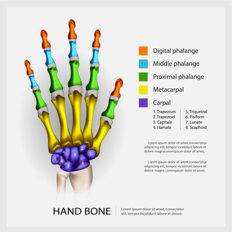
This vibrant illustration is a color-coded representation of the bones of the human hand. Each bone type is assigned a specific color, making it easy to distinguish between them.
The distal phalanges are highlighted in orange. These are the bones at the tips of the fingers and thumb, and they support the fingernails and help us to manipulate small objects.
The middle phalanges are colored in blue. These are present in all the fingers except for the thumb, which does not have a middle segment.
The proximal phalanges, shown in green, are closest to the hand and connect to the metacarpals. They form the base of the fingers and are part of the knuckles when you make a fist.
The metacarpal bones are depicted in yellow. These are the long bones inside the main area of the hand, which connect the carpal bones of the wrist to the finger bones.
The carpal bones, represented in purple, form the wrist and are arranged in two rows at the base of the hand. These bones are crucial for wrist motion and stability.
The carpal bones are individually named in the illustration:
- The trapezium is at the base of the thumb, allowing for the thumb’s unique range of motion.
- The trapezoid is found next to the trapezium and is the smallest carpal bone in the distal row.
- The capitate is the largest carpal bone, located at the center of the wrist.
- The hamate is recognizable by its hook-like projection on the palmar side of the hand.
- The triquetrum is involved in complex movements of the wrist.
- The pisiform is a small, pea-shaped carpal bone that articulates with the triquetrum.
- The lunate bone is centrally located within the proximal row and articulates with the radius bone of the forearm.
- The scaphoid bone is situated near the base of the thumb and is one of the most frequently fractured carpal bones.
Understanding the hand’s bone structure is fundamental in various fields, from anatomy and medicine to ergonomics and prosthetics design. The hand’s complex structure allows for a remarkable range of motion and the ability to perform precise tasks.
Finger Anatomy
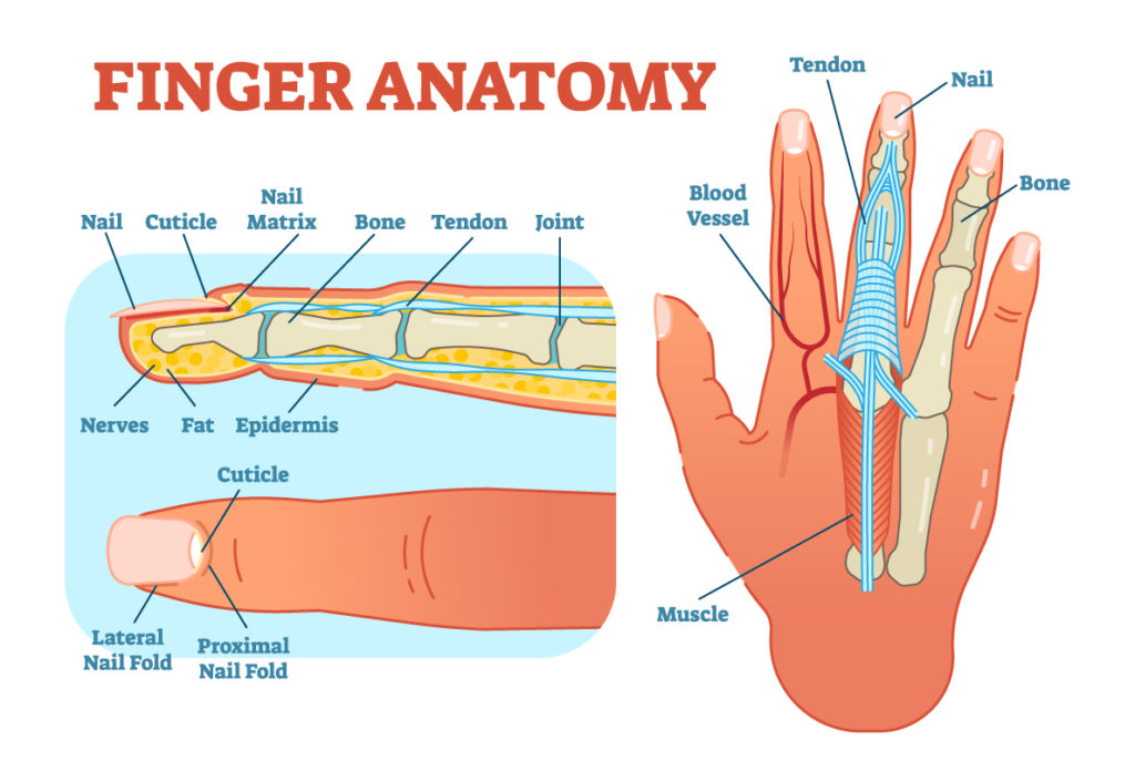
The diagram presents a detailed look at finger anatomy, dissecting the layers and structures that make up a finger.
On the left, a cross-section of a fingertip is depicted. Starting from the top layer, we see the nail, which is a hard keratinous structure providing protection to the fingertip. Below the nail is the nail matrix, which is the tissue under the nail that it grows from. The cuticle is the skin at the base of the nail, which acts as a seal protecting the nail matrix from infection.
Beneath the nail and cuticle, the bone of the fingertip provides the rigid structure to the finger. Surrounding the bone is a network of tendons, which are fibrous connective tissues attaching muscle to bone, allowing for movement. Joints are where two bones meet and are essential for finger flexibility.
On the sides, we see the lateral and proximal nail folds, which are skin folds that outline the nail on three sides. Underneath the skin, there is a layer of fat providing cushioning, followed by nerves responsible for the sensation in the finger. The epidermis, the outermost layer of skin, provides a protective barrier.
On the right side of the diagram, a whole finger is shown with the internal structures visible. The tendons run along the length of the finger, leading down to the muscles in the hand and forearm that control finger movement. Blood vessels are shown adjacent to the tendons, supplying the finger with oxygen and nutrients, and removing waste products.
This comprehensive view demonstrates how the bones, muscles, tendons, nerves, and blood vessels work together to provide structure, sensation, and movement to the finger, all of which are protected by the outer layers of skin and nail.
Anatomy of the Wrist
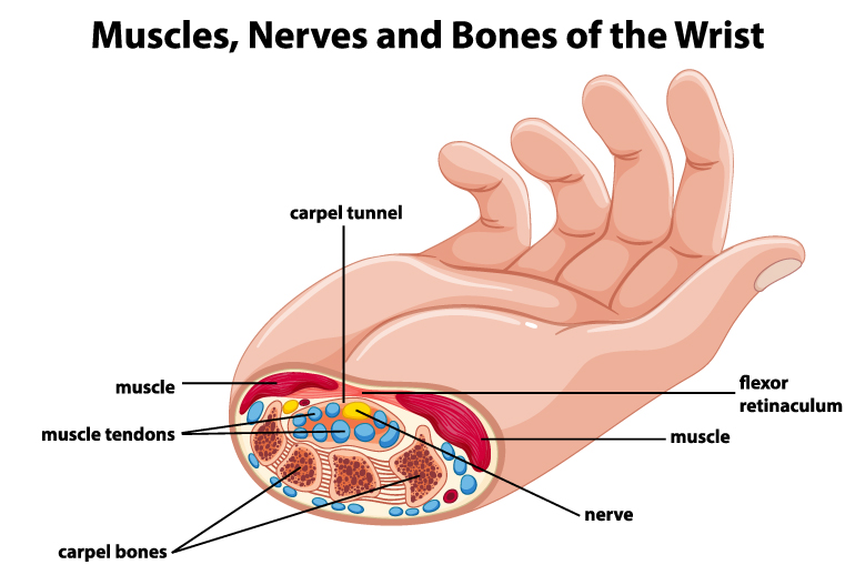
The image provides a cross-sectional view of the wrist, revealing the muscles, nerves, and bones that comprise this complex area.
Central to the image is the carpal tunnel, a narrow channel in the wrist through which the median nerve and several tendons pass. This tunnel is formed by the carpal bones on the bottom and the flexor retinaculum, a strong fibrous band, on the top.
The carpal bones are the small bones that make up the wrist and form the floor of the carpal tunnel. They are arranged in two rows and provide a sturdy base for the hand.
Muscle tendons, which appear as bundled cords, run through the carpal tunnel. These tendons belong to the muscles that flex the fingers and thumb. Their movement is facilitated by the synovial sheath, a lubricating membrane that surrounds the tendons.
The median nerve, depicted in yellow, also traverses the carpal tunnel. It provides sensation to the palm side of the thumb, index, middle, and part of the ring fingers, as well as motor functions to some small muscles at the base of the thumb.
Surrounding the carpal tunnel, we see muscles of the hand and forearm. These muscles control a variety of movements from gross grasp to fine motor skills.
Above the cross-sectional view, the image shows the muscles as they would appear when the skin is intact, giving context to the underlying anatomical structures.
Understanding the relationship between these elements is important for diagnosing and managing conditions like carpal tunnel syndrome, where pressure on the median nerve can lead to pain, tingling, and weakness in the hand and arm.
Carpal Tunnel Anatomy
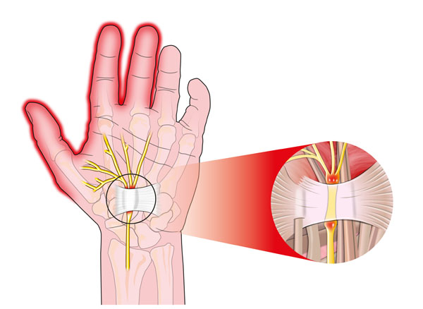
The image illustrates the intricate network of nerves and blood vessels within the palm of the hand.
The central focus is the carpal tunnel, a narrow passageway in the wrist encased by bones and ligaments. Through this tunnel, the median nerve and several tendons pass from the forearm into the hand. The median nerve is responsible for sensation in the thumb, index, middle, and part of the ring finger, as well as motor functions in some small muscles of the hand.
Surrounding the median nerve, we see the tendons that connect muscles of the forearm to the bones of the hand and fingers, allowing for movement. These tendons are encased in sheaths to reduce friction as they slide back and forth during hand movements.
The inset on the right side of the image provides a closer look at the carpal tunnel, highlighting the median nerve and tendons within it. We can observe the compact nature of this space, which is why any swelling or inflammation here can lead to carpal tunnel syndrome, a condition that causes pain, tingling, and numbness in the hand and arm.
The blood vessels running alongside the tendons supply the hand with oxygenated blood and nutrients, which is vital for the health and function of the tissues.
Understanding this anatomy is crucial for diagnosing and treating conditions related to nerve compression and circulatory issues within the hand.
Anatomical Terms and Definitions
| Term | Definition |
|---|---|
| Carpal Bones | The bones that form the wrist, arranged in two rows: the proximal row (scaphoid, lunate, triquetrum, pisiform) and the distal row (trapezium, trapezoid, capitate, hamate). The hamate is noted for its hook-like projection. |
| Carpal Tunnel | A narrow passageway in the wrist encased by bones and ligaments, through which the median nerve and several tendons pass from the forearm into the hand. |
| Cuticle | The skin at the base of the nail, acting as a seal protecting the nail matrix from infection. |
| Distal Phalanges | The bones at the tips of the fingers and thumb, supporting the fingernails and helping manipulate small objects. |
| Flexor Retinaculum | A strong fibrous band that forms the top of the carpal tunnel. |
| Hook of Hamate | A hook-like projection on the hamate bone, part of the carpal bones in the wrist. |
| Metacarpal Bones | The long bones inside the main area of the hand, connecting the carpal bones of the wrist to the finger bones. |
| Middle Phalanges | The bones present in the center of each finger, except for the thumb, which lacks a middle segment. |
| Nail Matrix | The tissue under the nail from which it grows. |
| Phalanges | The bones of the fingers. Each finger has three phalanges – proximal, middle, and distal, except for the thumb, which has only two – proximal and distal. |
| Proximal Phalanges | The bones closest to the hand, forming the base of the fingers and part of the knuckles. |
| Sesamoid Bones | Small bones typically found within tendons, varying in number and placement, commonly found at the base of the thumb in the hand. |
| Synovial Sheath | A lubricating membrane that surrounds muscle tendons to reduce friction during movement. |
| Tendons | Fibrous connective tissues that attach muscle to bone, allowing for movement. |
