
Table of Contents
Facial Muscle Anatomy
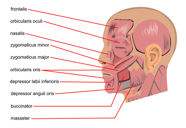
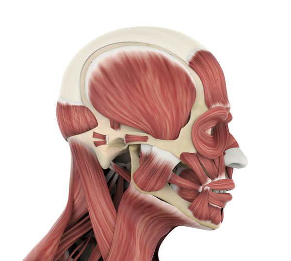
This image illustrates several of the facial muscles, which are primarily responsible for facial expression. Starting from the top, the frontalis muscle covers the forehead. Its main function is to raise the eyebrows and wrinkle the forehead.
The orbicularis oculi muscle encircles the eye socket. It acts to close the eyelids and is involved in the various facial expressions such as blinking and winking.
Moving to the mid-face, the nasalis muscle is located over the bridge of the nose. It compresses the bridge and depresses the tip of the nose, playing a significant role in expressions like flaring the nostrils.
Below this muscle, we have the zygomaticus minor and the zygomaticus major, both of which originate from the zygomatic bone. The zygomaticus minor helps to elevate the upper lip, contributing to expressions such as smiling. The zygomaticus major is larger and also elevates and draws the upper lip outward.
Near the mouth, the orbicularis oris acts like a sphincter for the mouth, allowing actions like puckering the lips and closing the mouth.
On the lower side of the face, the depressor labii inferioris is responsible for lowering the bottom lip, while the depressor anguli oris draws the corners of the mouth downward and laterally, which is often associated with frowning or sadness.
The buccinator muscle is situated deep in the cheek. It pulls the cheek against the teeth and is used in actions like blowing and chewing.
Finally, the masseter muscle is a thick, rectangular muscle at the angle of the jaw. This muscle is a primary mover in mastication (chewing), elevating the mandible to close the mouth and clench the teeth.
Together, these muscles not only allow us to make a wide array of facial expressions but also have functional roles in eating and breathing.
Neck Muscle Anatomy
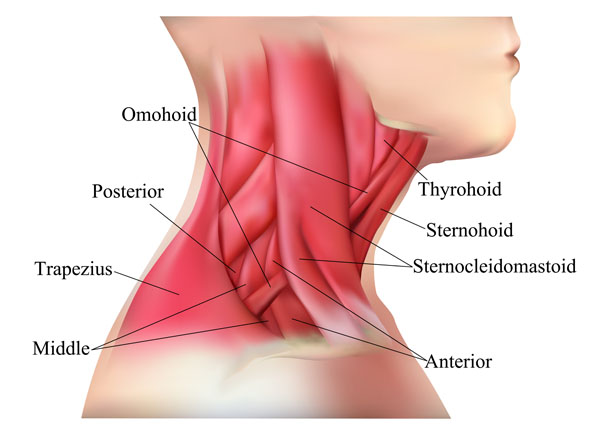
The image presents a lateral view of the neck highlighting various muscles that support neck movements, swallowing, and other functions.
The trapezius muscle is one of the major muscles of the back and is partially visible here at the back of the neck. It extends from the occipital bone to the lower thoracic vertebrae and to the scapula, aiding in moving the shoulder blades and supporting arm movements.
In front of the trapezius, we have the sternocleidomastoid muscle, which is easily recognizable due to its prominent position and function. It runs obliquely across the side of the neck, originating from the sternum and clavicle and inserting at the mastoid process of the temporal bone. This muscle is involved in rotating the head to the opposite side and flexing the neck. When both sternocleidomastoid muscles act together, they flex the neck or elevate the sternum, aiding in forced inhalation.
Deep to the sternocleidomastoid, we find the omohyoid muscle. It is a slender muscle that extends from the superior border of the scapula to the hyoid bone and is involved in lowering the hyoid bone and larynx during swallowing.
Below the omohyoid, the image shows the thyrohyoid muscle, which lies beneath the sternohyoid and is a small muscle that stretches between the thyroid cartilage and the hyoid bone. This muscle depresses the hyoid and elevates the larynx, which occurs during swallowing.
The sternohyoid muscle is a narrow, strap-like muscle in the midline of the neck, superficial to the thyrohyoid. It originates from the manubrium of the sternum and clavicle and inserts on the hyoid bone. Its primary action is to depress the hyoid bone after it has been elevated during swallowing or speaking.
These muscles, in addition to their primary functions, also contribute to the contour of the neck and play a role in various physiological activities like speaking, breathing, and positioning the head.
Neck Muscles: Posterior View
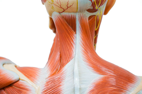
In this anatomical image, we observe a detailed view of the musculature of the neck from a posterior perspective. The most prominent muscle visible here is the trapezius, which extends across the back of the neck and shoulders. The trapezius muscle is divided into three parts: the superior or upper part, which supports the weight of the arm; the middle part, which retracts the scapula; and the inferior or lower part, which rotates and depresses the scapula.
Just beneath the lower border of the trapezius, we can see the omohyoid muscle. This is a unique muscle due to its two bellies connected by an intermediate tendon. The omohyoid depresses the hyoid bone and is involved in swallowing.
Moving to the anterior aspect, we find the sternocleidomastoid muscle, which is one of the most prominent and easily visible muscles at the front of the neck. This muscle runs obliquely across the side of the neck, starting from the sternum and clavicle and inserting at the mastoid process of the temporal bone. It acts to rotate and flex the neck.
Beneath the sternocleidomastoid, we see the sternohyoid muscle, a narrow muscle running vertically along the midline. It is one of the infrahyoid muscles, and its function is to depress the hyoid bone, which is important during the swallowing process.
The thyrohyoid muscle, located superior to the sternohyoid, is a small muscle running between the thyroid cartilage and the hyoid bone. Its function is to decrease the distance between the thyroid cartilage and the hyoid bone, also participating in swallowing and speech.
These muscles play vital roles in the movement and stabilization of the neck, as well as in functions such as swallowing and speaking. The arrangement of these muscles is crucial for the structural integrity and functional movement of the head and neck.
Nerves and Structures of the Head and Neck
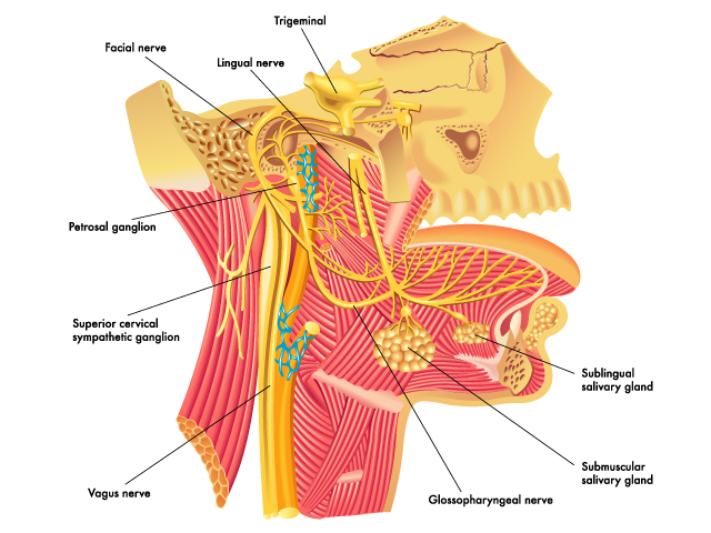
This diagram illustrates the intricate network of nerves and structures within the head and neck, focusing on the cranial nerves and associated ganglia.
The trigeminal nerve is prominently featured here, which is the main sensory nerve of the face and motor nerve for the muscles of mastication.
The facial nerve is also shown, which controls the muscles of facial expression, and provides taste sensations from the anterior two-thirds of the tongue and oral cavity.
We see the lingual nerve, which is a branch of the mandibular division of the trigeminal nerve, providing sensory innervation to the front two-thirds of the tongue.
The petrosal ganglion represented here is part of the glossopharyngeal nerve, which is involved in taste and other functions of the posterior third of the tongue, and contributes to the innervation of the parotid salivary gland via the otic ganglion.
The superior cervical sympathetic ganglion is part of the sympathetic nervous system, responsible for the ‘fight or flight’ response. It innervates the eye and blood vessels in the head, among other structures.
The vagus nerve, the longest of the cranial nerves, controls a range of functions including heart rate, gastrointestinal peristalsis, sweating, and quite a few muscle movements in the mouth, including speech (via the larynx).
The glossopharyngeal nerve has a mixed function, including taste from the posterior one-third of the tongue and innervation of the stylopharyngeus muscle, which elevates the pharynx and larynx during swallowing and speech.
The sublingual salivary gland and submandibular salivary gland are shown, which produce saliva that enters the mouth beneath the tongue and at the floor of the mouth, respectively.
Together, these nerves and ganglia contribute to the complex functions of sensation, taste, salivation, swallowing, and the regulation of blood pressure and heart rate, highlighting the complexity and coordination required for the normal functioning of the head and neck.
Head and Neck Muscles: Profile View
This image provides an illustrative view of the musculature of the head and neck. It shows a profile view of a skull overlaid with the muscles that are responsible for facial expression, mastication (chewing), and movements of the head.
The large muscle covering the top and side of the skull is the temporalis muscle, which is a fan-shaped muscle important for chewing. It originates from the temporal fossa and the fascia covering the temporal bone, and it inserts onto the coronoid process of the mandible (jawbone).
We can also see the orbicularis oris muscle encircling the mouth, which controls movements such as puckering and pursing of the lips. This muscle is vital for actions like speaking and expressing emotions.
The masseter muscle, one of the strongest muscles in the body, is shown on the side of the face, covering the lateral aspect of the mandible. It is crucial for elevating the jaw to close the mouth, and it plays a significant role in mastication.
Other muscles visible in this image include those around the eye socket. For example, the orbicularis oculi muscle, which is responsible for closing the eyelids, is shown.
Additionally, we can observe the muscles of the neck, including portions of the sternocleidomastoid muscle, which runs down the side of the neck and is involved in rotating and flexing the head.
These muscles are intricately connected to the bones of the skull and mandible, and their coordinated contractions enable us to chew, speak, and make a wide range of facial expressions.
Nerve and Vascular Network: Side View
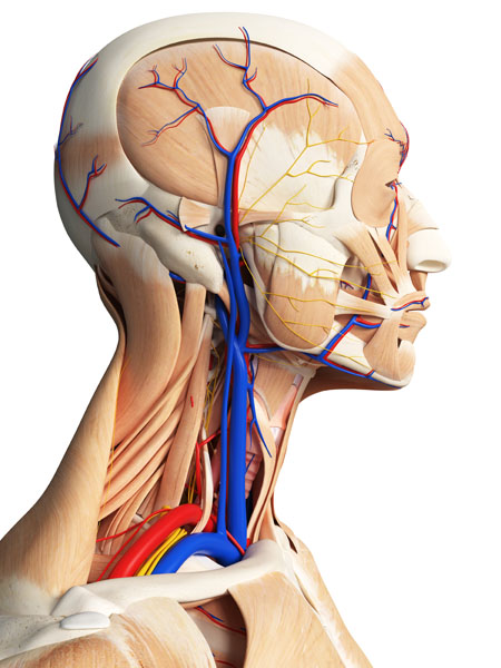
This detailed image presents a profile view of the head and neck, highlighting both the musculature and the vascular network. The blue vessels are veins, which carry deoxygenated blood back to the heart, while the red vessels are arteries, which transport oxygenated blood from the heart to nourish the tissues.
In the head region, the intricate arterial and venous networks around the scalp and face are visible. These include the superficial temporal artery and vein, which can be seen branching across the temple area.
The facial artery and vein are also shown, with branches that supply and drain blood from the facial muscles and skin. These vessels are essential for providing the tissues of the face with oxygen and nutrients, and for removing metabolic waste.
Within the neck, the image shows the carotid arteries (common, external, and internal branches) traveling up each side of the neck. These are key pathways delivering blood to the brain, face, and neck. The internal jugular veins are also visible, running parallel to the carotid arteries, returning blood from the brain back towards the heart.
The musculature shown includes the sternocleidomastoid muscles of the neck, which are prominent and play a role in rotating and flexing the head. We can also see some of the muscles responsible for facial expression, although they are not the main focus of this image.
Overall, the image captures the complexity of the anatomical structures in the head and neck and the vital role of the circulatory system in maintaining the functions of these areas.
Blood Supply of the Head and Neck
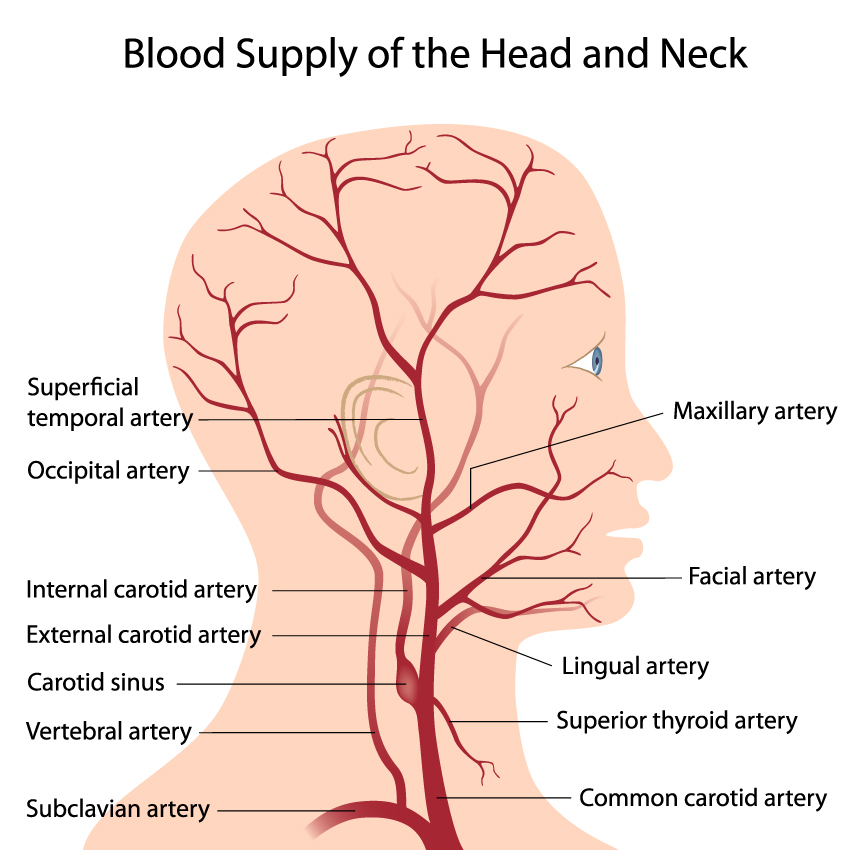
The image illustrates the arterial blood supply to the head and neck. The primary arteries labeled here originate from the aorta, branch off in the neck, and then spread throughout the head and upper neck.
The common carotid artery is a major artery that runs up the neck and divides into the internal and external carotid arteries. The internal carotid artery supplies blood to the brain, while the external carotid artery branches out to supply the face, scalp, and neck.
The external carotid artery further divides into several branches:
- The superior thyroid artery supplies the thyroid gland and parts of the larynx.
- The lingual artery provides blood to the tissues of the tongue.
- The facial artery supplies blood to the superficial face, including the oral and nasal cavities.
- The maxillary artery is a major supplier of deep facial structures, including the teeth and muscles of mastication.
- The superficial temporal artery ascends to supply the scalp and portions of the face and ear.
Also indicated is the occipital artery, which primarily supplies blood to the posterior scalp.
The vertebral artery, a branch of the subclavian artery, is shown entering the transverse foramina of the cervical vertebrae. It will ascend through the neck and enter the cranium to supply the posterior part of the brain.
Lastly, the subclavian artery is depicted as the continuation of the aortic arch that supplies blood to the arms, with some branches supplying the thoracic wall and other parts of the body.
These arteries, along with their branches, ensure that the head and neck receive an adequate supply of oxygenated blood, which is essential for the functioning of the brain, facial muscles, and sensory organs.
Cranial Nerves and Vascular Network: Lateral View
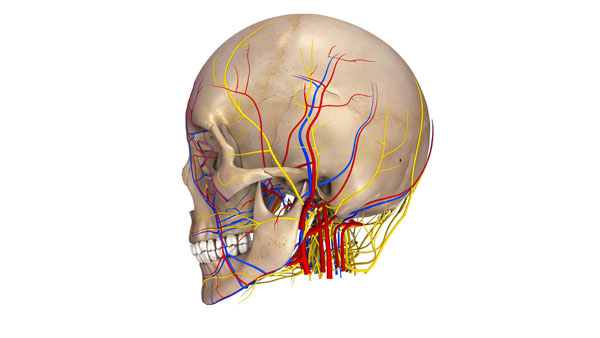
The image displays a lateral view of a human skull with an overlay of the cranial nerves and blood vessels. The cranial nerves are color-coded in yellow, and the blood vessels are color-coded in red and blue, indicating arteries and veins, respectively.
The cranial nerves are part of the peripheral nervous system and emerge directly from the brain. This image likely depicts several of the twelve cranial nerves, which are responsible for sensory and motor functions of the head and neck. Some of the nerves could be the trigeminal nerve, which is responsible for facial sensation and motor functions such as biting and chewing, or the facial nerve, which controls the muscles of facial expression.
The red blood vessels are arteries, which carry oxygenated blood away from the heart to the brain and the facial tissues. The blue vessels are veins, which carry deoxygenated blood back to the heart. The arterial system in the head typically includes branches of the internal and external carotid arteries, while the venous system includes the superficial and deep veins of the head and neck that drain into the internal jugular veins.
This complex network of nerves and vessels is crucial for providing the brain with nutrients and oxygen and for carrying out the brain’s commands to the facial muscles and sensory organs.
Cranial Nerves and Vascular Network: Posterior
This image provides a posterior view of the skull with a visualization of the cranial nerves and blood vessels. The cranial nerves, depicted in yellow, are key components of the peripheral nervous system and are responsible for transmitting sensory information and motor commands between the brain and various areas of the head and neck.
The red vessels represent the arterial supply, which carries oxygen-rich blood to the cranial tissues, and the blue vessels represent the venous drainage, carrying deoxygenated blood away from the cranial cavity back to the heart.
The layout of the nerves and vessels around the base of the skull is intricate, with multiple paths crossing over and branching out to supply and innervate specific regions. The distribution pattern reflects the body’s need to maintain a complex, tightly regulated network for proper physiological function. This network ensures that the brain receives the necessary oxygen and nutrients and that the sensory and motor functions are carried out effectively.
Anatomical Terms and Definitions
| Term | Definition |
|---|---|
| Buccinator | A muscle deep in the cheek that pulls the cheek against the teeth, used in actions like blowing and chewing. |
| Cranial Muscles | Muscles that are part of the epicranius, involved in scalp movement. |
| Depressor Anguli Oris | A muscle that draws the corners of the mouth downward and laterally, often associated with frowning or sadness. |
| Depressor Labii Inferioris | A muscle responsible for lowering the bottom lip. |
| Frontalis | A muscle covering the forehead, responsible for raising the eyebrows and wrinkling the forehead. |
| Levator Anguli Oris | A muscle that raises the corner of the mouth, involved in smiling. |
| Masseter | A thick, rectangular muscle at the angle of the jaw, primary mover in mastication (chewing), elevating the mandible to close the mouth and clench the teeth. |
| Mentalis | A muscle situated at the tip of the chin, responsible for protruding the lower lip as in a pout. |
| Nasalis | A muscle located over the bridge of the nose, compresses the bridge and depresses the tip of the nose, playing a significant role in expressions like flaring the nostrils. |
| Orbicularis Oculi | A muscle that encircles the eye socket, acting to close the eyelids, involved in facial expressions such as blinking and winking. |
| Orbicularis Oris | Acts like a sphincter for the mouth, allowing actions like puckering the lips and closing the mouth. |
| Platysma | A broad sheet-like muscle extending from the chest up the neck to the lower jaw, involved in the depression of the mandible and the skin of the neck, contributing to expressions of shock or horror. |
| Sternocleidomastoid | A prominent muscle running obliquely across the side of the neck, involved in rotating the head to the opposite side and flexing the neck. |
| Temporal Muscle | Located on the side of the head, involved in the elevation and retraction of the jaw. |
| Trapezius | A major muscle of the back partially visible at the back of the neck, extends from the occipital bone to the lower thoracic vertebrae and to the scapula, aiding in moving the shoulder blades and supporting arm movements. |
| Zygomaticus Major | Elevates and draws the upper lip outward, larger than zygomaticus minor and contributes to expressions such as smiling. |
| Zygomaticus Minor | Helps to elevate the upper lip, contributing to expressions like smiling. |
