
Table of Contents
Rhomboid muscles
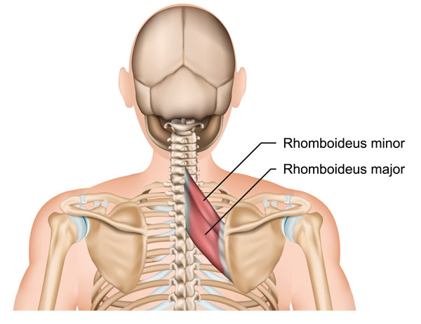
The illustration shows the upper back with a focus on two specific muscles: the Rhomboideus minor and the Rhomboideus major. These muscles, commonly referred to as the rhomboid muscles, are located between the spine and the shoulder blades, also known as the scapulae.
The Rhomboideus minor is situated superiorly to the Rhomboideus major. It originates from the nuchal ligaments and the spinous processes of the C7 and T1 vertebrae and inserts onto the medial border of the scapula, near its base. Its primary function is to stabilize the scapula by holding it close to the thoracic wall.
Directly beneath the Rhomboideus minor lies the Rhomboideus major. This muscle starts from the spinous processes of the T2 to T5 vertebrae and also attaches to the medial border of the scapula. The Rhomboideus major aids in retracting the scapula by pulling it towards the vertebral column, as well as elevating the medial border of the scapula.
Both muscles play crucial roles in the movement and stabilization of the shoulder, and they work together to coordinate the scapula’s motion, especially during actions that involve moving the arms backward or lifting them. These muscles are essential for maintaining good posture and shoulder alignment.
Erector Spinae Muscles
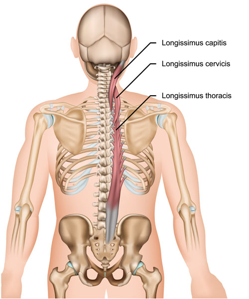
The image depicts the posterior view of the human torso, highlighting the Longissimus muscle group, which is part of the erector spinae muscles. These muscles are situated along the vertebral column and are critical in the movement and stabilization of the spine.
At the top, we see the Longissimus capitis, which runs from the upper thoracic and lower cervical vertebrae to the base of the skull. This muscle is involved in extending and rotating the head.
Below the Longissimus capitis is the Longissimus cervicis, stretching from the thoracic vertebrae up to the cervical vertebrae. Its function includes the extension and lateral flexion of the cervical spine.
Extending the length of the thoracic spine, we have the Longissimus thoracis, which is the longest part of the Longissimus group. It originates from the lumbar region and inserts into the lower thoracic vertebrae and ribs. The primary actions of the Longissimus thoracis include extending the vertebral column and maintaining an erect posture.
These muscles are paramount for various movements including back extension, lateral flexion, and contributing to the rotation of the spine. They also play a significant role in maintaining proper posture and are engaged during many daily activities, such as lifting and bending.
Levator Scapulae Muscle
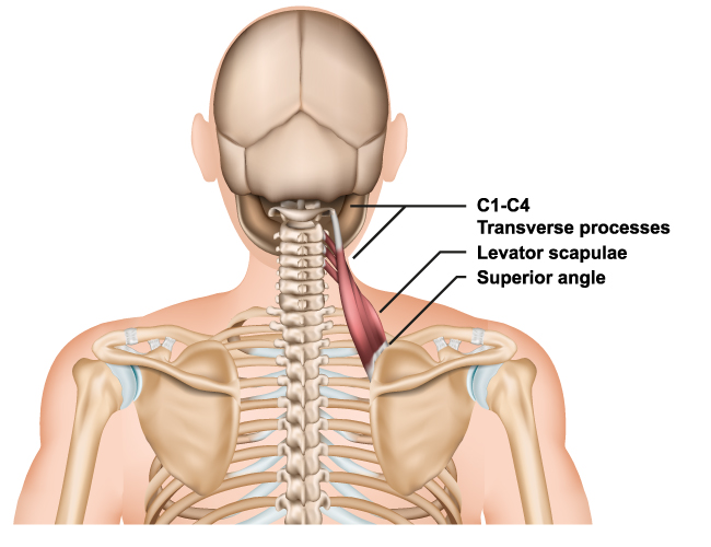
In this image, we are looking at the Levator scapulae muscle, alongside its anatomical position in relation to the cervical vertebrae and the scapula. The Levator scapulae is shown originating from the transverse processes of the first four cervical vertebrae, labeled C1-C4. This muscle extends downward, inserting at the superior angle of the scapula.
The Levator scapulae muscle plays a critical role in the movement of the scapula. It helps to elevate the scapula, which is essential during shoulder elevation movements, such as shrugging. Additionally, it assists in the downward rotation of the scapula when the arm is raised above the head, as well as in the tilting of the neck to the same side. Due to its position and function, tension or discomfort in this muscle can be associated with neck stiffness or pain, often exacerbated by poor posture or prolonged sitting.
Latissimus Dorsi Muscle
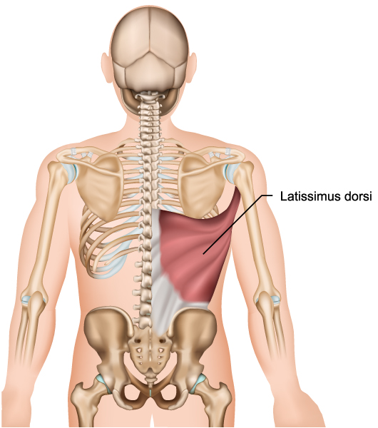
This illustration shows the posterior view of the torso, highlighting the Latissimus dorsi muscle, one of the largest muscles in the back. The Latissimus dorsi is a broad, flat muscle that covers the lower back and stretches up to the mid-back, under the armpit, and across to the shoulder blade.
The Latissimus dorsi originates from the lower vertebrae of the spine, the iliac crest of the pelvis, the lower ribs, and, indirectly, from the scapula’s inferior angle. Its fibers then converge to form a tendon that inserts into the bicipital groove of the humerus.
The primary functions of the Latissimus dorsi include the internal rotation, adduction, and extension of the shoulder joint. It plays a significant role in movements such as pulling a weight toward the body, as seen in actions like rowing, and is also involved in forceful expiration as part of the respiratory process.
This muscle is key for various athletic movements and everyday activities that require the use of the upper limbs, especially those involving pulling forces. The Latissimus dorsi also contributes to the contour of the back and affects the posture.
Upper Back Muscle Anatomy
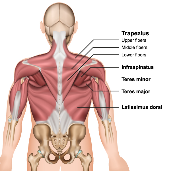
The image provides a detailed posterior view of the upper back muscles, highlighting several key muscle groups.
At the top, we have the Trapezius muscle, which is a large, triangular-shaped muscle that extends from the nape of the neck to the middle of the back and across to the shoulder. It’s divided into three parts: the upper fibers, middle fibers, and lower fibers. The upper fibers elevate the shoulder blade and extend the neck backward, the middle fibers retract the shoulder blade, and the lower fibers depress the shoulder blade.
Beneath the Trapezius, the Infraspinatus muscle is visible, which is a thick triangular muscle occupying the majority of the infraspinatous fossa on the posterior aspect of the scapula. It is one of the four rotator cuff muscles and functions to externally rotate the arm at the shoulder.
Below the Infraspinatus is the Teres minor, a narrow, elongated muscle of the rotator cuff. It aids in the rotation of the arm laterally and helps bring it toward the body.
Adjacent to the Teres minor is the Teres major, which is not part of the rotator cuff. It assists in the internal rotation and adduction of the arm.
Lastly, we see the Latissimus dorsi, the broad, sweeping muscle that extends from the lower back to the upper arm bone (humerus). It is responsible for the internal rotation, adduction, and extension of the shoulder.
These muscles are essential for a wide range of shoulder movements and contribute significantly to the posture and stability of the upper back and shoulder regions. They are also involved in various actions such as lifting, pulling, and rotating the arms.
Spinae Muscle Group
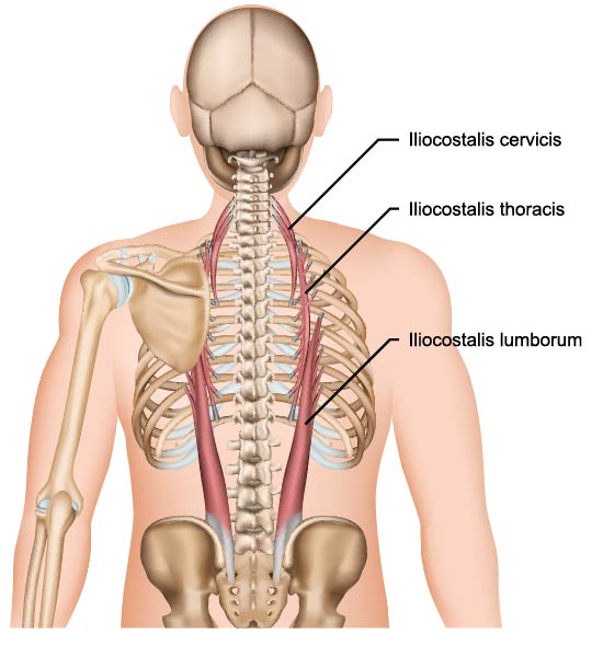
The illustration shows the Iliocostalis muscle, which is one of the three major muscles that make up the erector spinae muscle group in the back. The erector spinae muscles are critical for maintaining posture and controlling the movements of the spine.
The Iliocostalis is further divided into three distinct regions based on their location:
The Iliocostalis cervicis is located at the top and it extends from the angles of the lower cervical and upper thoracic ribs to the transverse processes of the cervical vertebrae. It is involved in the lateral flexion of the neck and assists with extension.
Below that is the Iliocostalis thoracis, which spans the lower thoracic region. It runs from the angles of the ribs to the transverse processes of the upper lumbar and lower thoracic vertebrae. This muscle helps in the extension and lateral flexion of the thoracic spine.
The Iliocostalis lumborum is the lowest part of the muscle and is the most lateral group of the erector spinae. It extends from the iliac crest, sacrum, and lumbar vertebrae to the lower ribs. It assists with bending the spine to the side and with the extension of the lumbar spine.
Together, these muscles work to support the spine during various movements, including bending, twisting, and extending. They are essential for the stabilization of the vertebral column and are engaged during many activities, such as lifting objects and standing upright.
Anatomical Terms and Definitions
| Term | Definition |
|---|---|
| Erector Spinae | A group of muscles and tendons which run nearly the entire length of the spine on the left and the right, from the sacrum, or sacral region, and hips to the base of the skull. They are primarily responsible for extending the spine, and maintaining posture and spine stability. |
| Iliocostalis | Part of the erector spinae muscle group, it is divided into cervicis, thoracis, and lumborum sections, extending from the lower back to the upper cervical region. Its main function includes aiding in the lateral flexion of the vertebral column. |
| Infraspinatus | A thick triangular muscle located on the back of the scapula and part of the rotator cuff, involved in lateral rotation of the arm at the shoulder. |
| Latissimus Dorsi | A large, flat muscle on the back that stretches to the sides, behind the arm, and is partially covered by the trapezius on the back near the midline. It is involved in the internal rotation, adduction, and extension of the shoulder. |
| Levator Scapulae | A muscle that originates from the transverse processes of the first four cervical vertebrae and inserts into the superior part of the scapula, functioning mainly to elevate the scapula. |
| Longissimus | Part of the erector spinae muscle group, running alongside the spine, it is involved in extending and laterally flexing the spine, and can also rotate the head. |
| Rhomboideus Major and Minor | Muscles located in the upper back between the spine and the scapula. They are involved in the retraction, elevation, and rotation of the scapula. |
| Serratus Posterior Inferior | A muscle of the human body, located at the junction of the thoracic and lumbar regions. It acts to depress the lower ribs, aiding in forced expiration. |
| Serratus Posterior Superior | Located at the upper back, it elevates the upper ribs, aiding in the process of inhalation. |
| Teres Major | A muscle of the upper limb and one of the six scapulohumeral muscles. It is mainly involved in the medial rotation and adduction of the arm. |
| Teres Minor | Part of the rotator cuff, this muscle assists in rotation of the arm away from the body and helps hold the humeral head in the glenoid cavity of the scapula. |
| Trapezius | A major muscle of the back, extending from the nuchal region of the skull to the lower thoracic vertebrae and laterally to the spine of the scapula. Its actions include moving, rotating, and stabilizing the scapula and extending the neck. |
