
Table of Contents
Basic Liver Anatomy
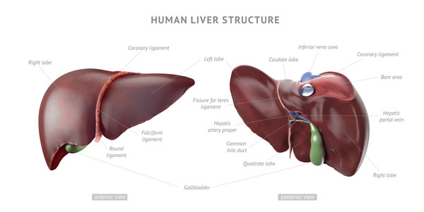
This image presents a dual view of the human liver, both from an anterior (front) and a posterior (back) perspective, with key structures labeled to guide understanding of its anatomy.
On the left side of the image, we see the anterior view of the liver. The liver is divided into two main lobes: the right and left lobes, with the right lobe being significantly larger. The falciform ligament, a sheet-like connective tissue structure, is clearly visible as it descends to attach the liver to the anterior abdominal wall and diaphragm, and demarcates the two lobes on the front. The round ligament, a remnant of the fetal umbilical vein, is also marked, appearing at the inferior edge of the falciform ligament.
The right side of the image illustrates the posterior view of the liver. Here, you can observe the caudate lobe, which is located on the posterior aspect of the right lobe and near the inferior vena cava. The inferior vena cava is labeled at the top, indicating its crucial role in transporting deoxygenated blood from the lower body back to the heart. The hepatic artery and the common bile duct are depicted adjacent to one another, often part of the portal triad, which also includes the portal vein (not distinctly labeled here). The gallbladder sits in a fossa beneath the liver, collecting and concentrating bile.
Additionally, the coronary ligaments, extensions of the peritoneum that help to suspend the liver in the abdominal cavity, are identified on both the right and left sides of the liver.
Overall, this image summarizes the external morphology of the liver and its associations with the vascular and biliary structures essential for its function. It underscores the liver’s role as a central organ in various physiological processes including metabolism, detoxification, and bile production.
External Liver Anatomy
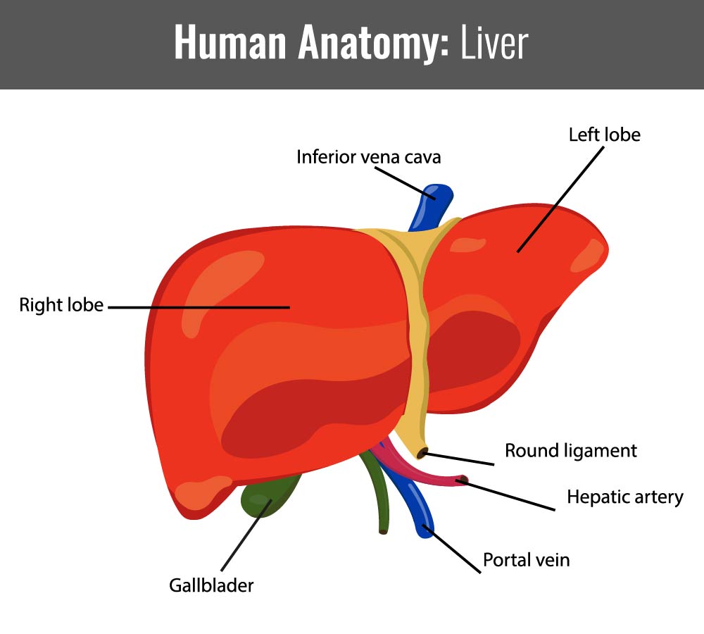
This image presents a labeled diagram of the human liver, an essential organ in the human body, focusing on its external anatomy and the major vessels associated with it. It’s a simplified, educational illustration, likely intended for students or patients to understand the basic structure of the liver and its connections to the circulatory and biliary systems.
The liver is shown in two distinct lobes: the larger right lobe and the smaller left lobe, which are typical anatomical distinctions. The color used is a bright red, likely intended to represent the rich blood supply rather than the actual color of the organ.
Several important blood vessels are labeled:
- The Inferior Vena Cava: This large vein is shown at the top of the liver, and it is responsible for carrying deoxygenated blood from the lower half of the body back to the heart.
- The Hepatic Artery: Colored in red, indicating that it carries oxygenated blood to the liver from the heart.
- The Portal Vein: Illustrated in blue, which is traditional for veins in medical illustrations, indicating that it brings nutrient-rich blood from the digestive organs to the liver for processing.
Below the liver, the Gallbladder is indicated, a small green structure that stores and concentrates bile produced by the liver. The bile is transported from the liver to the gallbladder through the bile ducts, which are not labeled in this particular diagram.
Additionally, the Round Ligament of the liver is marked. This ligament is a remnant of the fetal umbilical vein and runs along the free edge of the falciform ligament, dividing the left part of the liver into medial and lateral sections.
This diagram is a fundamental representation meant to convey how the liver is situated in the body relative to these vessels and structures, providing a clear visual guide to the liver’s anatomy and its connections within the circulatory and biliary systems. It encapsulates the liver’s role as a processing and filtering hub within the body’s physiology.
Illustration of the Liver Lobule
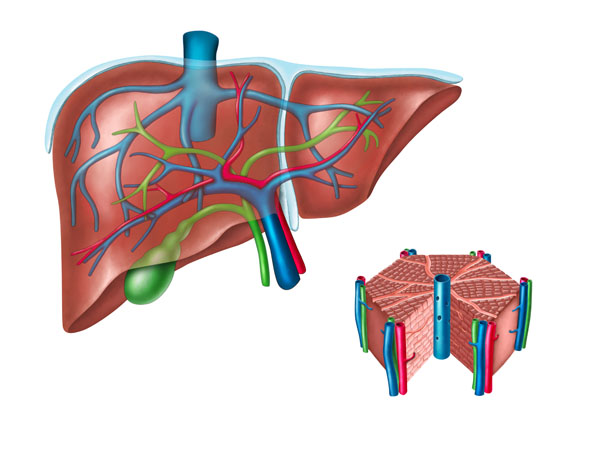
The image is showing a simplified representation of the human liver and its associated structures, alongside an enlarged detail of liver lobules, the functional units of the liver.
On the left, the liver is depicted in a stylized manner, with a semi-transparent quality allowing for the visualization of internal structures. The liver’s unique reddish-brown coloration is noted, and it’s shown in a somewhat three-dimensional perspective that gives a view of its largest lobe. The portal vein, which is usually depicted in blue, is shown entering the liver, signifying its role in delivering nutrient-rich blood from the gastrointestinal tract. Branching off from the portal vein are smaller vessels, which would distribute this blood within the liver.
The hepatic artery, typically represented in red, is shown providing oxygenated blood to the liver. Alongside these, the green structure likely represents the bile duct, which carries bile produced by the liver cells to the gallbladder and then to the small intestine to aid in digestion. These vessels and ducts are part of the hepatic portal triad, which also includes the hepatic portal vein and tiny branches of the hepatic artery.
On the right is an amplified view of liver lobules, the hexagonal units consisting of plates of hepatocytes (liver cells) arranged around a central vein. These lobules are connected to the blood vessels and bile ducts shown on the left, emphasizing the liver’s role in filtering blood, metabolizing nutrients and drugs, and secreting bile.
The image summarily communicates the liver’s vascular architecture and its functional microanatomy. The relationship between the macroscopic and microscopic anatomy highlights the process of blood inflow, filtering, and bile secretion, which are central to the liver’s role in metabolism and detoxification.
Microanatomy of the Liver Lobule Structure
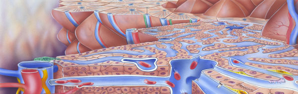
The image provided is an intricate illustration of the microanatomy of the liver, specifically a close-up view of the liver’s lobular structure and the blood flow within it.
The central focus is the hepatic lobules, which are the functional units of the liver. Each lobule is hexagonal in shape and consists of plates of hepatocytes (liver cells) radiating out from a central vein. This central vein is the endpoint for blood processed by the hepatocytes, and it drains into the hepatic veins which then join the inferior vena cava, carrying blood away from the liver back to the heart.
Surrounding each lobule, there are portal triads at each corner. These triads are composed of a branch of the portal vein (in blue), a branch of the hepatic artery (in red), and a bile duct (in green). The portal vein carries nutrient-rich blood from the gastrointestinal tract, and the hepatic artery supplies oxygen-rich blood to the lobules. This mixture of blood enters the lobules through sinusoids, small channels that are lined by fenestrated endothelium, which allows for the efficient transfer of substances between the blood and the hepatocytes.
The bile ducts collect bile produced by the hepatocytes, which is then transported to the gallbladder or directly to the duodenum. The intricate network of bile ducts is visible as a branching green structure.
This highly detailed illustration effectively conveys the complexity of the liver’s internal structure. The liver’s role in filtering blood, metabolizing various substances, and producing bile is dependent on this intricate arrangement of vessels and cells, and the image captures the organized yet complex nature of this essential organ.
Diagram of the Liver Lobule
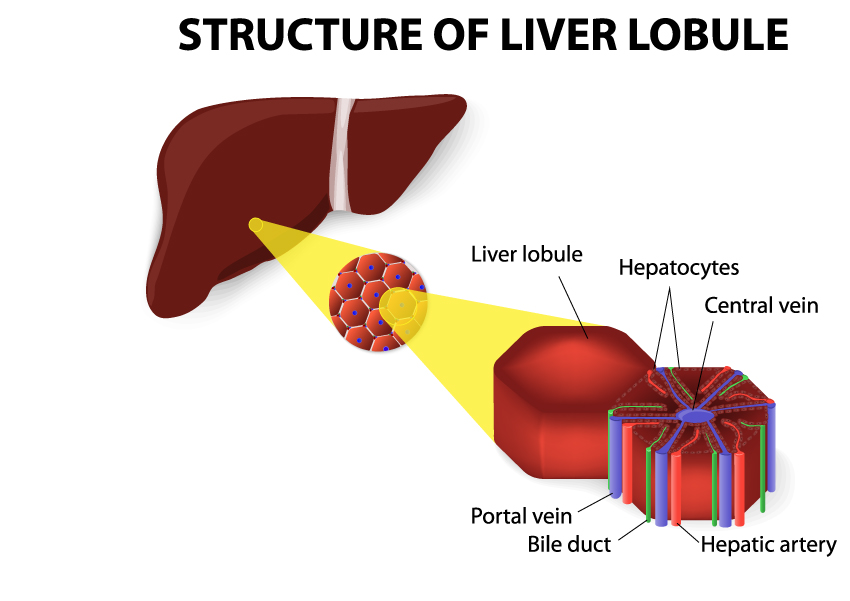
This image is a diagrammatic representation of the structure of a liver lobule, which is the fundamental functional unit of the liver. The illustration simplifies and magnifies the structure to emphasize the key components and their spatial relationships.
The main part of the image shows a hexagonally-shaped liver lobule with a central vein running through its core. This central vein collects the blood after it has been processed by the liver cells and channels it towards the hepatic veins and eventually into the inferior vena cava.
The lobule is composed of numerous hepatocytes (liver cells), which are depicted as small red units arranged in rows radiating outward from the central vein. These hepatocytes are responsible for the liver’s various functions, including detoxification, metabolism, and bile production.
At the corners of the lobule, we see representations of the portal triad, which consists of branches of the portal vein (in blue), bile duct (in green), and hepatic artery (in red). The portal vein brings nutrient-rich blood from the digestive tract, the hepatic artery supplies oxygenated blood, and the bile duct collects bile secreted by the hepatocytes.
The depiction of the liver lobule next to an image of the liver itself helps to contextualize where these lobules are located within the organ. It underscores the idea that while the liver is a single large organ, it is composed of many such units, each performing the essential functions of the liver. This image conveys the concept of how blood and bile flow through the liver’s microscopic architecture to carry out complex physiological processes.
This image is an infographic depicting the anatomy of the right eye as viewed from above, providing a comprehensive look at both the external and internal structures.
Starting with the external parts, the upper portion shows the eye lid, which protects the eye and helps distribute tear fluid across the surface of the eye. The lacrimal caruncle is a small, pink, globular nubbin of flesh located at the inner corner of the eye, which contains glands that produce a portion of the tear film. Adjacent to this is the tear duct, also known as the nasolacrimal duct, which drains tears from the eye into the nasal cavity.
Moving to the internal anatomy, the sclera is the white, fibrous outer layer of the eyeball; its main function is to provide structure, strength, and flexibility to the eye. The cornea is the transparent, dome-shaped surface that covers the front of the eye and, together with the lens, helps to refract and focus light onto the retina.
The iris is the colored part of the eye that surrounds the pupil, the central opening that regulates the amount of light entering the eye. The lens sits just behind the iris and pupil, and it is held in place by suspensory ligaments attached to the ciliary body, which contains the muscle that alters the shape of the lens for focusing, a process known as accommodation.
Between the cornea and the lens are the anterior and posterior chambers, which are filled with a clear fluid called the aqueous humor. This fluid maintains intraocular pressure and provides nutrients to the avascular structures of the eye.
The vitreous body is the clear gel that fills the space between the lens and the retina, helping to maintain the spherical shape of the eye and offering support to the retina. The retina itself is the thin layer of tissue that lines the back of the eye, containing the photoreceptor cells that convert light into electrical signals.
The fovea centralis, located in the center of the macula of the retina, is responsible for sharp central vision. The hyaloid canal is a small channel that runs through the vitreous body, which in the fetal eye allows the passage of nutrients from the optic nerve to the developing lens.
Finally, the optic nerve is depicted exiting the back of the eye. This nerve is responsible for transmitting visual information from the retina to the brain. Accompanying the optic nerve are the retinal blood vessels, which supply the retinal tissue with nutrients and oxygen.
The muscles illustrated are the lateral rectus muscle, which moves the eye outward away from the nose, and the medial rectus muscle, which moves the eye inward towards the nose.
Overall, the infographic serves as an educational tool, providing a clear and detailed visual representation of the various components of the eye and their respective roles in the complex process of vision.
Diagram of Interrelated Structures
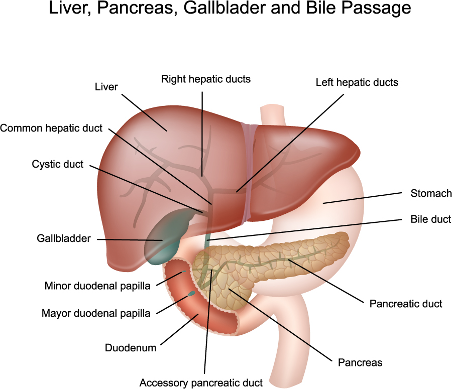
This diagram offers a detailed look at the interrelated structures of the liver, pancreas, gallbladder, and the bile passage system. It’s a simplified representation intended to help understand how these organs work together in the digestive process.
The liver is shown at the top, with its right and left hepatic ducts labeled. These ducts collect bile from their respective sides of the liver and converge to form the common hepatic duct. Below the liver, the gallbladder is depicted; it’s a small, pouch-like organ where bile is stored and concentrated. The cystic duct connects the gallbladder to the common hepatic duct, and together they form the common bile duct, which is responsible for delivering bile to the small intestine.
The pancreas is illustrated beneath the liver and gallbladder, extending horizontally across the image. This organ produces digestive enzymes that are transported to the duodenum through the pancreatic duct. The diagram shows the pancreatic duct joining the common bile duct at the major duodenal papilla, which is the point where both bile and pancreatic enzymes enter the duodenum, the first section of the small intestine.
An accessory pancreatic duct is also labeled, which is an additional duct that some people have, opening into the duodenum at the minor duodenal papilla, slightly above the major papilla.
The relationship between these structures is vital for digestion. Bile produced by the liver and stored in the gallbladder emulsifies fats, while pancreatic enzymes help in the digestion of proteins, fats, and carbohydrates. The precise arrangement ensures that these substances are released into the duodenum where digestion continues.
This visual explanation helps illustrate the anatomy and cooperation of organs involved in the digestive process, emphasizing their locations and connections to each other.
Gallbladder Pathology
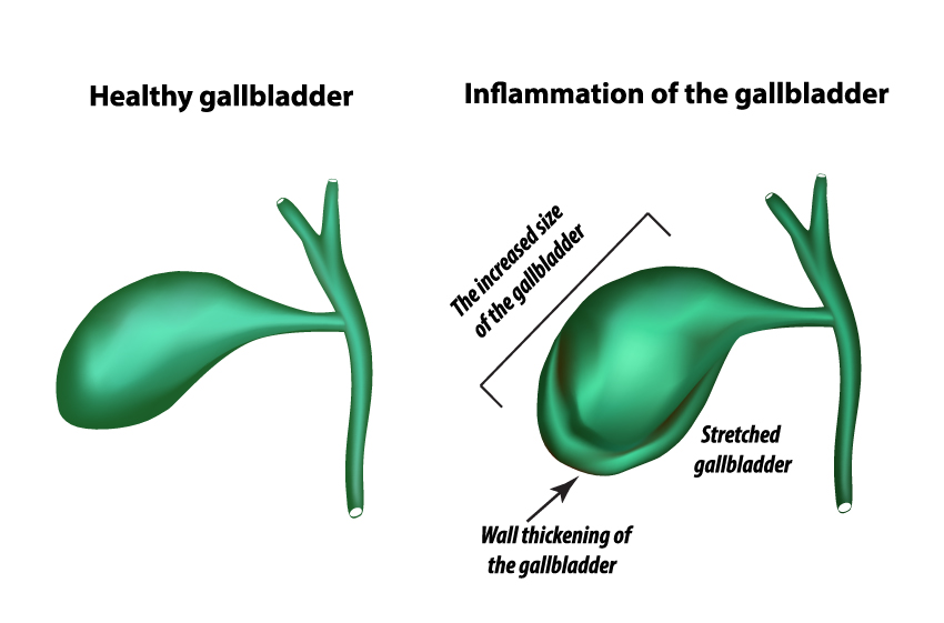
The image compares two states of the gallbladder: on the left is a healthy gallbladder, and on the right is a gallbladder experiencing inflammation.
The healthy gallbladder is shown as a smooth, pear-shaped organ with a narrow cystic duct extending from it. This is the normal appearance, indicating that the gallbladder is likely functioning properly, storing and concentrating bile that is produced by the liver.
In contrast, the right side of the image shows an inflamed gallbladder. It is visibly larger and distorted compared to the healthy one, with a label pointing out the increased size of the gallbladder. Additionally, there’s a noticeable bulge labeled as “stretched gallbladder,” suggesting that the organ is filled, possibly with bile or fluid due to obstruction or infection. The wall of the gallbladder is labeled as thickened, which is a common response to irritation or inflammation and can be a sign of conditions like cholecystitis.
The comparison between the two states provides an educational illustration of how gallbladder inflammation can manifest physically. This could be useful for medical students or patients to visualize and understand the changes that occur during gallbladder inflammation.
Gallstone Pathology
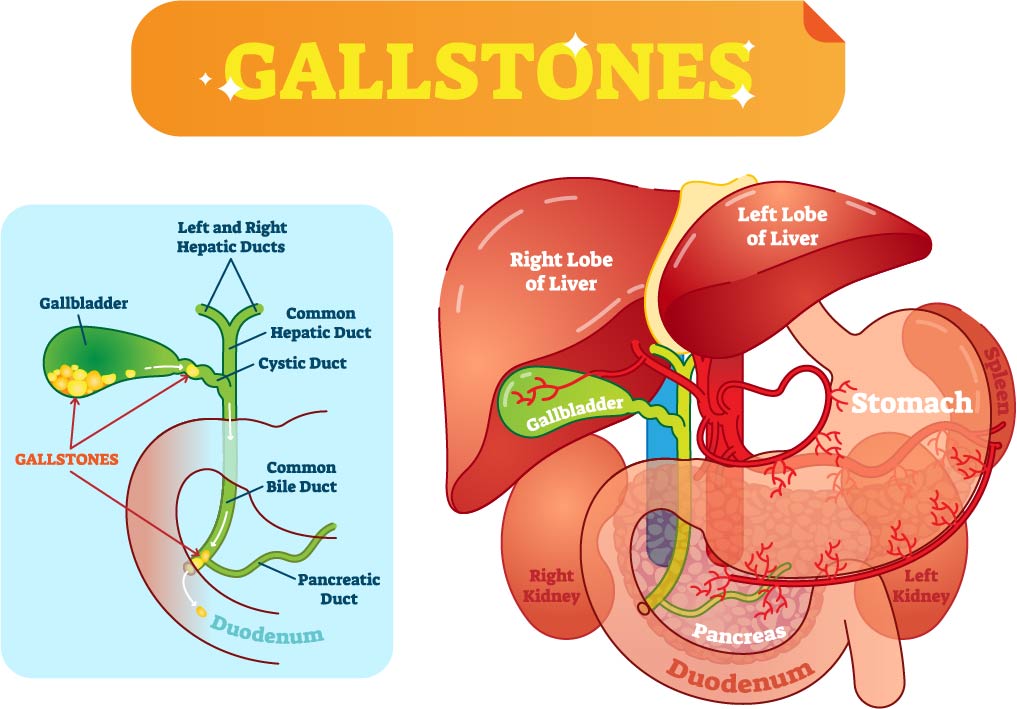
This vibrant image provides an educational overview of gallstones and their location in relation to the liver, gallbladder, and bile ducts.
On the left side of the image, there’s a focused illustration of the gallbladder and the associated bile ducts. The gallbladder is shown in green, and inside it, there are yellow formations labeled as gallstones. These are solid particles that form from bile cholesterol or bilirubin in the gallbladder. The bile ducts are illustrated as tubes that transport bile from the liver (via the left and right hepatic ducts) to the duodenum. The hepatic ducts merge into the common hepatic duct, which then joins the cystic duct from the gallbladder to form the common bile duct. The location of the gallstones suggests that they could obstruct the flow of bile, potentially leading to a medical condition requiring treatment.
On the right, the image depicts a semi-transparent view of the human abdomen, with the liver, stomach, pancreas, duodenum, and kidneys. The liver is shown with its two main lobes, and the gallbladder is nestled underneath the liver’s right lobe. The transparency of the liver allows for the visualization of the internal vasculature, emphasizing the organ’s blood supply and connection to the gallbladder and bile ducts. The stomach is positioned anteriorly to the pancreas, and the duodenum wraps around the pancreas. Both kidneys are also visible, with the right kidney situated just below the liver and the left kidney near the spleen and pancreas, indicating their relative positions in the body.
Anatomical Terms and Definitions
| Term | Definition |
|---|---|
| Bile Duct | A green structure that carries bile from the liver cells to the gallbladder and then to the small intestine to aid in digestion. |
| Central Vein | The endpoint for blood processed by hepatocytes in a liver lobule, draining into the hepatic veins. |
| Common Bile Duct | Delivers bile to the small intestine, formed by the convergence of the cystic duct and the common hepatic duct. |
| Falciform Ligament | A sheet-like connective tissue that divides the liver into two main lobes and attaches it to the anterior abdominal wall and diaphragm. |
| Gallbladder | A small green structure below the liver that stores and concentrates bile produced by the liver. |
| Gallstones | Solid particles that form from bile cholesterol or bilirubin in the gallbladder, potentially obstructing the flow of bile. |
| Hepatic Artery | Provides oxygenated blood to the liver, typically represented in red. |
| Hepatic Lobules | Hexagonal units consisting of plates of hepatocytes arranged around a central vein, the functional units of the liver. |
| Hepatic Portal Triad | Consists of the hepatic portal vein, tiny branches of the hepatic artery, and bile ducts. |
| Hepatocytes | Liver cells responsible for filtering blood, metabolizing nutrients and drugs, and secreting bile. |
| Inferior Vena Cava | A large vein carrying deoxygenated blood from the lower half of the body back to the heart. |
| Pancreatic Duct | Transports digestive enzymes from the pancreas to the duodenum. |
| Portal Triads | Located at each corner of a liver lobule, composed of a branch of the portal vein, a branch of the hepatic artery, and a bile duct. |
| Portal Vein | Usually depicted in blue, delivering nutrient-rich blood from the gastrointestinal tract to the liver. |
| Round Ligament | A remnant of the fetal umbilical vein running along the free edge of the falciform ligament. |
| Sinusoids | Small channels in liver lobules, lined by fenestrated endothelium for efficient transfer of substances between the blood and hepatocytes. |
