
Table of Contents
Types of Ovarian Cysts
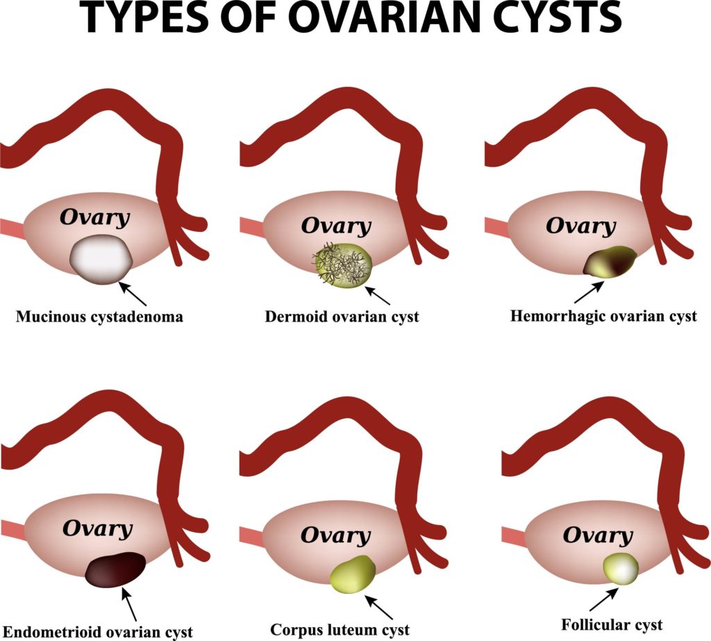
The image displays six types of ovarian cysts, each with a visual representation of its appearance on or within the ovary.
Mucinous cystadenoma is depicted as a large, smooth cyst within the ovary, filled with a sticky, gelatinous material. These cysts can grow quite large and may cause the ovary to move out of its usual position in the pelvis.
The dermoid ovarian cyst, also known as a mature cystic teratoma, is shown containing various types of tissue, such as hair, fat, or even teeth. These cysts arise from cells that have the ability to differentiate into various tissue types.
A hemorrhagic ovarian cyst is represented with a reddish-brown coloration, indicating the presence of blood. These cysts occur when a very small vessel in the cyst wall breaks, and the blood enters the cyst.
The endometrioid ovarian cyst, often associated with endometriosis, resembles a dark, blood-filled space and is formed when endometrial tissue grows in the ovaries.
The corpus luteum cyst is shown as a small, yellowish structure, which forms from the corpus luteum after ovulation. Sometimes these cysts can fill with blood or fluid.
Finally, the follicular cyst is depicted as a simple, clear cyst, representing an unruptured follicle that has continued to grow and fill with fluid beyond the normal size.
These visualizations are useful for understanding the differences between various types of ovarian cysts, their structures, and potential contents. Ovarian cysts are fluid-filled sacs that can form in the ovaries during a woman’s reproductive years. While many ovarian cysts are benign and may resolve on their own, some may cause symptoms and require medical intervention.
Polycystic Ovarian Syndrome (PCOS)
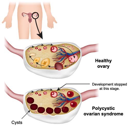
The image presents a comparison between a healthy ovary and an ovary affected by Polycystic Ovarian Syndrome (PCOS).
The top half of the image shows a healthy ovary, characterized by the presence of normal follicles at various stages of development. Each follicle contains an oocyte, which is a developing egg cell. The largest follicle, often called the dominant follicle, is preparing for ovulation. Blood vessels and connective tissue support the structure and function of the ovary.
The bottom half illustrates an ovary affected by PCOS. This condition is characterized by the presence of multiple cysts. These cysts are actually immature follicles that have not undergone proper development or ovulation. They often appear as a “string of pearls” on medical imaging. The text “Development stopped at this stage” indicates that these follicles have failed to mature and, as a result, remain as cysts within the ovary. This can lead to hormonal imbalances, irregular menstrual cycles, and sometimes difficulties with fertility.
The illustration serves as an educational tool to depict the typical appearance and differences between a healthy ovary and one with PCOS, aiding in the understanding of how this syndrome affects ovarian function.
Breast Pathology
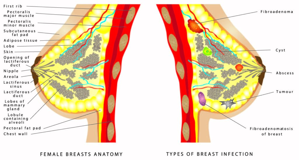
The image is divided into two sections, with the left side detailing the anatomy of the female breast and the right side illustrating various types of breast conditions.
On the left, the anatomy of the female breast is shown in cross-section. At the top, we see the first rib, which provides structural support. Below this is the pectoralis major muscle, a thick, fan-shaped muscle that contributes to the contour of the chest. Beneath it lies the pectoralis minor muscle, a thinner muscle that lies underneath the pectoralis major. The subcutaneous adipose tissue, or fat pad, is depicted surrounding the breast tissue, contributing to the size and shape of the breast. The skin covers the outside of the breast, and beneath it is the opening of the lactiferous ducts at the nipple. The areola is the pigmented area surrounding the nipple. Within the breast, we see the lobules of mammary gland containing alveoli, which are the milk-producing glands. These lobules are connected to the nipple via the lactiferous ducts.
On the right, various breast conditions are illustrated. A fibroadenoma is shown as a benign tumor that is solid, smooth, and noncancerous. A cyst, a fluid-filled sac within the breast tissue, is also depicted. An abscess, which is a collection of pus that has built up within the tissue of the breast, is indicated. The image also shows a tumor, which can be benign or malignant, and fibroadenomatosis of the breast, a condition characterized by the presence of multiple fibroadenomas.
The image serves an educational purpose, demonstrating both the normal structure of the breast as well as common conditions that can affect breast tissue. It’s useful for understanding the complex anatomy of the breast as well as recognizing the diversity of conditions that can present in this part of the body.
Types of Uterine Fibroids
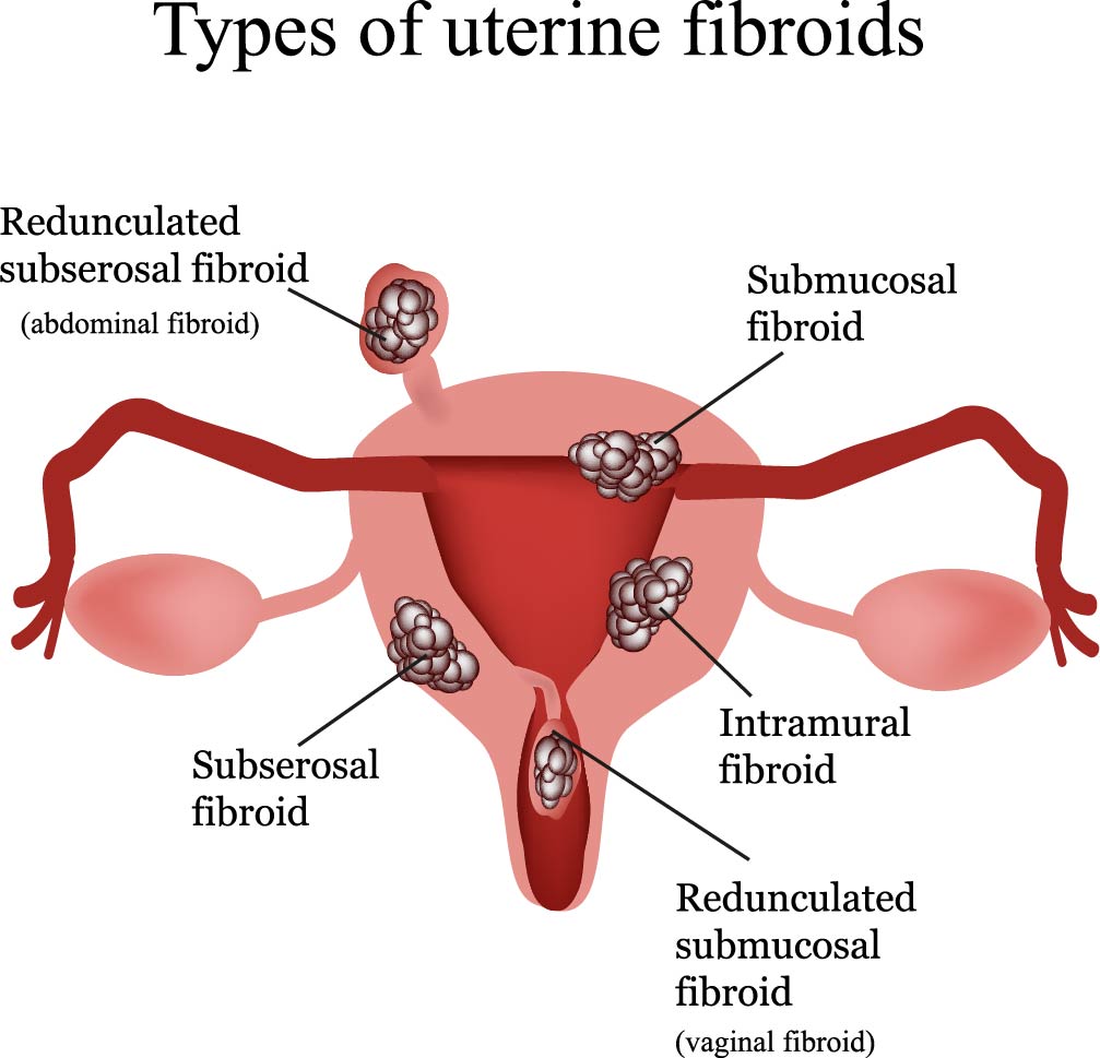
The image presents an overview of the different types of uterine fibroids, which are noncancerous growths of the uterus that often appear during childbearing years.
Subserosal fibroids are depicted as extending to the outside of the uterus. They project from the outer surface of the uterus into the pelvic cavity.
Intramural fibroids are shown within the muscular wall of the uterus. These are the most common type of fibroid and can lead to an enlargement of the uterus as they grow.
Submucosal fibroids are located just under the interior surface of the uterus, within the endometrial cavity. These can cause heavy menstrual bleeding and trouble conceiving.
Redunculated subserosal fibroid, also referred to as an abdominal fibroid, is a subserosal fibroid that develops a stem, a slender base that supports the tumor.
Redunculated submucosal fibroid, sometimes called a vaginal fibroid, is a submucosal fibroid that also develops a stem, making it appear to be hanging within the uterine cavity or outside the uterus.
This illustration helps in understanding where fibroids can develop within the uterus and the potential impact on the organ’s structure and function. These growths are typically benign and may not always cause symptoms, but they can sometimes lead to complications such as pain and heavy menstrual bleeding.
Female Reproductive Structures
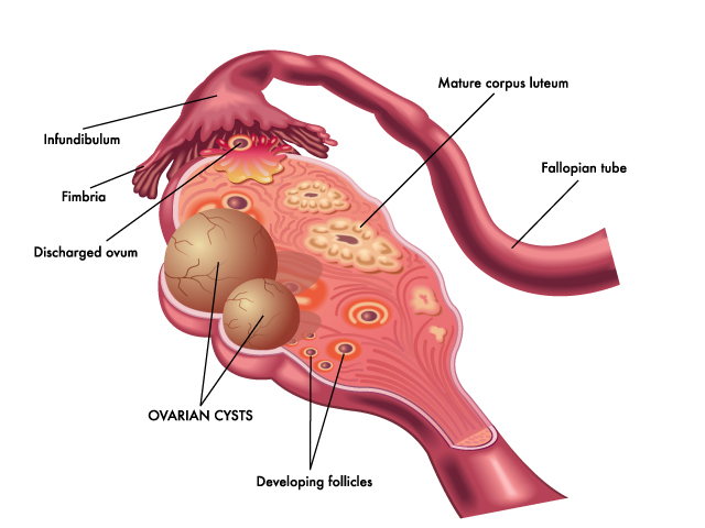
The illustration provides a cross-sectional view of a human ovary and its relation to the fallopian tube, highlighting various reproductive structures and processes.
The fallopian tube, also known as the uterine tube, is displayed arching over the ovary. The infundibulum is the funnel-shaped end of the fallopian tube near the ovary, and the fimbriae are finger-like projections at the end of the infundibulum that help guide the ovum into the tube.
Visible on the ovary is a discharged ovum, an egg that has been released from a follicle during ovulation and is now ready for potential fertilization.
Developing follicles are also shown within the ovary. These are fluid-filled sacs that contain immature oocytes (eggs). They grow and mature in a cyclical pattern in response to hormone signals.
Highlighted are ovarian cysts, which are fluid-filled sacs on the ovary. They often form during the process of ovulation. Depending on the type, they can be a normal part of the menstrual cycle or represent a pathological condition.
Finally, the mature corpus luteum is indicated. This is a temporary endocrine structure that forms from the remnants of a follicle after it has released its ovum. The corpus luteum secretes hormones such as progesterone, which help maintain the uterine lining for potential implantation of a fertilized egg.
This image is commonly used for educational purposes to explain the anatomy and function of the ovary, as well as the normal processes of ovulation and potential issues such as cyst formation.
