
Table of Contents
Basic Female Reproductive System Anatomy
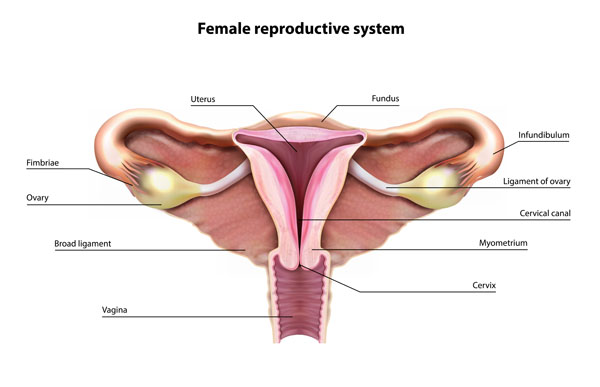
This image is an anatomical illustration of the female reproductive system, specifically focusing on the structure of the uterus and its connecting components.
At the center, we have the uterus, a muscular organ where fetal development occurs. The uppermost part of the uterus is called the fundus, which is the rounded top portion. Extending laterally from the fundus are the fallopian tubes, ending in fimbriae, which are finger-like projections that capture the ovum released from the ovary during ovulation.
Below the uterus, we see the cervix, which is the lower, narrow part of the uterus that opens into the vagina. The cervical canal runs through the cervix and allows passage between the uterus and vagina.
The myometrium, labeled on the right side of the uterus, refers to the thick, muscular layer of the uterus that is responsible for contractions during childbirth and menstruation.
The broad ligament is a wide fold of peritoneum that connects the sides of the uterus to the walls and floor of the pelvis, providing support to the uterus.
The ovaries, which are small, oval-shaped glands located on either side of the uterus, are responsible for producing eggs (ova) and the hormones estrogen and progesterone.
Lastly, the ligament of the ovary, not to be confused with the suspensory ligament of the ovary (which is not shown in this image), is a fibrous cord that connects the ovary to the uterus. This structure supports the ovary in its proper place within the pelvic cavity.
This illustration is commonly used in educational settings to explain the anatomy and function of the female reproductive system.
Detailed View of the Female Pelvic Anatomy
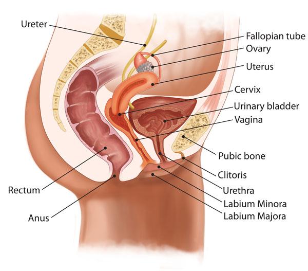
This image provides a detailed view of the female pelvic anatomy, showing both the reproductive and urinary systems, as well as other related structures.
Starting from the top, the ureter is shown, which is a duct that carries urine from the kidneys to the urinary bladder. The urinary bladder is depicted just above the vagina, responsible for storing urine before it is excreted through the urethra.
The reproductive system includes the ovary, which is an egg-producing reproductive organ. It is connected to the uterus via the fallopian tube, through which the egg travels to reach the uterus. The uterus is a muscular organ that houses and nourishes a fertilized egg during pregnancy. Below the uterus is the cervix, which extends into the vagina, the muscular canal leading to the external body.
The image also illustrates the external genitalia, including the clitoris, a small, sensitive protrusion that is comparable to the male penis in terms of nerve endings and sensitivity. The urethra, the tube that allows urine to exit the body, is positioned below the clitoris. Flanking the vaginal opening are the labia minora and labia majora, which are folds of skin that protect the internal genital structures.
Below the reproductive system, the rectum is shown leading to the anus, which is the terminal end of the digestive tract.
Finally, the pubic bone is noted at the bottom, which is part of the pelvis and provides structural support to the pelvic region.
This comprehensive illustration serves educational purposes, highlighting the anatomy and relationship between various pelvic structures in the female body.
Menstrual Cycle
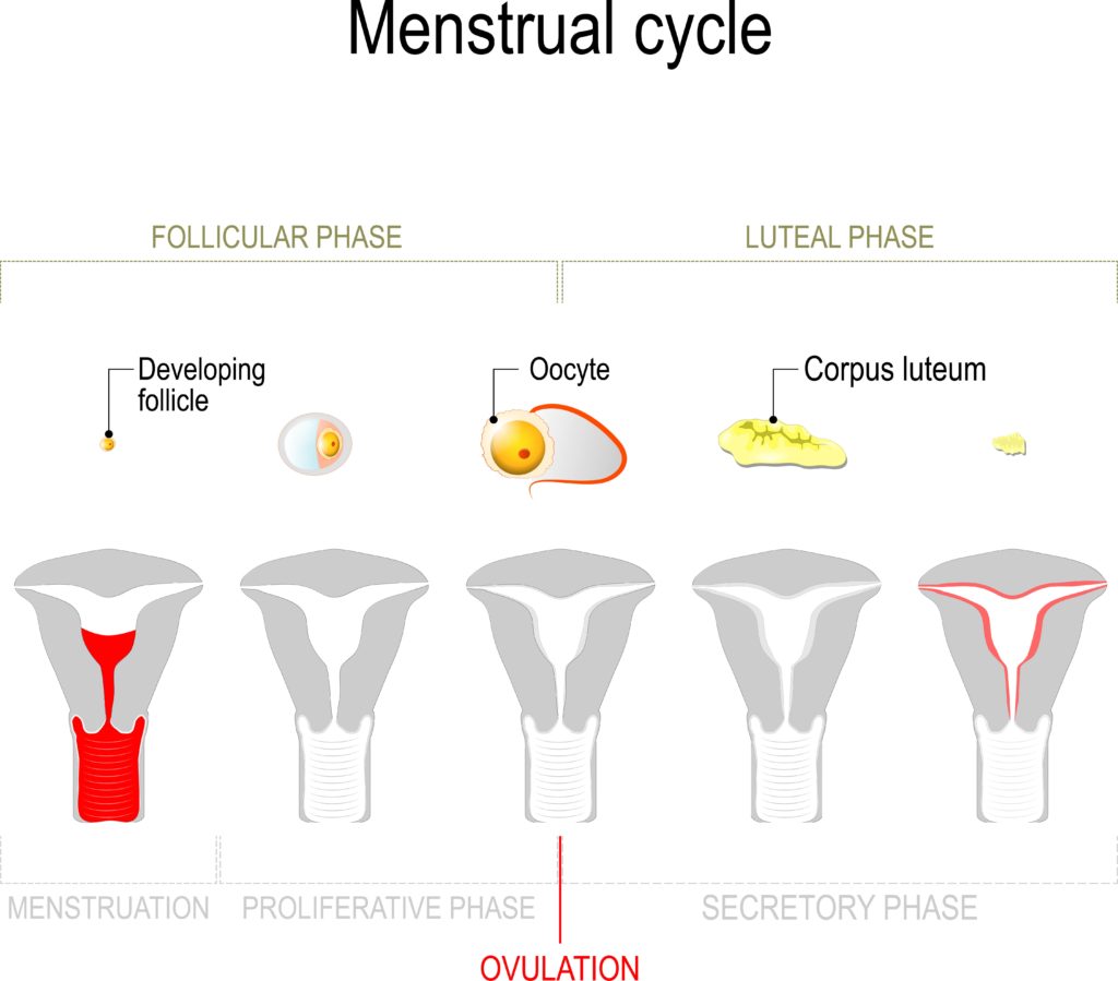
The image provides a simplified overview of the menstrual cycle, segmented into different phases and illustrated with changes in the ovary and uterus.
The menstrual cycle is divided into two main phases: the follicular phase and the luteal phase. These phases are marked by distinct hormonal changes and are separated by ovulation.
During the follicular phase, a developing follicle in the ovary is shown, which contains an immature egg cell, or oocyte. This phase is characterized by the growth and maturation of the ovarian follicles before one is released during ovulation.
Ovulation is indicated by the release of the oocyte from the dominant follicle. This is a critical event in the menstrual cycle and is marked by the red line in the timeline.
Following ovulation, the luteal phase begins, during which the ruptured follicle transforms into the corpus luteum. The corpus luteum is a temporary endocrine structure in the ovary that secretes hormones, primarily progesterone, to prepare the endometrium for a potential pregnancy.
In the uterus, the menstrual cycle is demonstrated in three stages: menstruation, the proliferative phase, and the secretory phase. Menstruation is the shedding of the uterine lining, depicted in red, which occurs if the oocyte is not fertilized.
The proliferative phase is marked by the rebuilding of the endometrium, stimulated by estrogen from the developing follicles in the ovary.
Finally, the secretory phase is characterized by the thickening of the endometrium under the influence of progesterone from the corpus luteum, preparing the uterus for implantation of a fertilized egg.
This image serves as an educational tool to illustrate the dynamic changes that occur in both the ovary and the uterus throughout the menstrual cycle.
Order of Changes in the Ovary
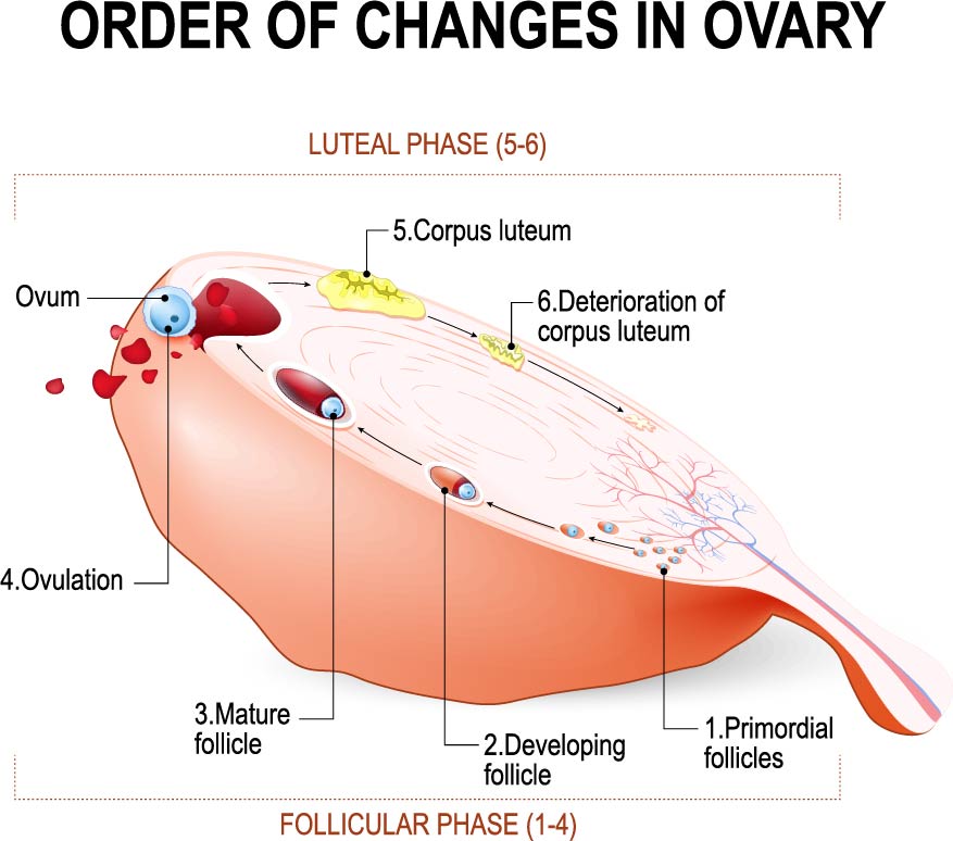
The image illustrates the sequential changes that occur in the ovary during the menstrual cycle, which is divided into the follicular phase and the luteal phase.
The follicular phase is represented by steps 1 to 4:
- Primordial follicles: These are the small, immature follicles that every female is born with. They lie dormant until they are recruited for development.
- Developing follicle: This shows a follicle in the process of maturation, which involves growth and accumulation of fluid.
- Mature follicle: When the follicle is fully mature, it bulges from the surface of the ovary, ready for ovulation.
- Ovulation: This is the moment when the mature follicle releases its ovum (egg) into the fallopian tube.
The luteal phase encompasses steps 5 and 6:
- Corpus luteum: After the ovum is released, the empty follicle transforms into the corpus luteum, which secretes hormones such as progesterone and estrogen to support the potential early stages of pregnancy.
- Deterioration of corpus luteum: If fertilization does not occur, the corpus luteum degenerates, leading to a decrease in hormone levels, which in turn triggers menstruation.
This image serves as an educational tool for understanding the ovarian cycle’s complexity and the hormonal coordination required for reproduction.
Process of Fertilization
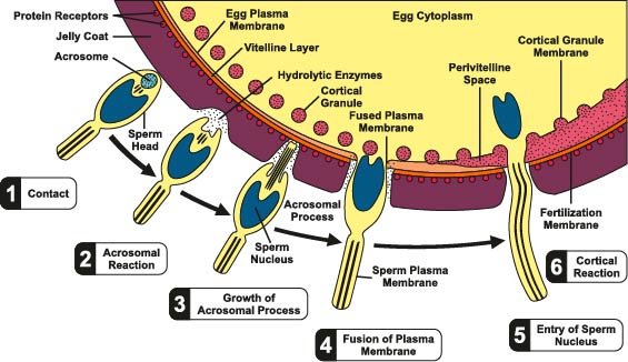
The image depicts the process of fertilization, highlighting the sequential interactions between a sperm cell and an ovum (egg).
Contact: The sperm initially comes into contact with the egg’s protective layer, known as the jelly coat.
Acrosomal Reaction: The sperm’s acrosome, a cap-like structure filled with enzymes, releases its contents upon contact with the egg. These hydrolytic enzymes will digest the jelly coat allowing the sperm to penetrate the egg.
Growth of Acrosomal Process: The enzymes create a pathway through the jelly coat, and the sperm extends an acrosomal process that penetrates the egg’s layers toward the plasma membrane.
Fusion of Plasma Membrane: The acrosomal process reaches the egg plasma membrane, and the membranes of the sperm and egg fuse. This allows the sperm to enter the egg.
Entry of Sperm Nucleus: Once the sperm enters the egg, its nucleus is transported into the egg cytoplasm, ready to merge with the egg’s genetic material.
Cortical Reaction: Following the fusion of the sperm and egg membranes, the egg releases the contents of its cortical granules into the perivitelline space (the space between the plasma membrane and vitelline layer). This reaction changes the egg membrane’s properties, preventing further sperm from binding and ensuring that only one sperm fertilizes the egg, a mechanism known as polyspermy block.
These steps are critical for successful fertilization and represent the beginning of a new organism’s development. The illustration serves as a detailed educational tool to explain the complex process of fertilization at a cellular level.
Development of a Human Embryo
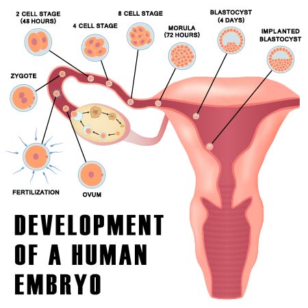
The image outlines the early stages of human embryonic development following fertilization.
The sequence begins with fertilization, where the sperm cell and ovum merge to form a zygote, the first cell of a new individual.
Following fertilization, the zygote undergoes a series of cell divisions:
- 2-cell stage (48 hours): The zygote divides for the first time, resulting in a two-cell embryo.
- 4-cell stage: The cells continue to divide, doubling to four cells.
- 8-cell stage: The division process repeats, resulting in an eight-cell embryo.
- Morula (72 hours): By this stage, the embryo consists of approximately 16 cells and resembles a mulberry, which is why it’s called a morula.
- Blastocyst (4 days): The morula continues to divide and starts to form a cavity, becoming a blastocyst. This stage precedes implantation.
Finally, the image shows the blastocyst implanting into the uterine wall, which is necessary for the establishment of pregnancy.
This image is a valuable educational resource, depicting the early stages of life from a single cell to a developing organism ready to grow within the uterine environment.
Hormonal Control of Ovulation
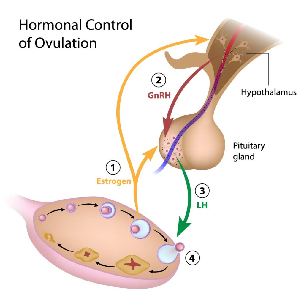
The image illustrates the hormonal regulation of ovulation in the female reproductive system.
Estrogen: Produced by developing follicles in the ovary, estrogen is the hormone responsible for the growth and maintenance of female sexual characteristics. It also plays a critical role in the menstrual cycle. High levels of estrogen stimulate the hypothalamus to release hormones that act on the pituitary gland.
GnRH (Gonadotropin-releasing hormone): This hormone is released by the hypothalamus in response to increased estrogen levels. GnRH triggers the pituitary gland to secrete gonadotropins.
LH (Luteinizing hormone): The pituitary gland releases LH in response to GnRH. A surge in LH levels leads to ovulation, the release of an egg from the ovary.
Ovulation: Triggered by the LH surge, ovulation is the process where a mature egg is released from the ovarian follicle into the fallopian tube, where it can be fertilized by sperm.
This sequence of hormonal interactions is known as the hypothalamic-pituitary-gonadal (HPG) axis. It is a critical system for the regulation of reproduction. The illustration serves as a simplified guide to understand how hormones interact to control the ovulatory cycle.
Cardinal Movements of Fetal Head During Labor and Delivery
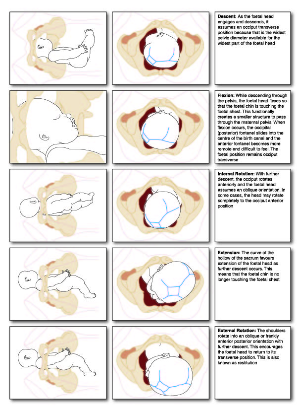
The sequence of images depicts the cardinal movements of fetal head during labor and delivery. These movements are a series of maneuvers that the fetal head undergoes in its passage through the birth canal during the process of natural childbirth.
Descent: The fetal head begins to move down from the uterus into the pelvis. The head typically enters the pelvis in an occiput transverse position, meaning the back of the head (occiput) is facing one of the mother’s hips.
Flexion: As the head descends, it flexes, causing the chin to move toward the fetal chest. This action decreases the presenting diameter of the head, allowing it to pass more easily through the pelvis.
Internal Rotation: The fetal head rotates internally so that the occiput moves from transverse to anterior, aligning the fetal head with the mother’s pelvic outlet.
Extension: As the head passes under the pubic symphysis (the joint at the front of the pelvis), it extends so that the face, forehead, and then the top of the head can emerge from the birth canal.
External Rotation (Restitution): After the head is delivered, it rotates externally to realign the head with the shoulders, which have entered the pelvis in the transverse position and will undergo internal rotation themselves to align for delivery.
These images serve as a visual guide for understanding the dynamic and complex process of childbirth, demonstrating the movements that facilitate the passage of the fetus through the birth canal.
Anatomical Terms and Definitions
| Term | Definition |
|---|---|
| Acrosome | A cap-like structure over the nucleus of a sperm cell, containing enzymes crucial for fertilization as it helps the sperm penetrate the egg. |
| Blastocyst | A stage in early human embryonic development that consists of a fluid-filled cavity and precedes implantation into the uterine wall. |
| Broad Ligament | A wide fold of peritoneum connecting the sides of the uterus to the walls and floor of the pelvis, providing support to the uterus. |
| Cervix | The lower, narrow part of the uterus that opens into the vagina, with a canal that allows passage between the uterus and vagina. |
| Cleavage | A series of rapid cell divisions following fertilization, resulting in the embryo's progression from a zygote to a multicellular structure without growth in size. |
| Corpus Luteum | A temporary endocrine structure in the ovary that secretes hormones, primarily progesterone, to prepare the endometrium for a potential pregnancy. |
| Endometrium | The lining of the uterus, which thickens during the menstrual cycle in preparation for possible implantation of an embryo. |
| Fallopian Tubes | Tubes extending from the uterus to the ovaries, facilitating the passage of eggs from the ovaries to the uterus. |
| Fertilization | The process by which a sperm cell and an ovum (egg) merge to form a zygote, initiating embryonic development. |
| Fibroadenoma | A benign, solid, noncancerous tumor in the breast. |
| Follicle | A fluid-filled sac in the ovary that contains an immature egg (oocyte); it grows and matures in response to hormonal signals during the menstrual cycle. |
| Fundus | The rounded upper part of the uterus. |
| GnRH | Gonadotropin-releasing hormone; released by the hypothalamus to stimulate the pituitary gland to secrete gonadotropins, which influence the menstrual cycle and ovulation. |
| LH | Luteinizing hormone; released by the pituitary gland in response to GnRH, with a surge leading to ovulation. |
| Morula | An early stage in the embryo development consisting of approximately 16 cells, resembling a mulberry, following the cleavage stage and preceding the blastocyst stage. |
| Myometrium | The thick, muscular layer of the uterus responsible for contractions during childbirth and menstruation. |
| Ovarian Cysts | Fluid-filled sacs on the ovary, often forming during the process of ovulation and varying in type, from functional cysts related to the menstrual cycle to pathological cysts. |
| Ovary | A reproductive organ in females that produces eggs (ova) and hormones, such as estrogen and progesterone. |
| Ovulation | The release of an egg from the ovary into the fallopian tube, where it can be fertilized by sperm. |
| Polycystic Ovarian Syndrome (PCOS) | A condition characterized by the presence of multiple cysts on the ovaries, leading to hormonal imbalances, irregular menstrual cycles, and fertility issues. |
| Spermatogenesis | The process of sperm development, from spermatids to fully mature sperm cells. |
| Ureter | A duct that carries urine from the kidneys to the urinary bladder. |
| Uterus | A muscular organ in the female reproductive system where fetal development occurs; consists of the fundus, body, and cervix. |
| Zygote | The first cell of a new individual formed immediately after the sperm and egg nuclei merge during fertilization. |
The image outlines the early stages of human embryonic development following fertilization.
The sequence begins with fertilization, where the sperm cell and ovum merge to form a zygote, the first cell of a new individual.
Following fertilization, the zygote undergoes a series of cell divisions:
- 2-cell stage (48 hours): The zygote divides for the first time, resulting in a two-cell embryo.
- 4-cell stage: The cells continue to divide, doubling to four cells.
- 8-cell stage: The division process repeats, resulting in an eight-cell embryo.
- Morula (72 hours): By this stage, the embryo consists of approximately 16 cells and resembles a mulberry, which is why it’s called a morula.
- Blastocyst (4 days): The morula continues to divide and starts to form a cavity, becoming a blastocyst. This stage precedes implantation.
Finally, the image shows the blastocyst implanting into the uterine wall, which is necessary for the establishment of pregnancy.
This image is a valuable educational resource, depicting the early stages of life from a single cell to a developing organism ready to grow within the uterine environment.
