
Table of Contents
Alzheimer's Disease
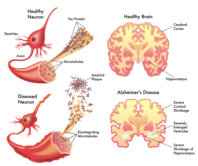
The image provides a comparative view of a healthy neuron and brain versus those affected by Alzheimer’s disease.
In the top left, a healthy neuron is shown with dendrites, which receive signals from other neurons, and an axon, which sends signals to other neurons. The axon’s internal support structure, the microtubules, are intact, and tau proteins, which stabilize these microtubules, are in their normal state.
Below, a diseased neuron is depicted with abnormalities typical in Alzheimer’s disease. The microtubules are disintegrating, leading to the collapse of the neuron’s transport system. Tau proteins have become abnormal and form neurofibrillary tangles, which interfere with the neuron’s function and lead to cell death. Amyloid plaques, which are clumps of protein fragments that accumulate outside of neurons, are also present. These plaques and tangles disrupt communication between neurons and are hallmarks of Alzheimer’s disease.
On the top right, a healthy brain section shows normal-sized ventricles (fluid-filled spaces in the brain) and a well-preserved hippocampus, which is involved in memory formation.
The bottom right illustrates the brain affected by Alzheimer’s disease, showing severe cortical shrinkage due to the loss of neurons and synapses. The ventricles are enlarged as a result of brain tissue loss, and there is severe shrinkage of the hippocampus, reflecting the memory deficits characteristic of Alzheimer’s disease.
This visual comparison is an educational tool for understanding the structural changes in the brain and neurons associated with Alzheimer’s disease, which underlie the symptoms of memory loss, cognitive decline, and changes in behavior and personality.
Ischemia Physiology
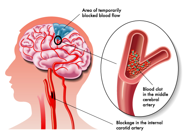
The image illustrates a medical condition related to the cerebral circulation, specifically a blockage in the brain’s blood supply. On the left side, a profile of a human head is shown with the brain exposed, highlighting the vascular system within it. A marked area on the brain indicates a region of temporarily blocked blood flow, suggesting ischemia, where blood supply (and thus oxygen) is restricted.
The close-up view on the right provides a detailed look at the blockage within a blood vessel. Here, it shows a blood clot lodged in the middle cerebral artery, which is a branch of the internal carotid artery that supplies a significant portion of the brain’s outer layer. The blockage in the internal carotid artery itself can lead to a significant reduction in blood flow to the brain, potentially resulting in a stroke.
This blockage can cause an ischemic stroke, which occurs when an artery to the brain is blocked, preventing oxygen and nutrients from reaching a portion of the brain. Brain cells in the affected area can die, resulting in permanent damage. Immediate medical attention is critical to restore blood flow and minimize brain damage.
The illustration is designed to educate on the serious impact of cerebrovascular accidents and the importance of understanding the vascular anatomy of the brain for both prevention and treatment of such events.
Skull Pain Perception
Here is a sequence of images depicting different aspects of the brain’s anatomy and functions. However, without specific instructions for the latest image, I’ll give a general explanation based on what the previous images have covered:
The illustrations provided have detailed various brain structures, from the cerebral lobes to the blood vessels supplying the brain. They’ve also depicted comparisons between a healthy brain and one affected by Alzheimer’s disease, demonstrating the characteristic changes such as cortical shrinkage and ventricle enlargement. Additionally, they’ve shown the impact of a stroke, where a blood clot can block an artery and interrupt blood flow, potentially leading to an infarcted area in the brain.
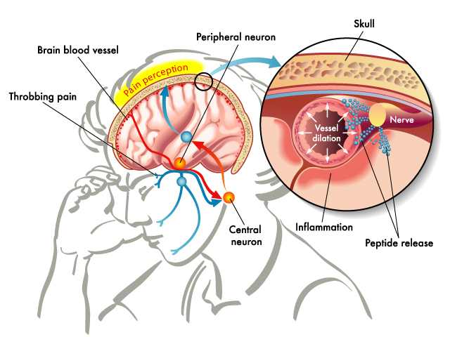
In terms of neural function, the images have highlighted how neurons work in healthy states and how they are affected by disease. They’ve also provided insight into the mechanism of pain perception within the brain, illustrating how signals from peripheral neurons are interpreted as pain by central neurons in the brain.
Each image serves as a visual education tool, demonstrating the complexity of the brain’s structure and the importance of its blood supply, the intricacies of neuronal pathways, and the profound effects of neurological diseases. These images are typically used in educational settings to help students and healthcare professionals visualize and better understand the complex workings of the brain.
Brain Stroke
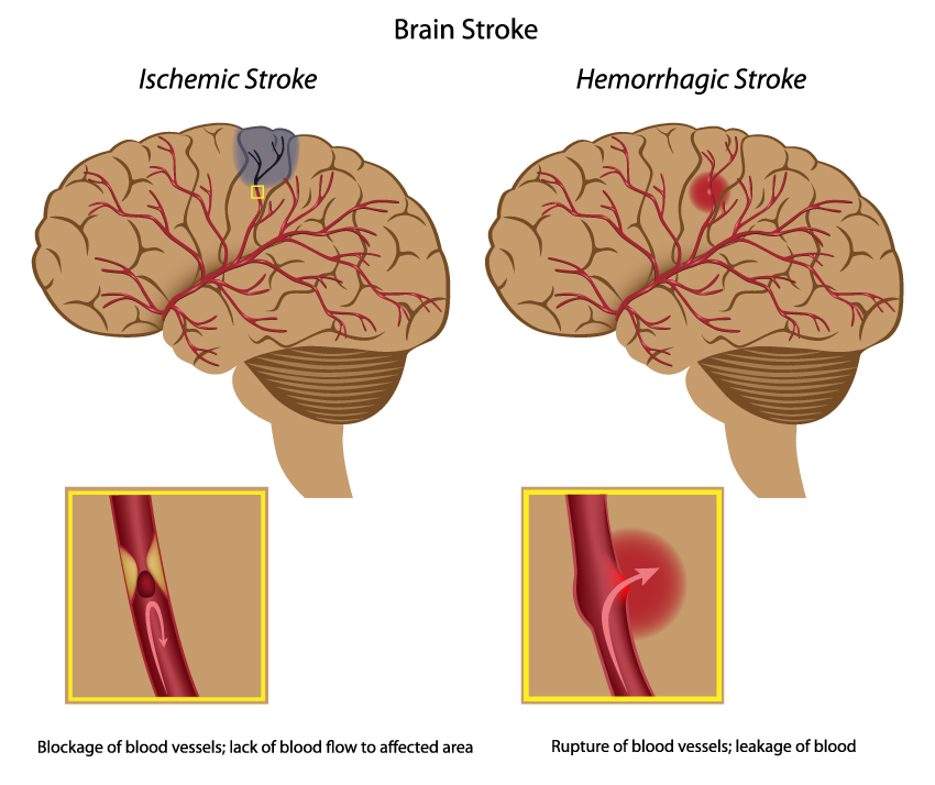
The image illustrates the two main types of strokes: ischemic and hemorrhagic.
On the left, an ischemic stroke is depicted, characterized by the blockage of blood vessels, leading to a lack of blood flow to the affected area of the brain. This blockage is often due to a blood clot, and the region of the brain that is deprived of blood becomes damaged or dies, which is illustrated by the blue shaded area in the brain.
On the right, a hemorrhagic stroke is shown, where there is a rupture of blood vessels, causing leakage of blood into the brain. This leaked blood can cause damage to brain cells, and the pressure buildup from the leaked blood can lead to further injury. The area of bleeding is indicated by the red shading in the brain.
In the smaller insets, close-up views of the blood vessels show the specific pathology of each type of stroke. For the ischemic stroke, a clot is obstructing the inside of the blood vessel. For the hemorrhagic stroke, there is a break in the vessel wall with blood escaping into the surrounding tissue.
Understanding the differences between these two types of strokes is crucial for medical diagnosis and treatment. Ischemic strokes, which are more common, often require treatment to remove the blockage, while hemorrhagic strokes may require surgery to repair the damaged blood vessels and alleviate pressure caused by the bleeding.
Meningitis

The image is an informative illustration about meningitis, an inflammation of the membranes (meninges) surrounding the brain and spinal cord. The diagram provides a cross-sectional view of the brain, showing the inflammation in the dura mater, arachnoid, and pia mater.
It also lists the pathogenic agents that can enter through the blood and cause this condition. Meningitis can be caused by various infectious agents, including bacteria, viruses, fungi, and parasites.
The symptoms of meningitis are grouped into different categories in the image:
- Systematic symptoms include high fever, seizures, stiff neck, sleepiness, or difficulty waking up.
- Central symptoms are headaches and altered mental status.
- Eye-related symptoms include sensitivity to light.
- Ear symptoms can include phonophobia or an extreme sensitivity to sound.
- Skin symptoms may present as paleness, spots, or rash.
- Muscular symptoms include severe muscle pain.
- Stomach-related symptoms are nausea and vomiting.
The complications of meningitis are severe and can include hearing loss, memory difficulties, learning disabilities, brain damage, gait problems, seizures, kidney failure, shock, and even death.
This visual aid is crucial for understanding the seriousness of meningitis and the importance of early detection and treatment to prevent these potential complications.
Meningeal Layers and Subarachnoid Space
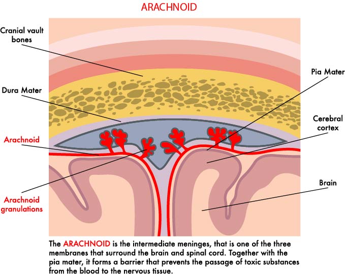
The illustration provides a cross-sectional view of the meningeal layers and subarachnoid space within the human head, detailing the relationship between the meninges, blood vessels, and brain tissue.
At the topmost layer, we see the cranial vault bones, which form the skull’s protective casing. Directly beneath the bone is the dura mater, the outermost and toughest of the three meningeal layers that envelop the brain and spinal cord.
The arachnoid layer is depicted just below the dura mater. It is named for its web-like appearance and is the middle meningeal layer. This layer is important for providing a protective barrier and is separated from the pia mater by the subarachnoid space, which is filled with cerebrospinal fluid (CSF).
The arachnoid granulations (or villi) are shown protruding into the dura mater. These structures are responsible for the reabsorption of CSF into the venous blood system, allowing for the drainage of CSF from the subarachnoid space into the venous circulation.
Beneath the arachnoid, the subarachnoid space is visible, which houses the CSF and acts as a cushion for the brain, providing it with mechanical protection.
The innermost layer is the pia mater, a delicate membrane that closely adheres to the surface of the brain, following its contours and blood vessels into the sulci, or grooves, of the cerebral cortex. The pia mater works in conjunction with the other meningeal layers to protect and encapsulate the central nervous system.
This visual aid serves to explain the protective and supportive roles of the meningeal layers, particularly the arachnoid membrane, as well as the circulation of cerebrospinal fluid within the brain’s ventricular system. It emphasizes the importance of these structures in maintaining the brain’s environment and protecting it from physical damage and chemical imbalances.
