
Table of Contents
Basic Brain Anatomy
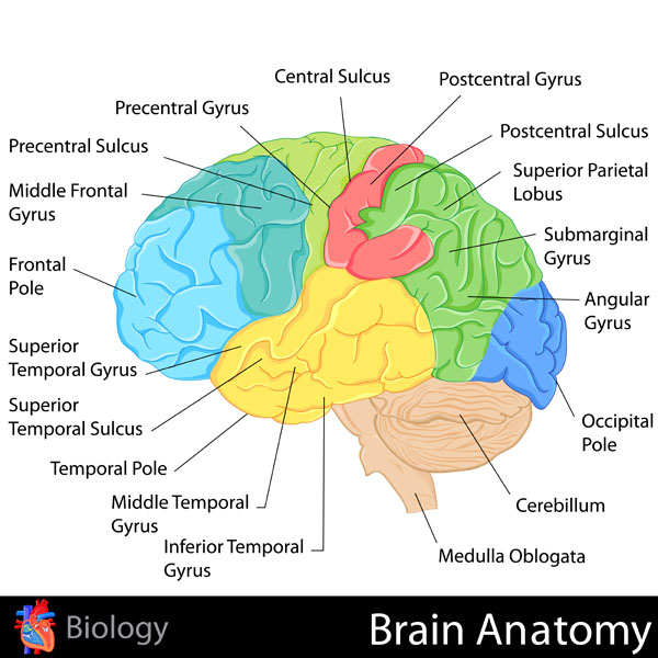
This image displays the lateral aspect of the human brain, highlighting the cerebral cortex’s lobes and some key gyri and sulci, as well as other significant brain structures. The central sulcus, a prominent landmark, separates the frontal lobe, shown in blue, from the parietal lobe, shown in green. Anterior to the central sulcus, the precentral gyrus, known as the primary motor cortex, is responsible for voluntary motor function. Posterior to the central sulcus, the postcentral gyrus represents the primary somatosensory cortex, essential for processing tactile information.
The frontal lobe, responsible for complex cognitive functions, includes the middle frontal gyrus and the frontal pole. The superior temporal gyrus and the temporal pole are part of the temporal lobe, shown in yellow, which is involved in auditory processing and memory. The middle and inferior temporal gyri are also part of the temporal lobe and play roles in object recognition and semantic memory.
Beneath the temporal lobe, the cerebellum, depicted in orange, is critical for motor control and coordination. The brainstem, which includes the medulla oblongata, controls many autonomic functions and connects the brain to the spinal cord.
The superior parietal lobule, angular gyrus, and submarginal gyrus are part of the parietal lobe and are involved in spatial orientation and perception. The occipital lobe, shown in brown, includes the occipital pole and is the primary visual processing center.
This diagram serves as a valuable educational tool to understand the topographical organization of the human brain and the basic functions associated with its different regions.
Thermoregulation Process
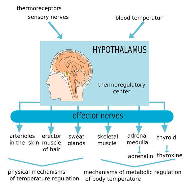
The image presents a schematic representation of the human body’s thermoregulation process, which is the mechanism by which the body maintains its core internal temperature. At the center of this process is the hypothalamus, which is located in the brain and acts as the thermoregulatory center. It receives input about the body’s temperature from two main sources: thermoreceptors, which detect temperature changes, and blood temperature, which is monitored internally.
The hypothalamus then processes this information and activates effector nerves to initiate responses to adjust the body’s temperature. These responses are divided into physical mechanisms of temperature regulation and mechanisms of metabolic regulation of body temperature:
Physical mechanisms involve:
- Arterioles in the skin: The hypothalamus can cause these blood vessels to dilate or constrict, affecting heat loss or retention.
- Erector muscle of hair: The activation of these muscles can cause hair to stand up, trapping more heat close to the body.
- Sweat glands: The hypothalamus can stimulate sweating, which cools the body as the sweat evaporates off the skin surface.
Metabolic mechanisms involve:
- Skeletal muscle: The hypothalamus can trigger shivering, which is the rapid contraction of muscles to generate heat.
- Adrenal medulla: This gland can release adrenaline, which increases the metabolic rate and heat production.
- Thyroid: The hypothalamus can influence the thyroid gland to release thyroxine, which also increases the metabolic rate and heat production.
These systems work in concert to ensure that the body’s temperature remains within a narrow, optimal range, despite external temperature changes. This process is crucial for the proper functioning of enzymatic reactions and overall physiological processes.
Hypothalamus Anatomy
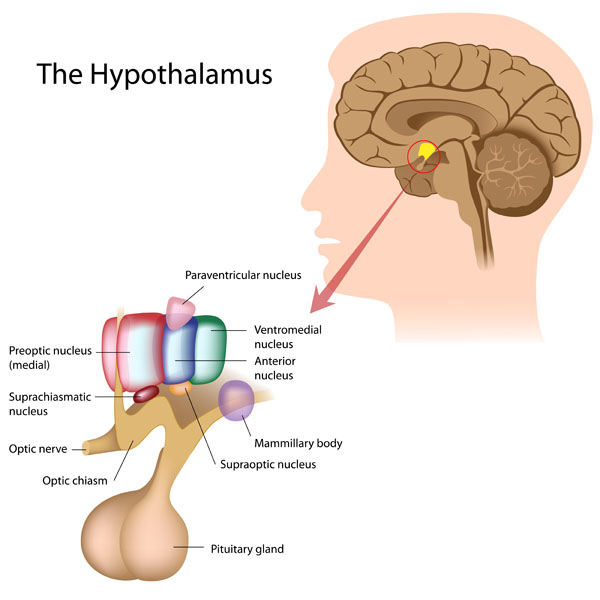
The image displays an anatomical overview of the hypothalamus and its relationship to the pituitary gland and surrounding brain structures. The hypothalamus is highlighted within the brain, indicating its position deep within the cerebral structure.
Several nuclei of the hypothalamus are color-coded and labeled:
- The paraventricular nucleus, which is involved in the regulation of several functions including the release of hormones from the pituitary gland, stress response, and appetite control.
- The ventromedial nucleus plays a critical role in satiety and body weight regulation.
- The anterior nucleus, which is implicated in temperature regulation.
- The suprachiasmatic nucleus, which is known as the body’s “master clock,” regulating circadian rhythms and the sleep-wake cycle.
- The preoptic nucleus (medial), which is involved in thermoregulation and sexual behavior.
Below the hypothalamus, the optic chiasm is shown where the optic nerves cross. The mammillary body, which is part of the limbic system, is involved in memory formation. The supraoptic nucleus, not to be confused with the suprachiasmatic nucleus, is important for the production of antidiuretic hormone (ADH), which regulates water balance in the body.
The image also shows the pituitary gland, often termed the “master gland,” as it secretes hormones that regulate many of the body’s functions, including growth, metabolism, and reproduction. The gland is connected to the hypothalamus, illustrating the close relationship between these two structures in hormone regulation and the endocrine system as a whole.
This educational illustration serves to show the complexity and the essential roles of the hypothalamus in maintaining homeostasis through its interactions with the endocrine and nervous systems.
Limbic System
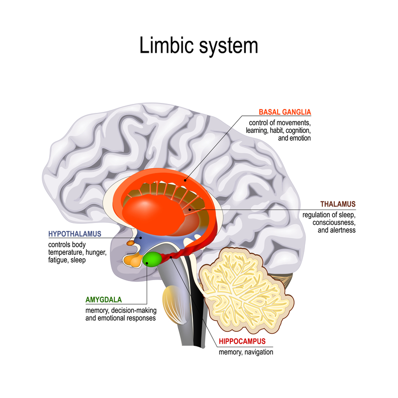
The image provides a detailed view of the limbic system in the human brain, a complex set of structures that play a key role in emotion, behavior, and memory, along with the basal ganglia and thalamus, which are closely associated with the limbic system functionally.
At the forefront of the limbic system, colored in red, is the cingulate gyrus, which is involved in emotional processing and regulation. Beneath it, the orange structure represents the fornix, a bundle of nerve fibers that carry signals from the hippocampus to the mammillary bodies and other parts of the brain.
The amygdala, shown in green, is a critical component of the limbic system associated with memory, decision-making, and emotional responses, including fear and pleasure. It is deeply embedded within the temporal lobe and has extensive connections with other brain regions.
Colored in blue, the hypothalamus sits below the thalamus and is not traditionally part of the limbic system but is closely connected to it. The hypothalamus controls body temperature, hunger, fatigue, sleep, and plays a significant role in the endocrine system, regulating the release of hormones.
On the bottom right, in yellow, is the hippocampus, vital for memory formation and spatial navigation. It’s one of the first regions affected in diseases such as Alzheimer’s, leading to the characteristic early memory loss.
The basal ganglia, located deep within the cerebral hemispheres and labeled in the upper right, are a group of nuclei involved in the control of voluntary motor movements, procedural learning, habit formation, cognition, emotion, and eye movements.
Finally, the thalamus, labeled in the upper right and not colored, acts as the brain’s relay station for sensory and motor signals (except for the sense of smell) and is also crucial for the regulation of sleep, consciousness, and alertness.
This illustration is an educational tool, simplifying the complex interrelations and functions of these brain regions to give a foundational understanding of the limbic system’s role in human behavior and emotional processing.
Cerebrospinal Fluid
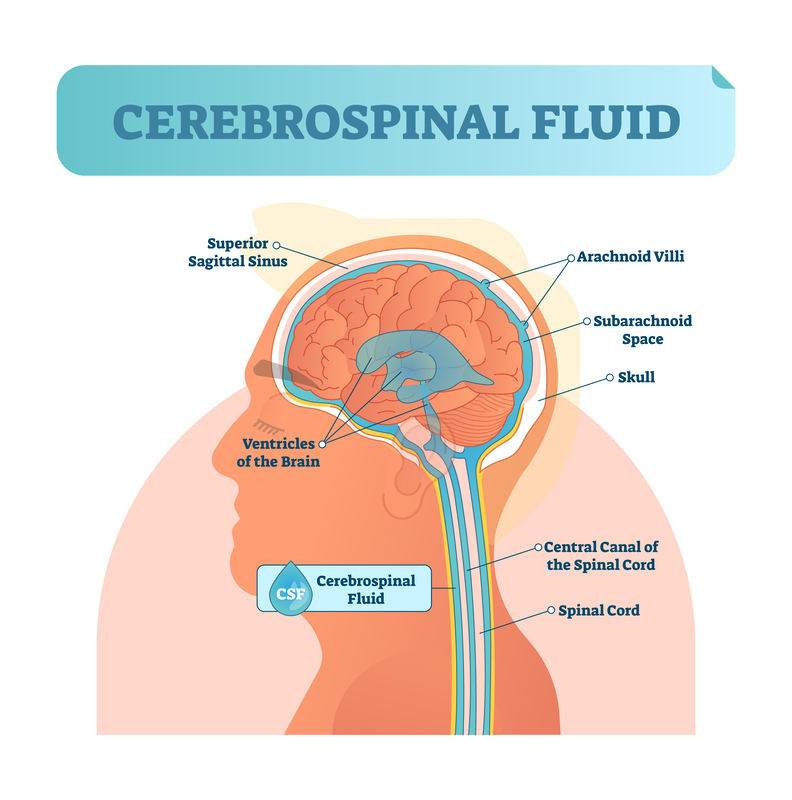
This image provides a clear visualization of the cerebrospinal fluid (CSF) in the central nervous system. The CSF is depicted in blue, highlighting its flow through and around the brain and spinal cord.
The brain’s ventricles, a series of interconnected, fluid-filled spaces, produce CSF which then circulates through these ventricles. From the lateral ventricles, the CSF flows into the third ventricle, then through the cerebral aqueduct into the fourth ventricle, and finally into the subarachnoid space surrounding the brain and spinal cord.
The superior sagittal sinus, a blood-filled space running along the top of the brain within the dura mater, is where the CSF is reabsorbed into the bloodstream via structures called arachnoid villi. These villi act as one-way valves to allow CSF to exit the subarachnoid space and enter the venous system.
Surrounding the central nervous system is the subarachnoid space, where the CSF provides a cushioning effect, protecting the brain and spinal cord from trauma. This space is located between the arachnoid mater and the pia mater, two of the three membranes that cover and protect the brain and spinal cord.
The central canal of the spinal cord is also filled with CSF and runs the length of the spinal cord. The spinal cord itself, protected by the vertebral column, extends from the base of the skull to the lower back and is the main pathway for transmitting information between the brain and the body.
Cerebrospinal fluid is essential for the homeostasis of the central nervous system. It provides buoyancy to the brain, effectively reducing its weight and preventing the brain from being crushed under its own weight. It also serves to transport nutrients to the brain and remove waste products.
The illustration effectively demonstrates the crucial role of CSF in brain and spinal cord function and protection.
Meningeal Layers and Subarachnoid Space
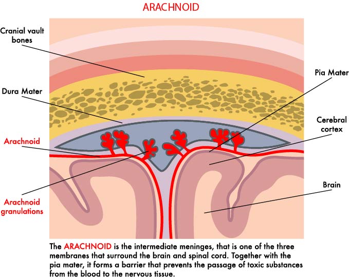
The illustration provides a cross-sectional view of the meningeal layers and subarachnoid space within the human head, detailing the relationship between the meninges, blood vessels, and brain tissue.
At the topmost layer, we see the cranial vault bones, which form the skull’s protective casing. Directly beneath the bone is the dura mater, the outermost and toughest of the three meningeal layers that envelop the brain and spinal cord.
The arachnoid layer is depicted just below the dura mater. It is named for its web-like appearance and is the middle meningeal layer. This layer is important for providing a protective barrier and is separated from the pia mater by the subarachnoid space, which is filled with cerebrospinal fluid (CSF).
The arachnoid granulations (or villi) are shown protruding into the dura mater. These structures are responsible for the reabsorption of CSF into the venous blood system, allowing for the drainage of CSF from the subarachnoid space into the venous circulation.
Beneath the arachnoid, the subarachnoid space is visible, which houses the CSF and acts as a cushion for the brain, providing it with mechanical protection.
The innermost layer is the pia mater, a delicate membrane that closely adheres to the surface of the brain, following its contours and blood vessels into the sulci, or grooves, of the cerebral cortex. The pia mater works in conjunction with the other meningeal layers to protect and encapsulate the central nervous system.
This visual aid serves to explain the protective and supportive roles of the meningeal layers, particularly the arachnoid membrane, as well as the circulation of cerebrospinal fluid within the brain’s ventricular system. It emphasizes the importance of these structures in maintaining the brain’s environment and protecting it from physical damage and chemical imbalances.
Corpus Callosum Structure
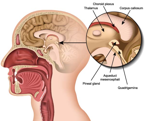
This image is a sagittal section of the human head, providing an internal view of the brain and some of its key structures.
Highlighted in the illustration is the corpus callosum, the thick band of nerve fibers that connects the two cerebral hemispheres, allowing for communication between them. Above it lies the choroid plexus, which is a network of cells that produce cerebrospinal fluid (CSF), vital for cushioning the brain and maintaining a stable environment within the central nervous system.
The thalamus, a dual lobed mass of gray matter, is visible beneath the corpus callosum. The thalamus acts as the main relay station for sensory impulses to the cerebral cortex. It plays a crucial role in the processing and transmission of sensory information to the appropriate higher brain centers.
Beneath the thalamus is the pineal gland, a small endocrine gland that secretes the hormone melatonin, which regulates sleep patterns.
Also shown is the aqueduct mesencephali (also known as the cerebral aqueduct), a narrow channel that connects the third and fourth ventricles of the brain, allowing for the passage of CSF.
The quadrigemina, part of the tectum in the midbrain, comprises the superior and inferior colliculi, which are responsible for visual and auditory reflexes respectively.
The image also provides a view of the nasal and oral cavities, illustrating the relationship of the brain to these structures in the head.
This visual aid serves an educational purpose, detailing the anatomical positioning of these various brain structures and their relative positions to each other, which is important for understanding their functions within the central nervous system.
Protective Membranes
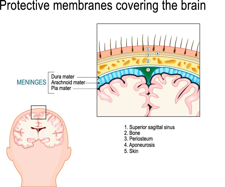
The image is an educational illustration that explains the layers of protection surrounding the human brain. At the top, there is a detailed cross-section that labels the layers from the outermost to the innermost, starting with the skin and ending with the brain surface.
The outermost layer, labeled as ‘5’, is the skin, which provides the first line of defense against external environmental factors. Below the skin is ‘4’, the aponeurosis, which is a layer of fibrous tissue that helps anchor the skin to the tissues underneath it.
Beneath the aponeurosis is ‘3’, the periosteum, a dense layer of vascular connective tissue that envelops the bones of the skull. The periosteum contains blood vessels that supply bone cells with nutrients.
Under the periosteum is ‘2’, the bone, specifically the skull bones, which form a rigid protective housing for the brain.
The superior sagittal sinus, marked as ‘1’, is a venous sinus located within the layers of the dura mater. It plays a critical role in draining venous blood from the brain to the internal jugular vein.
The layers of the meninges are highlighted on the lower part of the cross-section. The dura mater is the tough and durable outermost layer of the meninges that provides a protective covering for the brain. The arachnoid mater is the middle layer, and it is depicted as a thin, web-like structure that cushions the central nervous system. Between the arachnoid mater and the pia mater lies the subarachnoid space, which is filled with cerebrospinal fluid (CSF).
The innermost layer is the pia mater, which is a thin, delicate membrane that closely adheres to the brain’s contours. It is not labeled in the diagram but can be inferred as the layer directly covering the brain tissue.
Below the cross-section, the image shows an outline of a head with an inset box indicating the area of the cross-section above, providing context for the location of these protective layers. This visual aid is commonly used to teach about the meninges and the protective coverings of the brain.
Optic Chiasm
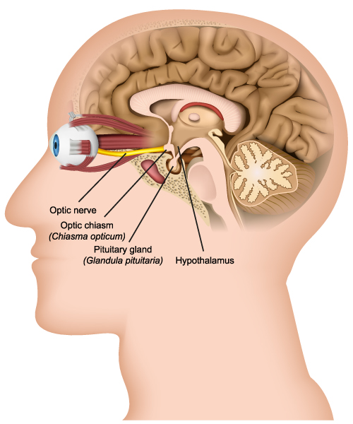
The image provides a detailed sagittal section of the human head, showcasing the relationship between the eye, the optic nerve, and parts of the brain.
The optic nerve is depicted as it extends from the back of the eye, carrying visual information from the retina to the brain. It then meets the optic chiasm (Chiasma opticum), which is a critical juncture where the optic nerves from both eyes partially cross. This structure allows visual information from both eyes to be processed on both sides of the brain, enabling binocular vision.
The pituitary gland (Glandula pituitaria), often referred to as the “master gland,” is shown beneath the optic chiasm. This gland produces hormones that regulate a variety of bodily functions, including growth, metabolism, and reproductive processes.
The hypothalamus, located just above the pituitary gland, is a crucial component of the brain for maintaining homeostasis. It regulates various autonomic functions, such as temperature control, thirst, hunger, sleep, and emotional activity.
This illustration is particularly useful for understanding the anatomical positioning and connectivity of the optic nerve, optic chiasm, pituitary gland, and hypothalamus, as well as their proximity to each other. These structures’ interrelation is essential for functions such as vision, hormone production, and the regulation of the body’s internal environment.
Cerebellum Anatomy
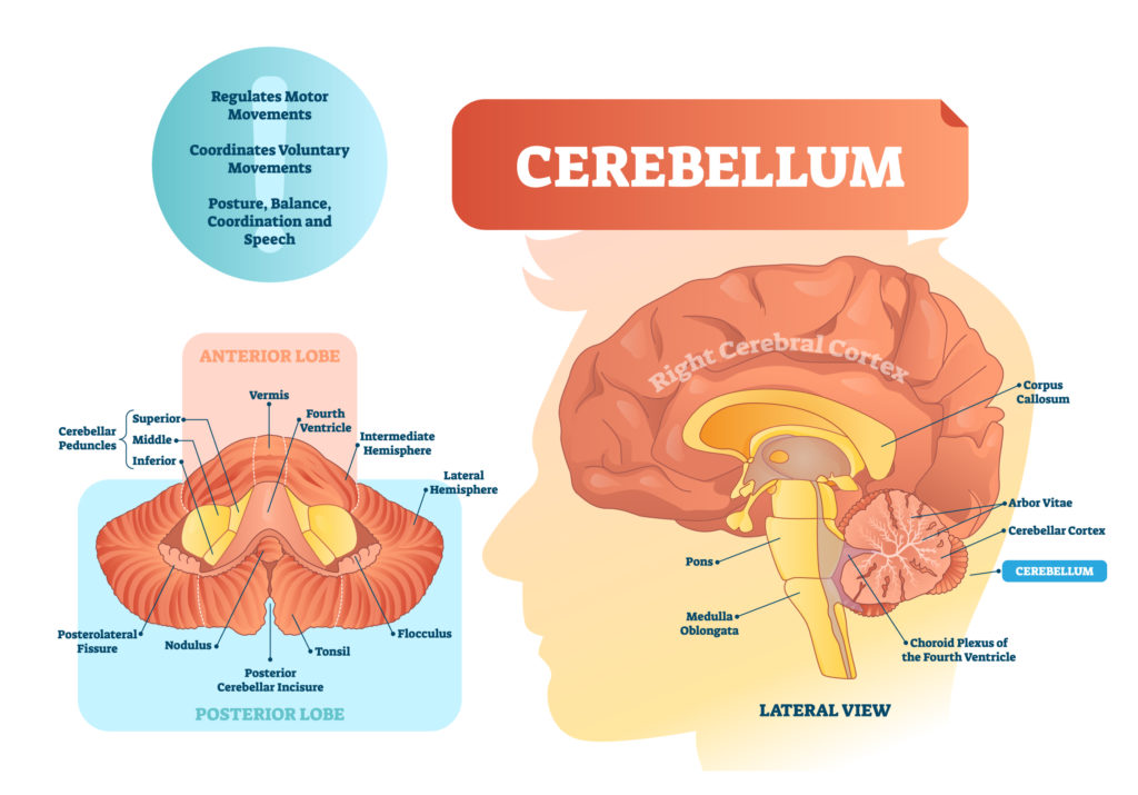
The image provides a comprehensive view of the cerebellum and its functional regions, as well as a lateral view of the brain showing the position of the cerebellum relative to other structures.
On the left side of the image, the cerebellum is divided into the anterior and posterior lobes, separated by the primary fissure. The cerebellum is responsible for regulating motor movements, coordinating voluntary movements, and maintaining posture, balance, and speech. Within the anterior lobe, the illustration shows the cerebellar peduncles, which are nerve tracts that communicate between the cerebellum and other parts of the brain, as well as the spinal cord. The superior, middle, and inferior cerebellar peduncles are labeled to indicate their relative positions.
The nodulus and the tonsil, which are parts of the cerebellar structure involved in processing vestibular inputs for balance, are also shown, along with the flocculus, which is involved in motor control. The fourth ventricle of the brain, a fluid-filled cavity that helps protect the brain from trauma and transports cerebrospinal fluid, is also depicted.
On the right side, the lateral view of the brain shows the cerebellum at the back of the head, underneath the larger cerebrum. It’s connected to the brainstem via the pons, which is involved in motor control and sensory analysis. The medulla oblongata is also shown; it regulates vital functions such as breathing and heart rate.
The illustration also features the arbor vitae, which refers to the distinctive tree-like arrangement of white matter in the cerebellum. This structure is crucial for the coordination of signals in the cerebellum. The cerebellar cortex is the outer layer of the cerebellum, which processes information from the spinal cord and other parts of the brain.
Overall, this illustration serves as an educational tool, providing insights into the anatomy and functions of the cerebellum, as well as its connections to the central nervous system.
Lobes and Brain Functions
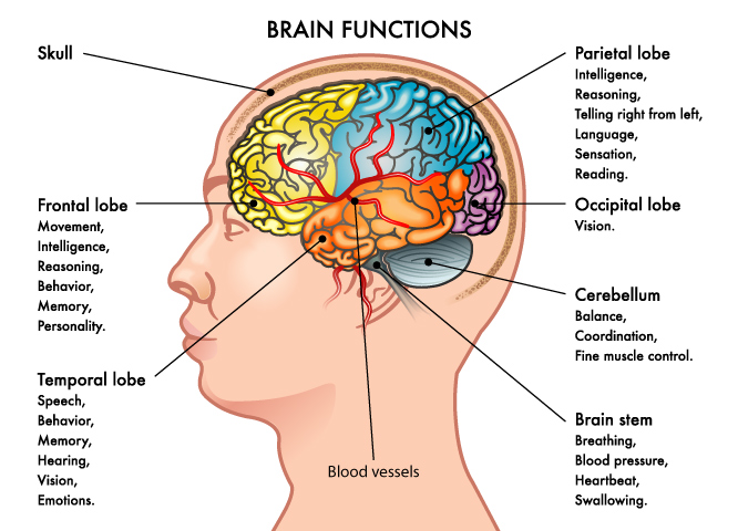
The image is a lateral view of the human head, depicting a color-coded brain with labels indicating various lobes and their associated functions. Each lobe of the brain is responsible for different cognitive and sensory activities:
The frontal lobe, colored in blue, is associated with movement, intelligence, reasoning, behavior, memory, and personality. This lobe is critical for voluntary motor activity, decision-making, and moderating social behavior.
The temporal lobe, shown in green, is involved with speech, behavior, memory, hearing, vision, and emotions. It plays a significant role in processing auditory information and is also important for the encoding of memory.
The parietal lobe, illustrated in yellow, is linked to intelligence, recognizing the difference between right and left, language, sensation, and reading. It integrates sensory information from various modalities, particularly determining spatial sense and navigation.
The occipital lobe, in red, is primarily responsible for vision. It processes visual data and helps in the recognition of shapes and colors.
The cerebellum, shown in purple, located at the back of the brain beneath the occipital lobes, is essential for balance, coordination, and fine muscle control. It is involved in the regulation and coordination of movement, posture, and balance.
The brain stem, which is not color-coded in the image, controls basic life functions such as breathing, blood pressure, heartbeat, and swallowing.
The image also indicates the skull, which encases and protects the brain, and the blood vessels, which supply the brain with necessary nutrients and oxygen.
Overall, this visual aid helps in understanding the basic functions attributed to different parts of the brain and how they contribute to the overall functioning of the human body.
Frontal Lobe Anatomy
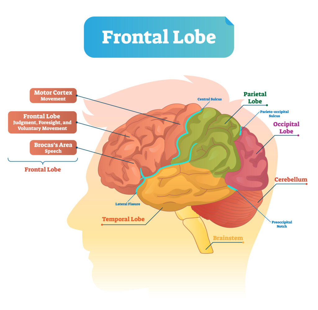
The image illustrates a lateral view of the human brain, highlighting the frontal lobe and other major areas with their associated functions.
The frontal lobe, prominently shaded and labeled, is responsible for higher cognitive functions such as judgment, foresight, and voluntary movement. Within this lobe, the motor cortex is indicated, which controls movement. Also noted in the frontal lobe is Broca’s area, which is crucial for speech production.
The central sulcus, a prominent groove on the surface of the brain, is marked, separating the frontal lobe from the parietal lobe. The parietal lobe, associated with processing sensory information such as touch, temperature, and pain, is labeled above the central sulcus.
Just behind the parietal lobe, the occipital lobe is identified, which is primarily involved in visual processing. The image also shows the cerebellum at the back of the brain, beneath the occipital lobe, which is responsible for balance and coordination of movement.
The temporal lobe is situated below the frontal and parietal lobes, marked by the lateral fissure. It plays a key role in auditory processing, memory, and integrating sensory input.
Finally, the brainstem is depicted below the cerebellum, which controls many basic life functions such as breathing, heart rate, and blood pressure.
The image serves as an educational tool to outline the basic regions of the brain and to give an overview of the primary responsibilities of each area, particularly emphasizing the role of the frontal lobe in complex behaviors and motor function.
Blood Supply of the Brain
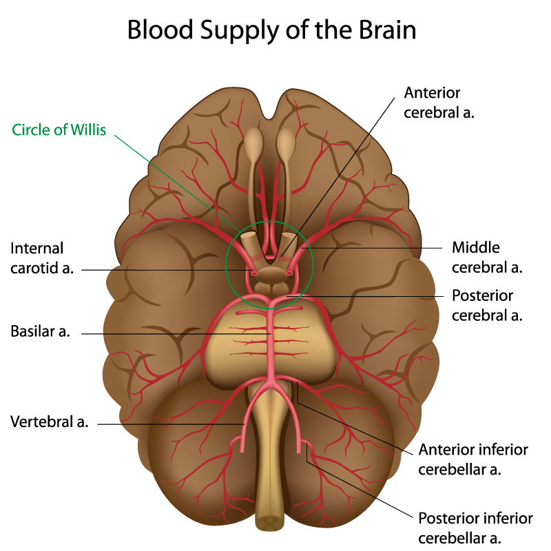
The illustration provides a detailed depiction of the brain’s blood supply. Central to this vascular arrangement is the Circle of Willis, a ring-like arterial structure that sits at the base of the brain and provides collateral blood flow between the anterior and posterior circulation of the brain.
Arteries that contribute to the Circle of Willis include:
- The internal carotid arteries, which ascend into the brain from the neck and branch into smaller arteries, including the anterior and middle cerebral arteries.
- The anterior cerebral arteries, which travel upward and forward from the Circle of Willis, supply the medial portions of the frontal lobes and superior medial parietal lobes.
- The middle cerebral arteries, the largest branches of the internal carotid, course laterally into the lateral sulcus (Sylvian fissure) and supply the lateral aspects of the frontal, parietal, and temporal lobes.
- The posterior cerebral arteries, which arise from the basilar artery, supply blood to the occipital lobes, the undersides of the temporal lobes, and various deep brain structures.
The basilar artery, formed by the union of the two vertebral arteries, ascends along the brainstem and provides branches to the brainstem and cerebellum before dividing into the posterior cerebral arteries.
The vertebral arteries, not visible in this illustration, enter the skull through the foramen magnum and merge to form the basilar artery. They are part of the posterior circulation, which supplies the posterior part of the brain.
The anterior and posterior inferior cerebellar arteries, branches of the basilar and vertebral arteries, supply blood to the cerebellum.
This network of arteries ensures the brain receives a constant, uninterrupted blood supply, which is essential for its functions. The Circle of Willis also plays a critical role in compensating for blockages or narrowings in the major arterial supply routes to the brain, which can help prevent strokes and other cerebrovascular events.
Anatomical Terms and Definitions
| Term | Definition |
|---|---|
| Adrenal Medulla | Gland that releases adrenaline, increasing metabolic rate and heat production. |
| Amygdala | Brain region involved in emotional responses, memory, and decision-making. |
| Angular Gyrus | Parietal lobe structure involved in spatial orientation and perception. |
| Anterior Nucleus | Hypothalamic area implicated in temperature regulation. |
| Anterior Pituitary Lobe | Pituitary gland part that produces various hormones for physiological processes. |
| Arbor Vitae | Tree-like arrangement of white matter in the cerebellum, crucial for signal coordination. |
| Arterioles | Blood vessels in the skin influenced by the hypothalamus to affect heat loss or retention. |
| Basal Ganglia | Group of nuclei in the brain involved in control of voluntary motor movements and various cognitive processes. |
| Blood Temperature | Internal monitor of body's temperature, influencing hypothalamic response. |
| Brainstem | Includes medulla oblongata and pons, controlling basic life functions. |
| Central Sulcus | Groove separating frontal and parietal lobes of the brain. |
| Cerebellar Cortex | Outer layer of the cerebellum, processing information from the spinal cord and brain. |
| Cerebellar Peduncles | Nerve tracts communicating between the cerebellum, brain, and spinal cord. |
| Cerebellum | Structure at the base of the brain crucial for balance, coordination, and fine motor control. |
| Cerebral Aqueduct | Narrow channel connecting the third and fourth ventricles of the brain, allowing passage of CSF. |
| Cerebral Cortex | Outer layer of the cerebrum involved in complex cognitive functions. |
| Cingulate Gyrus | Limbic system structure involved in emotional processing and regulation. |
| Corpus Callosum | Band of nerve fibers connecting the left and right cerebral hemispheres. |
| Frontal Lobe | Brain region responsible for complex cognitive functions, including reasoning and planning. |
| Hippocampus | Vital for memory formation and spatial navigation. |
| Hypothalamus | Brain region controlling autonomic functions and hormone release. |
| Inferior Temporal Gyrus | Temporal lobe structure playing a role in object recognition. |
| Medulla Oblongata | Part of the brainstem regulating vital bodily functions. |
| Middle Cerebral Arteries | Branches of the internal carotid artery supplying the lateral aspects of the brain. |
| Middle Frontal Gyrus | Part of the frontal lobe involved in cognitive functions. |
| Occipital Lobe | Primary visual processing center of the brain. |
| Paraventricular Nucleus | Hypothalamic nucleus involved in hormone release and stress response. |
| Parietal Lobe | Processes sensory information like touch and spatial awareness. |
| Pituitary Gland | The "master gland" regulating various bodily functions through hormone release. |
| Postcentral Gyrus | Represents the primary somatosensory cortex, essential for processing tactile information. |
| Precentral Gyrus | Known as the primary motor cortex, responsible for voluntary motor function. |
| Superior Temporal Gyrus | Part of the temporal lobe involved in auditory processing and memory. |
| Temporal Lobe | Involved in processing auditory information and memory. |
| Thalamus | Acts as the brain’s relay station for sensory and motor signals. |
| Thyroid | Gland influenced by the hypothalamus to regulate metabolic rate and heat production. |
| Ventromedial Nucleus | Hypothalamic nucleus critical for satiety and body weight regulation. |
