
Table of Contents
Basic Leg Muscle Anatomy
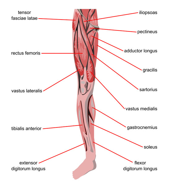
This illustration provides a detailed view of the major muscles in the human leg. At the top, near the hip joint, we have the tensor fasciae latae, a muscle that assists in stabilizing and moving the hip and knee. The iliopsoas, actually comprised of two muscles, the iliacus and psoas major, is important for hip flexion. Next, we see the pectineus, which is involved in hip flexion and adduction, and the adductor longus, which also serves to adduct the thigh.
The gracilis muscle, a long, thin muscle running down the inner thigh, is responsible for hip adduction and helps in knee flexion. Overlaying the thigh, we have the sartorius, the longest muscle in the human body, which crosses both the hip and knee joints, contributing to flexion, abduction, and lateral rotation of the hip, as well as flexion of the knee.
On the front of the thigh, the rectus femoris, part of the quadriceps group, is visible; it’s unique in that it crosses both the hip and knee joints and aids in extending the knee and flexing the thigh at the hip. Flanking the rectus femoris are the vastus lateralis and medialis, both part of the quadriceps muscle group as well, with roles primarily in knee extension.
Moving to the lower leg, the tibialis anterior is located on the front of the shin and is crucial for dorsiflexion and inversion of the foot. On the outer side of the lower leg is the extensor digitorum longus, which helps extend the toes and dorsiflex the foot.
Conversely, on the back of the leg, the calf muscles are prominent, including the gastrocnemius, which is the larger and more superficial muscle involved in plantarflexing the ankle and flexing the knee. Beneath it lies the soleus, which also aids in plantarflexion but is primarily active while walking or standing. Lastly, the flexor digitorum longus is presented, a muscle that flexes the toes and helps in plantar flexion of the foot.
Each muscle plays a vital role in various movements and stabilization of the leg, with some crossing multiple joints and thus contributing to a complex range of actions.
Lower Leg Anatomy: Tibialis Muscles
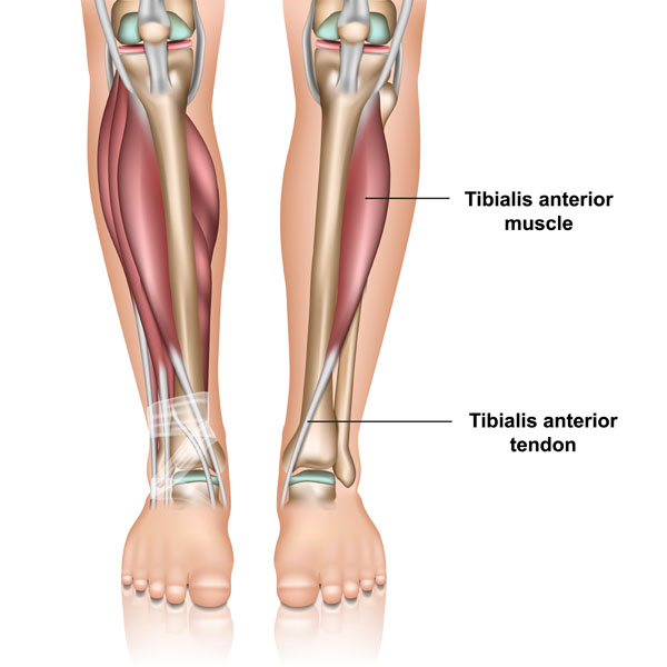
The image showcases the anatomy of the lower leg, focusing on the tibialis anterior muscle and its associated tendon. On the left, we observe the tibialis anterior muscle in its anatomical position. This muscle originates in the upper two-thirds of the lateral surface of the tibia, which is the larger and medial of the two bones in the lower leg.
The tibialis anterior muscle is responsible for dorsiflexion, which is the action of raising the foot upwards towards the shin, and inversion, which is the movement of turning the sole of the foot inward. This muscle is an important stabilizer for the ankle when walking or running.
The illustration on the right provides a more medial view, showing the pathway of the tibialis anterior muscle as it descends down the leg. The muscle tapers into a tendon as it approaches the ankle. The tibialis anterior tendon is clearly visible as it travels over the ankle joint to insert onto the medial cuneiform and first metatarsal bones of the foot.
This tendon is crucial in transferring the force generated by the tibialis anterior muscle to the foot, facilitating movement. The tendon must pass through a compartment where it is held in place against the bones of the ankle by a thick band of connective tissue known as the retinaculum. This sheath ensures that the tendon does not bowstring away from the ankle during dorsiflexion and inversion motions. Proper function of the tibialis anterior muscle and tendon is essential for activities such as walking, climbing stairs, and any motion that requires lifting the foot at the ankle joint.
Lower Leg Muscle and Bone Anatomy
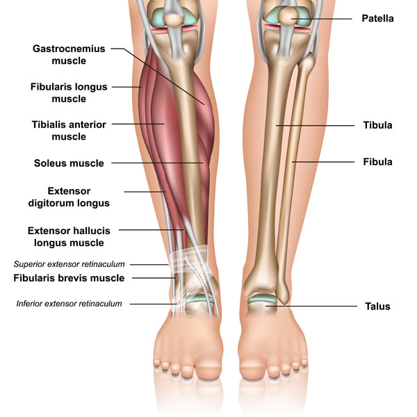
The image presents a detailed anatomical view of the lower leg, identifying various muscles and bones. On the left side, we can see the gastrocnemius muscle, which is the larger muscle forming the calf. It is responsible for the plantar flexion of the foot at the ankle joint and flexing the leg at the knee joint. Underneath the gastrocnemius is the soleus muscle, another plantar flexor of the foot that is not visible from the surface.
The fibularis longus, also known as the peroneus longus, is shown on the lateral aspect of the leg. This muscle assists in plantar flexion and eversion of the foot. Deep to the fibularis longus is the tibialis anterior muscle, which is a key muscle for dorsiflexion and inversion of the foot.
We also see the extensor digitorum longus, which extends the toes and dorsiflexes the foot, and the extensor hallucis longus, which extends the big toe and also aids in dorsiflexion of the foot. The fibularis brevis muscle is positioned beneath the fibularis longus and also functions to evert and plantar flex the foot.
The superior extensor retinaculum and the inferior extensor retinaculum are bands of fibrous tissue that hold the tendons close to the ankle as they pass from the leg into the foot.
On the right side, the image provides a posterior view of the leg, showing the patella, or kneecap, at the top, which is a small bone that sits in front of the knee joint. The tibia and fibula, the two long bones of the lower leg, are labeled, with the tibia being the larger and more medial bone and the fibula being thinner and positioned laterally.
Lastly, the talus is noted at the lower end of the image; it is one of the tarsal bones in the ankle that articulates with both the tibia and fibula to form the ankle joint, playing a critical role in the movement and stability of the foot.
Understanding the relationship between these muscles and bones is essential for grasping how movement at the ankle and foot is achieved and how these structures contribute to stability and locomotion.
Upper Thigh Posterior Muscles
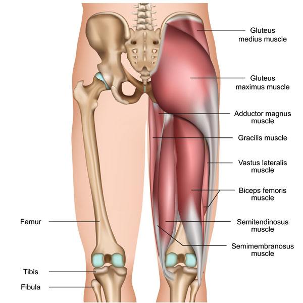
The image provides a lateral view of the right hip and thigh, detailing both the muscular and skeletal structures. At the top, we can see the bones of the pelvic girdle, with the femur, or thigh bone, articulating with the pelvis at the hip joint.
The large, superficial muscle covering most of the gluteal region is the gluteus maximus, which is the largest muscle in the body. It is primarily responsible for the extension, outward rotation, and abduction of the hip joint. Superior and anterior to the gluteus maximus is the gluteus medius muscle, which lies beneath the maximus and is crucial for hip abduction and stabilization of the pelvis during walking.
The adductor magnus muscle is a large, triangular muscle located on the medial side of the thigh. It works to adduct, flex, and medially rotate the thigh. The gracilis muscle, also located on the medial side of the thigh, is a long, strap-like muscle that assists in adduction of the thigh, flexion, and medial rotation of the leg at the knee joint.
On the lateral side of the thigh, the vastus lateralis muscle, part of the quadriceps group, can be seen. It is one of the primary muscles involved in extending the knee. The quadriceps group, which includes the vastus lateralis, is the main extensor muscle of the knee, critical for standing, walking, and running.
Moving down to the posterior aspect of the thigh, we can observe the biceps femoris muscle, which is part of the hamstrings. It has two heads and is involved in knee flexion and hip extension. Deep to the biceps femoris are the semitendinosus and semimembranosus muscles, also part of the hamstring group. These muscles have similar functions to the biceps femoris, including knee flexion, hip extension, and medially rotating the lower leg when the knee is flexed.
At the bottom of the image, we have the tibia and fibula, the two bones of the lower leg. The femur, tibia, and fibula are all long bones that support the weight of the body and contribute to the mobility and stability of the leg.
Understanding the interaction between these muscles and bones is essential to comprehend how movements at the hip and knee joints are controlled and coordinated, and how these structures support locomotive and stabilizing functions of the lower body.
Biceps Femoris Muscles Isolated
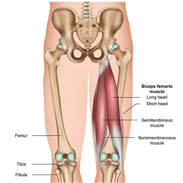
This image illustrates the posterior view of the human lower body, specifically focusing on the muscles of the posterior thigh, known as the hamstring group, along with the bones they interact with.
At the center, we see the biceps femoris muscle, which has two distinct parts: the long head and the short head. The long head originates at the ischial tuberosity of the pelvis and the short head originates from the femur. Both heads of the biceps femoris converge to insert on the head of the fibula and lateral aspect of the tibia. This muscle is involved in knee flexion and the lateral rotation of the lower leg when the knee is flexed, as well as extending the thigh at the hip.
Below the biceps femoris are the semitendinosus and semimembranosus muscles, also part of the hamstrings. The semitendinosus muscle, which lies more medially, originates from the ischial tuberosity as well and inserts on the medial aspect of the tibia. The semimembranosus, situated underneath the semitendinosus, also has its origin at the ischial tuberosity and inserts on the medial tibial condyle. Both of these muscles contribute to knee flexion, hip extension, and medially rotating the lower leg when the knee is flexed.
The skeletal structures in the image include the femur, which is the longest and strongest bone in the body forming the thigh; the tibia, the larger and more medial bone of the lower leg; and the fibula, the thinner bone located laterally to the tibia.
This muscular and skeletal arrangement in the posterior thigh and leg plays a crucial role in movements such as walking, running, and jumping, providing power and stability to the lower limbs.
Thigh Anterior Muscle and Bone Anatomy
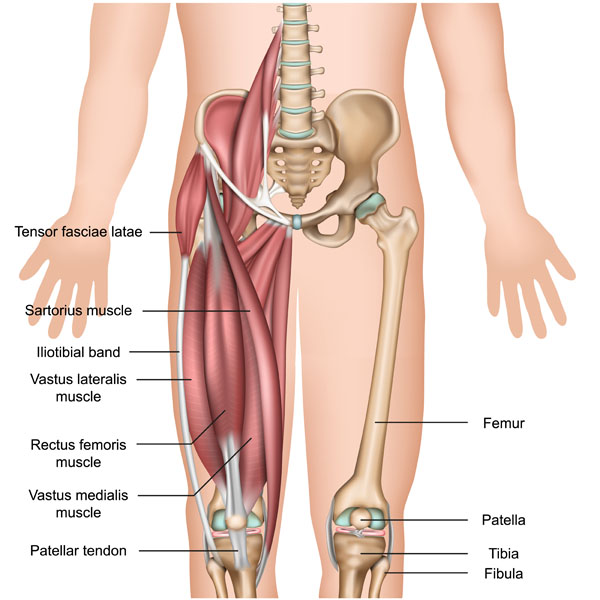
This image depicts the anterior view of the muscular and skeletal anatomy of the human thigh and knee region. The tensor fasciae latae muscle, situated on the lateral aspect of the hip, works to stabilize the pelvis in walking and running and is involved in hip abduction and medial rotation. Extending from this muscle is the iliotibial band, a long fibrous reinforcement of the fascia lata stretching from the iliac crest to the tibia, which is particularly important in lateral knee stabilization.
The sartorius muscle, the longest muscle in the human body, runs obliquely across the front of the thigh and acts to flex, abduct, and laterally rotate the hip, and also contributes to knee flexion. Underneath the sartorius, the rectus femoris muscle, one of the four quadriceps muscles, is visible. It is unique among the quadriceps as it crosses the hip joint and is involved in both hip flexion and knee extension.
Flanking the rectus femoris are the vastus lateralis and vastus medialis muscles, also part of the quadriceps group. The vastus lateralis is on the outer side of the thigh and the vastus medialis on the inner side. Both are powerful knee extensors and contribute to the overall function of the quadriceps, which is critical for activities like standing up, walking, and running.
At the knee, the patellar tendon connects the patella or kneecap to the tibia. The patella itself is shown at the front of the knee, enhancing the leverage of the quadriceps tendon which it continues as the patellar tendon.
The femur, the longest and strongest bone in the body, makes up the thigh, while the tibia and fibula are the two bones of the lower leg, with the tibia being the larger, medial bone that bears weight, and the fibula being the slender bone on the lateral side of the leg.
This complex interaction of muscles and bones allows for a wide range of movement and stability in the hip and knee joints, enabling locomotion and weight-bearing activities.
Calf Muscle Tear Pathology
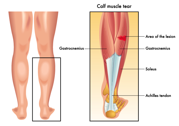
This image provides a comparison between a normal calf anatomy and a calf muscle tear. On the left, the posterior aspect of the lower legs is shown, with the gastrocnemius muscle visible under the skin. This muscle forms the bulk of the calf and is responsible for plantar flexing the foot at the ankle joint and flexing the leg at the knee joint.
The right side of the image zooms in to give a detailed look at the internal structure of the calf, highlighting a muscle tear. The gastrocnemius muscle is shown in red with an indicated area of the lesion or tear. Such tears are commonly referred to as a ‘calf strain’ and can be quite painful, causing bruising and swelling. The underlying soleus muscle, which also contributes to plantar flexion of the foot, is shown in blue. Both these muscles converge into the Achilles tendon, depicted in white, which is the thickest and strongest tendon in the body. The Achilles tendon inserts into the calcaneus, or heel bone, and is essential for walking, running, and jumping.
A tear in the gastrocnemius muscle can occur when the muscle is stretched beyond its capacity, often during sudden accelerations or changes in direction while running or jumping. Treatment typically involves rest, ice, compression, and elevation (RICE), and in more severe cases, may require physical therapy or surgical intervention.
Structure of a Long Bone
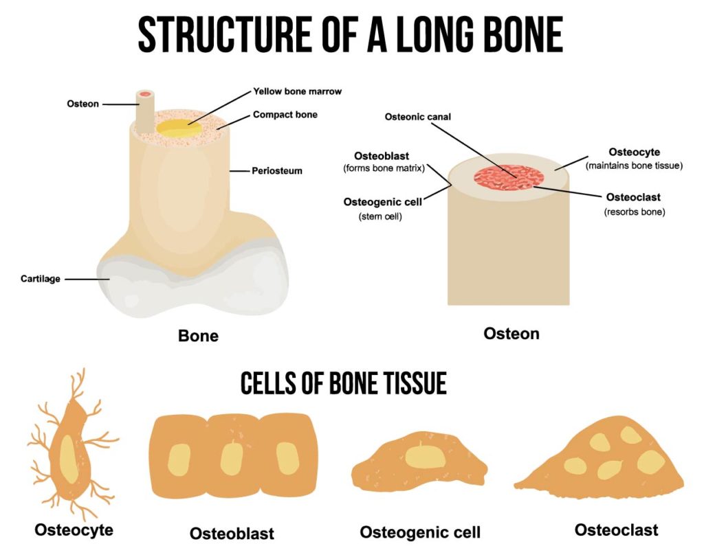
This educational diagram illustrates the structure of a long bone and the types of cells found in bone tissue.
The top section of the diagram depicts a longitudinal section and a cross-sectional view of a long bone. A long bone is characterized by an elongated shaft called the diaphysis, which is mainly composed of compact bone that provides strength and rigidity. The central cavity of the diaphysis is filled with yellow bone marrow, which is involved in the storage of fats.
Surrounding the compact bone is the periosteum, a dense layer of vascular connective tissue that covers the bone and serves as an attachment for muscles and tendons. Within the compact bone, we can see structures called osteons or Haversian systems, which are cylindrical structures that contain a central osteonic canal. These canals house blood vessels and nerves, crucial for bone viability and sensation.
The cross-sectional view shows the osteon more clearly, with concentric lamellae (layers of compact bone tissue) surrounding the osteonic canal. Embedded in the compact bone are osteocytes, mature bone cells responsible for maintaining bone tissue.
The lower part of the diagram highlights the cells of bone tissue, providing a closer look at the different cell types:
- Osteocytes are mature bone cells that maintain the bone matrix and reside in small cavities called lacunae.
- Osteoblasts are bone-forming cells that synthesize and secrete the bone matrix. Once osteoblasts become entrapped in the matrix they’ve secreted, they differentiate into osteocytes.
- Osteogenic cells are stem cells found in the periosteum and endosteum that can differentiate into osteoblasts.
- Osteoclasts are large cells that resorb or break down bone tissue, playing a critical role in bone remodeling and calcium homeostasis.
Together, these cells are involved in the ongoing process of bone remodeling, which involves the resorption of old bone and the formation of new bone, a dynamic process that allows bones to adapt to stress, repair themselves, and regulate mineral homeostasis in the body.
Anatomical Terms and Definitions
| Term | Definition |
|---|---|
| Adductor Longus | A muscle that serves to adduct the thigh and is involved in hip flexion. |
| Extensor Digitorum Longus | A muscle that helps extend the toes and dorsiflex the foot. |
| Gastrocnemius | The larger calf muscle involved in plantarflexing the ankle and flexing the knee. |
| Gluteus Maximus | The largest muscle in the body, responsible for the extension, outward rotation, and abduction of the hip joint. |
| Gracilis | A long, thin muscle running down the inner thigh, responsible for hip adduction and knee flexion. |
| Iliopsoas | Comprising the iliacus and psoas major muscles, important for hip flexion. |
| Pectineus | Involved in hip flexion and adduction. |
| Rectus Femoris | Part of the quadriceps group, unique for crossing both the hip and knee joints, aids in extending the knee and flexing the thigh at the hip. |
| Sartorius | The longest muscle in the human body, crossing both the hip and knee joints, contributing to flexion, abduction, and lateral rotation of the hip, as well as flexion of the knee. |
| Soleus | A muscle that aids in plantarflexion of the foot, primarily active while walking or standing. |
| Tensor Fasciae Latae | A muscle that assists in stabilizing and moving the hip and knee. |
| Tibialis Anterior | Located on the front of the shin, crucial for dorsiflexion and inversion of the foot. |
| Vastus Lateralis and Medialis | Part of the quadriceps muscle group, with roles primarily in knee extension. |
