
Table of Contents
Bone Pelvic Region Anatomy
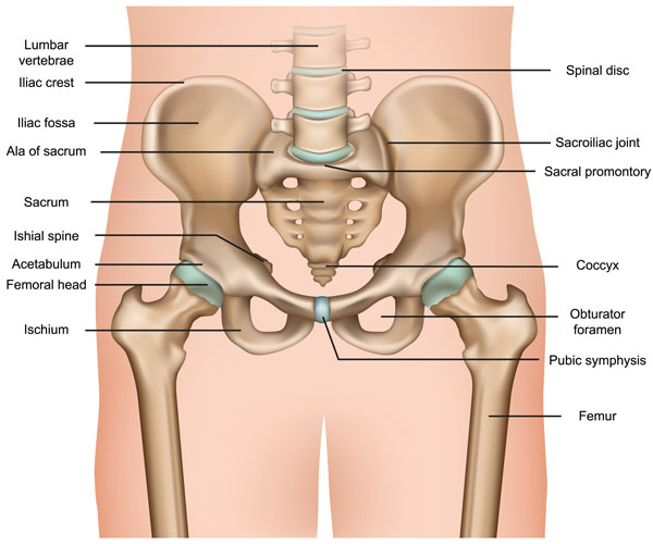
This image illustrates the human pelvic region and lower vertebral column in an anterior view, highlighting several key anatomical structures.
At the top, we see the lumbar vertebrae, which are the five largest and heaviest vertebrae located in the lower back between the thoracic vertebrae and the sacrum. Above the lumbar vertebrae is a spinal disc, which serves as a ligament that holds the vertebrae together while also allowing for slight mobility of the spine.
Flanking the lumbar vertebrae on either side are the iliac crests, which are the top borders of the ilium—the largest bone of the pelvis. These crests form the superior border of the greater pelvis and provide attachment for the iliacus, abdominals, and other muscles.
Below the iliac crest, we find the iliac fossa, a large, smooth, concave surface on the internal side of the ilium. The ala of sacrum is the wing-like part on either side of the sacral base, connecting the sacrum to the ilia of the pelvis.
Centrally located is the sacrum, a large, triangular bone at the base of the spine and at the upper and back part of the pelvic cavity, where it is inserted like a wedge between the two hip bones. Its upper part connects with the last lumbar vertebra, and its lower part with the coccyx via the sacrococcygeal symphysis.
Just below the sacrum, we see the coccyx, commonly referred to as the tailbone. It’s the final segment of the vertebral column, comprising three to five fused vertebrae.
The pelvic girdle also features the sacroiliac joints, which are the joints between the sacrum and the ilium of the pelvis, connected by strong ligaments. The sacral promontory is a prominent bulge on the front side of the sacrum, marking part of the border of the pelvic inlet.
On the lateral aspects of the pelvis, we can identify the acetabulum, a concave surface that serves as the socket for the femoral head, creating the hip joint. The femoral head is the highest part of the thigh bone (femur), fitting into the acetabulum.
Adjacent to the acetabulum, the ischium forms the lower and back part of the hip bone. The ischial spine is a pointed protrusion from the ischium, which serves as a point of attachment for ligaments and muscles.
At the junction of the ischium and pubis bones of the pelvis, we find the obturator foramen, a large opening that allows for the passage of nerves and blood vessels to the legs.
In the center, just below the acetabulum, is the pubic symphysis, a cartilaginous joint that sits between and joins the left and right superior rami of the pubic bones.
Lastly, the femur, the longest and strongest bone in the human body, is shown extending downward from the hip joint.
Pelvic and Hip Muscular Anatomy
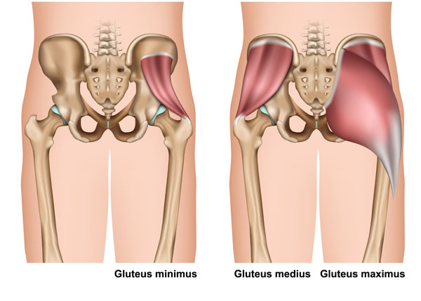
The image displayed shows the musculature of the human posterior pelvic and hip region with a focus on the gluteal muscles, which are key in the movement of the hip and thigh. On the left, we see the gluteus minimus, the smallest of the trio, lying directly beneath the gluteus medius. It is a fan-shaped muscle that originates on the outer surface of the ilium between the anterior and inferior gluteal lines and extends to the greater trochanter of the femur. Its primary function is to help with the rotation of the thigh outward and the abduction of the thigh, which means moving it away from the body’s central axis.
On the right, the image is layered to show both the gluteus medius and the gluteus maximus. The gluteus medius is partially covered by the gluteus maximus and is shown in a more subdued tone. It is a broad, thick, radiating muscle, situated on the outer surface of the pelvis. Its anterior fiber helps in flexing and medially rotating the thigh, while the posterior fibers extend and laterally rotate the thigh.
The gluteus maximus, shown in a darker red, is the largest and most superficial of the three gluteal muscles. It makes up a large portion of the shape and appearance of the hips. It extends from the sacrum, coccyx, and adjacent bones and fans out to attach to the femur and the iliotibial band of the fascia lata of the thigh. This powerful muscle is involved in a number of actions, including extending and laterally rotating the thigh, and it is a crucial muscle in walking, running, and climbing.
These muscles are essential not only for movement but also for the stability of the pelvis and hip joints. They are engaged in various movements of the lower body, including maintaining the trunk in the erect posture.
Hip Articular Capsule
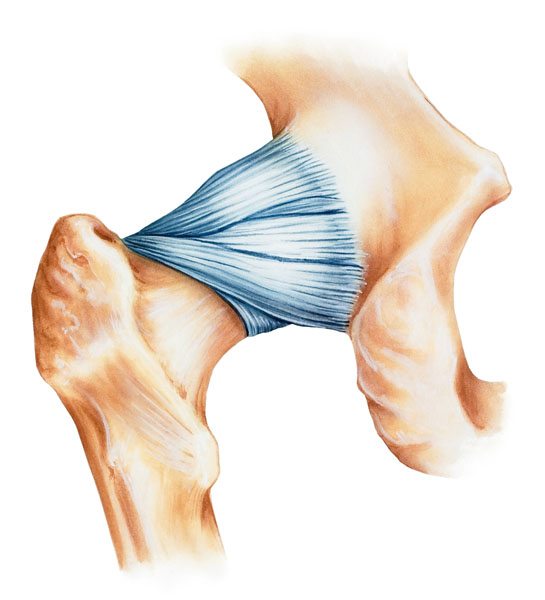
This illustration presents a detailed view of the hip joint with a focus on the articular capsule and the surrounding structures. The hip joint is a ball-and-socket synovial joint formed between the os coxa (hip bone) and the femur.
The central feature of this image is the articular capsule of the hip joint, which is a strong, fibrous structure that encircles the hip joint and holds it together. The capsule is composed of strong collagen fibers which appear as striations running longitudinally along the length of the capsule. This capsule provides stability to the hip joint while still allowing for a considerable range of motion.
Surrounding the capsule, we see the femoral head at the bottom right of the image, which is the ball part of the joint, fitting into the acetabulum of the hip bone. The femoral head has a smooth, rounded surface to articulate with the acetabulum, which is not visible in this view.
At the top left, we can observe part of the os coxa, specifically the area around the acetabulum, though the socket itself is covered by the articular capsule in this depiction. The acetabulum is a deep cup-like structure that accommodates the femoral head.
The hip joint is one of the most important joints in the body for weight-bearing activities such as standing, walking, and running. The articular capsule contributes significantly to the joint’s stability by resisting dislocation and providing a sealed environment for the joint fluid that lubricates the joint surfaces.
Muscles and Bursa of the Hip and Pelvis
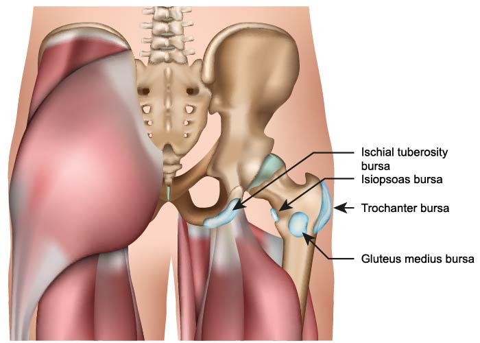
This image illustrates a lateral view of the human pelvis and proximal femur, focusing specifically on the bursae associated with the hip joint.
A bursa is a small fluid-filled sac that acts as a cushion to reduce friction between bones and the tendons or muscles around a joint. In this depiction, four bursae are indicated:
The ischial tuberosity bursa is located over the ischial tuberosity—the part of the pelvis that bears the weight when sitting. Bursae in this area minimize the friction for the gluteus maximus muscle as it moves over the ischial tuberosity.
The iliopsoas bursa is the largest bursa in the body. It lies between the iliopsoas muscle and the pelvic bone or the femoral head. It facilitates the smooth movement of the iliopsoas muscle during flexion and extension movements of the thigh.
The trochanteric bursa is found near the greater trochanter of the femur, which is the prominent bony point where the thigh bone turns inwards. The bursa serves to ease the movement of the tendons of the gluteus medius and minimus over the greater trochanter when the hip is in motion.
Lastly, the gluteus medius bursa is situated between the gluteus medius muscle and the greater trochanter. Similar to the trochanteric bursa, it helps in reducing the friction during the movement of the gluteus medius muscle over the femur.
The lumbar spine is seen at the top of the image, and the hip bone, including the ilium and the acetabulum, are shown. The femur is represented by the head, neck, and a portion of the shaft. The gluteus maximus muscle is visible in the background, covering the posterior aspect of the hip joint. The bursae play a crucial role in ensuring the smooth motion of these muscles and tendons over bony prominences.
Piriformis Muscles
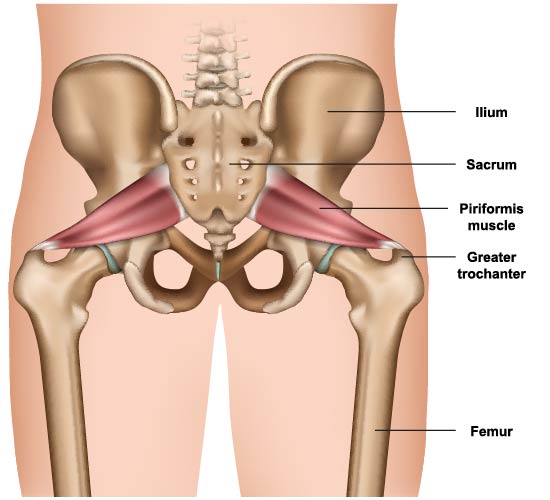
This illustration provides an anterior view of the pelvis, highlighting the piriformis muscles in relation to the pelvic bones and the proximal femur.
The piriformis muscles are shown in a deep red color, originating from the anterior surface of the sacrum—the triangular bone at the base of the spine—and inserting at the greater trochanter of the femur, which is the bony prominence on the upper thigh bone.
The ilium, part of the hip bones, is displayed on either side of the pelvis, forming the broad, wing-like structures on the upper part of the pelvis. The sacrum is the central part of the pelvic girdle and articulates with the ilium at the sacroiliac joints, which are not distinctly labeled here but are located where the ilium meets the sacrum.
The femur is the long bone of the thigh, and only the upper part is shown here. The greater trochanter is a significant landmark on the femur, serving as the point of attachment for the piriformis and several other muscles that are not depicted in this image.
The piriformis is an important muscle for several reasons: it helps to stabilize the hip joint, rotates the femur outward when the hip is extended, and abducts the femur (moves it away from the body) when the hip is flexed. Understanding the anatomy of the piriformis and its relationship with surrounding structures is important for diagnosing and treating conditions related to hip pain and sciatic nerve compression.
Iliopsoas Muscle Group
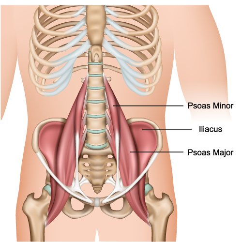
This image provides a view of the anterior lower torso with an emphasis on the iliopsoas muscle group, consisting of the psoas major, psoas minor, and iliacus muscles.
The psoas major muscles are large, thick, and triangular muscles that originate from the sides of the lumbar vertebrae and extend through the pelvis to attach to the lesser trochanter of the femur. The psoas major is the stronger and larger of the two psoas muscles, and it plays a key role in flexing the hip joint and lifting the upper leg towards the body.
Above the psoas major, the psoas minor muscles are depicted. They are much smaller and not as common, present in only about 50% of the population. When present, they run parallel to the psoas major muscles, originating from the lumbar vertebrae and inserting into the pelvis. The psoas minor assists in flexing the lower spine.
The iliacus muscles are broad, flat muscles that fill the iliac fossa on the inner surface of the ilium, part of the hip bones. They join the psoas major muscles to form the iliopsoas, which is the primary flexor of the thigh. This combined muscle group inserts into the lesser trochanter of the femur.
Together, the iliopsoas is crucial for walking, running, and standing upright, as it stabilizes the lower back and allows the hip to flex. When the body is in motion, these muscles work in coordination to control the movement of the hip and lower spine. The health and function of the iliopsoas muscle group are important for posture and locomotion.
Male Vs Female Pelvis Anatomy
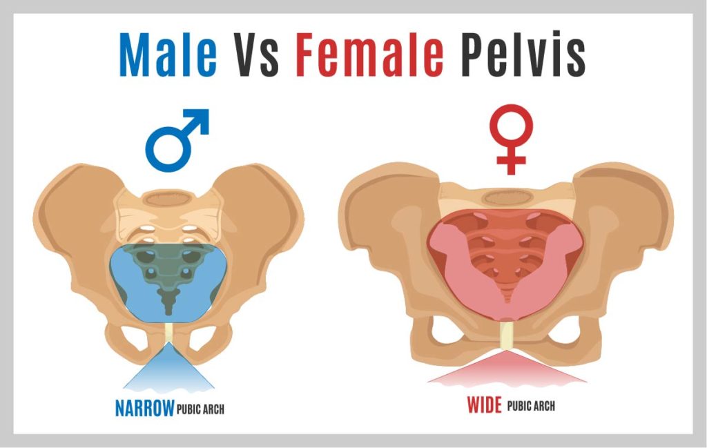
This image compares the structural differences between a male and a female pelvis. The male pelvis, on the left, is characterized by a narrower pubic arch – the angle formed below the pubic symphysis. This narrower arch is typically less than 90 degrees. The pelvic cavity itself appears more heart-shaped and the bone structures are more robust. These features are adapted for supporting a male’s generally heavier build and stronger musculature.
On the right, the female pelvis is depicted with a wider pubic arch, often greater than 90 degrees, which is one of the adaptations for childbirth. The pelvic inlet, the upper opening of the pelvis, is broader and more circular, providing more space in the pelvic cavity. This difference accommodates the needs of pregnancy and childbirth, allowing more room for the passage of a baby.
Additionally, the female pelvis shows more flared iliac wings and a shorter sacrum, further distinguishing it from the male pelvis. Overall, the female pelvis is wider and shallower, while the male pelvis is taller and more narrow.
These anatomical differences are a classic example of sexual dimorphism in human skeletal structure, where the physical differences between the sexes are related to biological and functional adaptations.
Ligaments of the Pelvic Girdle
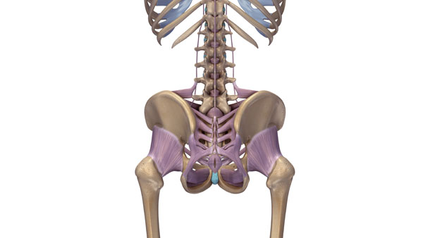
The image shows a posterior view of the human skeletal structure focusing on the ligaments of the pelvic girdle and lower spine in purple. The ligaments are the fibrous connective tissues that connect bones to other bones, providing stability and flexibility to the joints and skeletal structure.
Here, the purple structures illustrate the complex network of ligaments that support the sacroiliac joints, where the spine meets the pelvis, and the ligaments along the spine itself. These ligaments help stabilize the sacrum -- the shield-shaped bony structure at the base of the lumbar vertebrae and at the upper and back part of the pelvic cavity. They also support the connection between the sacrum and the ilium of the hip bones.
Additionally, the ligaments extend along the length of the spine, connecting the vertebrae to each other. They play a crucial role in maintaining the integrity of the spinal column, allowing for controlled movements and flexibility, and preventing dislocation and injury by limiting excessive movement.
The strength and elasticity of these ligaments provide a delicate balance that allows for movement while maintaining structure, making them vital components of the musculoskeletal system.
Sciatic Nerve Pathology
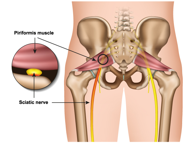
This image presents an anterior view of the pelvis with a focus on the piriformis muscle and its relationship with the sciatic nerve. The piriformis is a small muscle located deep in the buttock, behind the gluteus maximus. It runs from the lower spine to the top of the femur and functions to rotate the hip and turn the leg and foot outward.
The sciatic nerve is shown in yellow, highlighting its path as it runs beneath the piriformis muscle. This nerve is the longest and widest single nerve in the human body, extending from the lower end of the spinal cord, down the buttock and the back of the thigh, all the way to the foot.
The inset circle provides a magnified view, showing the close proximity of the sciatic nerve to the piriformis muscle. It is this relationship that can sometimes lead to piriformis syndrome, where the muscle irritates or compresses the sciatic nerve, causing pain, tingling, or numbness in the buttock and along the path of the nerve.
The pelvic bones, including the ilium and sacrum, provide the structure to which the piriformis muscle attaches. The anatomical illustration emphasizes the potential for neurological impingement where the sciatic nerve passes in close association with the piriformis muscle.
Anatomical Terms and Definitions
| Term | Definition |
|---|---|
| Acetabulum | A concave surface on the pelvic bone that serves as the socket for the femoral head, forming the hip joint. |
| Ala of Sacrum | The wing-like part on either side of the sacral base, connecting the sacrum to the ilia of the pelvis. |
| Coccyx | The final segment of the vertebral column, commonly referred to as the tailbone, comprising three to five fused vertebrae. |
| Femoral Head | The highest part of the thigh bone (femur), fitting into the acetabulum to form the hip joint. |
| Gluteus Maximus | The largest and most superficial of the gluteal muscles, involved in extending and laterally rotating the thigh, as well as in maintaining the trunk in an erect posture. |
| Gluteus Medius | A muscle partially covered by the gluteus maximus, involved in flexing, medially rotating, and abducting the thigh. |
| Gluteus Minimus | The smallest of the gluteal muscles, located beneath the gluteus medius, helping with thigh rotation and abduction. |
| Iliac Crest | The top border of the ilium, forming the superior border of the greater pelvis and providing attachment for muscles. |
| Iliac Fossa | A large, smooth, concave surface on the internal side of the ilium. |
| Ischial Spine | A pointed protrusion from the ischium, serving as a point of attachment for ligaments and muscles. |
| Iliopsoas | A combination of the iliacus and psoas major muscles, the strongest flexor of the thigh at the hip joint. |
| Lumbar Vertebrae | The five largest and heaviest vertebrae located in the lower back, between the thoracic vertebrae and the sacrum. |
| Obturator Foramen | A large opening created by the ischium and pubis bones of the pelvis, allowing for the passage of nerves and blood vessels to the legs. |
| Pelvic Girdle | The structure formed by the sacrum and the two hip bones (ilium, ischium, and pubis) on either side. |
| Pubic Symphysis | A cartilaginous joint that sits between and joins the left and right superior rami of the pubic bones. |
| Sacral Promontory | A prominent bulge on the front side of the sacrum, marking part of the border of the pelvic inlet. |
| Sacroiliac Joints | The joints between the sacrum and the ilium of the pelvis, connected by strong ligaments. |
| Sacrum | A large, triangular bone at the base of the spine and at the upper, back part of the pelvic cavity, inserted like a wedge between the two hip bones. |
