
Table of Contents
Vertebral Column
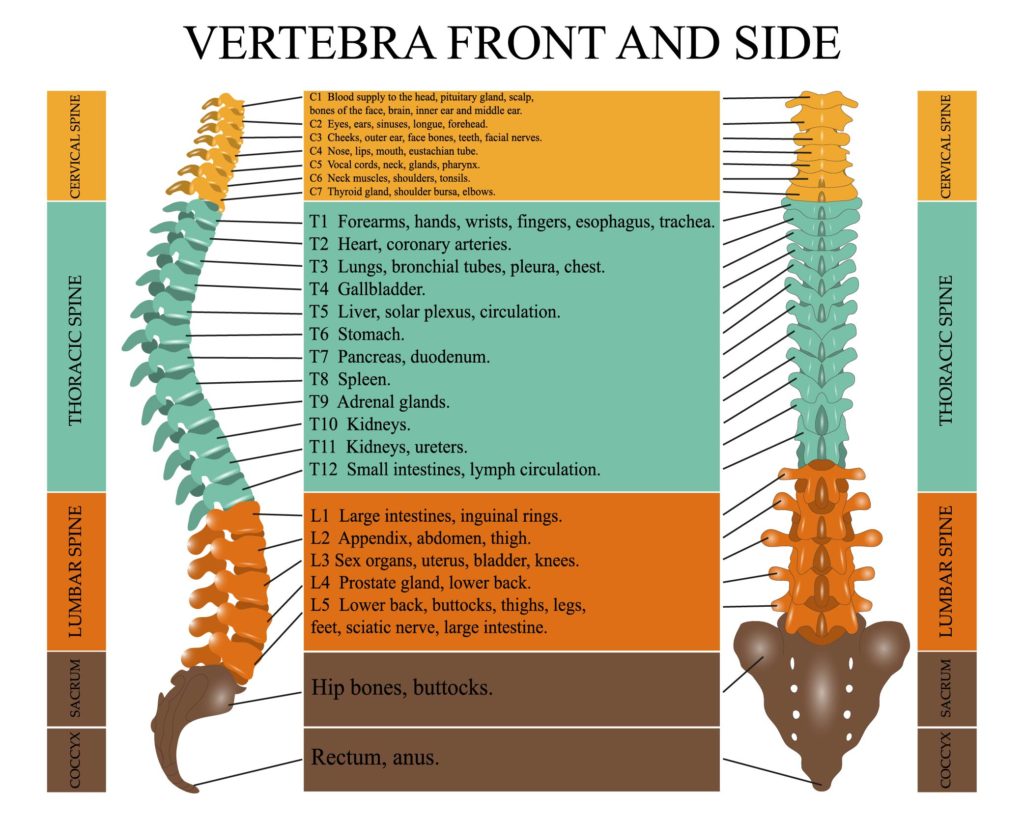
The illustration provided is an anatomical diagram showing the vertebral column from both the front (anterior) and side (lateral) views. It specifically highlights the different regions of the spine: cervical, thoracic, lumbar, sacral, and coccygeal. Each region is color-coded and labeled with the corresponding vertebrae from C1 to the coccyx. Alongside the vertebrae, there are annotations that link each spinal segment to various organs and body parts, indicating the areas potentially affected by the nerves that emanate from that segment of the spinal cord.
The cervical spine is at the top of the diagram, consisting of 7 vertebrae (C1-C7), associated with the head, neck, diaphragm, thyroid gland, and shoulders. This region of the spine is flexible, allowing for a wide range of head and neck movements.
The thoracic spine is below the cervical region and consists of 12 vertebrae (T1-T12). This region is more rigid, providing stability and supporting the rib cage. The nerves from these vertebrae are associated with many internal organs, such as the heart, lungs, gallbladder, and kidneys.
The lumbar spine follows with 5 vertebrae (L1-L5), associated with the lower back. This region bears the most weight and is thus more prone to injury. The nerves from the lumbar region innervate the lower abdominal muscles, hips, and legs.
Next is the sacral region, which is represented by a triangular bone made up of fused vertebrae, followed by the coccygeal region at the very bottom, often referred to as the tailbone.
The diagram also indicates the different curves in the spine: the cervical and lumbar regions have a lordotic curve, which means they curve inward toward the body, while the thoracic and sacral regions have a kyphotic curve, curving outward.
Such a diagram is useful in understanding the relationship between spinal health and overall well-being, as spinal problems can affect various organs and limbs due to the complex nature of nerve innervation.
Vertebral Column Anatomy
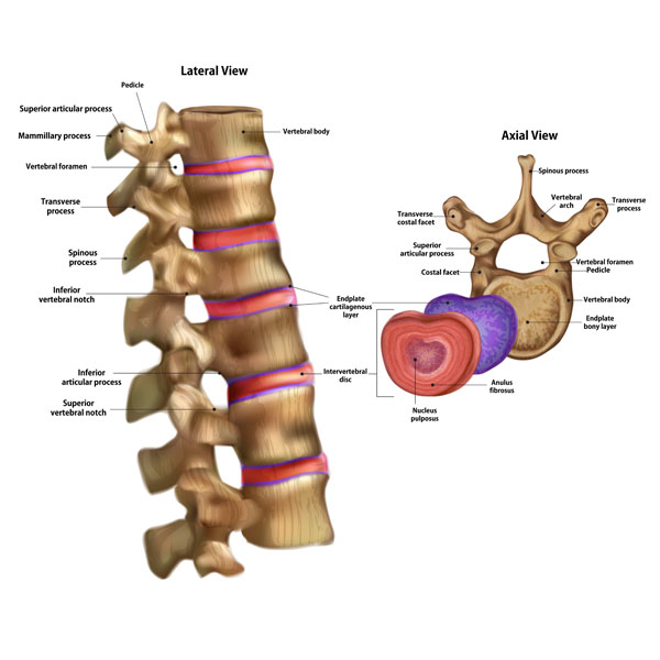
Here we see an illustration with two views of the human vertebral column, emphasizing the anatomy of individual vertebrae. On the left, we have a lateral view of a section of the vertebral column, and on the right, an axial view of a single vertebra.
Starting with the lateral view, we see a sequence of vertebrae aligned. Each vertebra consists of a vertebral body, which is the thick, disc-shaped anterior portion forming the robust, weight-bearing part. The vertebral foramen is a hollow space within each vertebra, and collectively, these form the vertebral canal through which the spinal cord passes. Projecting posteriorly from the vertebral body is a bony arch composed of pedicles and laminae, culminating in the midline with the spinous process, which can be felt through the skin down the back.
We also see the transverse processes, which extend laterally and serve as attachment sites for muscles and ligaments. The superior and inferior articular processes articulate with adjacent vertebrae to facilitate movement while maintaining stability. The notches above (superior vertebral notch) and below (inferior vertebral notch) the pedicles combine to form the intervertebral foramina, through which spinal nerves exit the spinal canal.
The intervertebral discs, which provide cushioning between the vertebrae, are depicted between the vertebral bodies. These discs allow for movement and act as ligaments to hold the vertebrae together. The endplate cartilage on the top and bottom of the vertebral bodies interfaces with the intervertebral discs, playing a role in the distribution of nutrients to the disc tissue.
Turning to the axial view on the right, we have a top-down look at a vertebra. Here, the vertebral body forms the bulk of the structure, with the vertebral foramen at its center providing passage for the spinal cord. The spinous process projects posteriorly, and the transverse processes extend laterally. The superior and inferior articular processes are more clearly visible in this view, showing where they would interlock with the adjacent vertebrae. The annulus fibrosus is the tough outer layer of the intervertebral disc, encircling the softer nucleus pulposus, which provides the disc its ability to absorb shock and maintain flexibility.
This detailed view of the vertebrae highlights the complex anatomy that allows for both stability and mobility in the spinal column, as well as the protection it provides to the spinal cord.
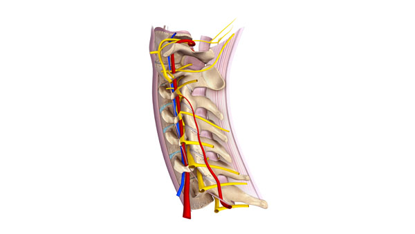
Structures Around the Vertebra
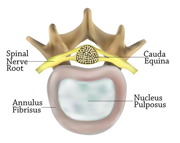
The image presents an axial view of a vertebra at the level where the spinal nerve root exits the vertebral column. It offers a closer look at the structures within and surrounding the vertebra.
Central to the image is the vertebral body, which is the large, oval-shaped structure providing the majority of the vertebra’s weight-bearing capability. Within the vertebral body, we see the nucleus pulposus, represented in a lighter shade, which is the inner core of the intervertebral disc. This gel-like substance allows the disc to absorb shock and provides the flexibility necessary for the spine’s movement.
Surrounding the nucleus pulposus is the annulus fibrosus, depicted as a thick ring. This is the tough, outer layer of the intervertebral disc, consisting of several layers of fibrocartilage. It contains the nucleus pulposus and distributes pressure evenly across the disc.
Above the vertebral body, the spinal nerve root is shown in yellow, exiting the vertebral column. This nerve root is part of the peripheral nervous system and carries motor, sensory, and autonomic signals between the spinal cord and the body.
Behind the spinal nerve root, the image labels the cauda equina, a bundle of spinal nerves and spinal nerve rootlets. Named for its resemblance to a horse’s tail, the cauda equina continues from the termination of the spinal cord down within the lumbar and sacral vertebral levels.
This image encapsulates crucial components of vertebral and neuroanatomy, emphasizing the relationships between the spinal cord, nerve roots, and the supportive structures of the intervertebral discs.
Intervertebral Disc Pressure Changes
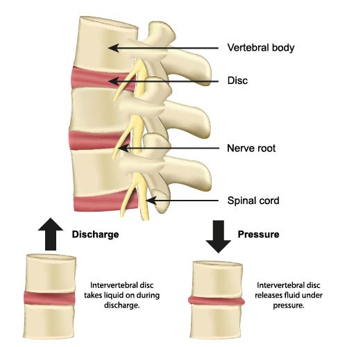
The illustration offers a simplified view of the vertebral column, emphasizing the intervertebral discs and their role in accommodating pressure changes. It offers a side view of the spine, where we can see several vertebral bodies stacked on top of each other with intervertebral discs between them, as well as the spinal cord and emerging nerve roots.
The intervertebral discs are key structures in the spine that serve as cushions between the individual vertebrae. The diagram illustrates two conditions of these discs: when they take in liquid during ‘discharge’ and when they release fluid under ‘pressure’. This dynamic process allows the discs to absorb shocks and maintain flexibility in the spinal column.
Under ‘discharge’, when the spine is unloaded or in a state of relaxation, the intervertebral discs can imbibe or take in water, which helps to maintain their height and elasticity. Conversely, under ‘pressure’, such as during standing, walking, or lifting, the discs release water. This mechanism is crucial for the discs’ ability to handle mechanical stress.
Additionally, the diagram shows the spinal cord running through the vertebral canal and the nerve roots branching out from it. The health and function of intervertebral discs are important not only for spinal mobility but also for the protection of these nerve structures.
Understanding the biomechanics of the intervertebral discs is important for recognizing how activities and posture can impact spinal health and for understanding the mechanisms behind conditions such as disc herniation, where a disc’s nucleus pushes out through a tear in the annulus fibrosus, potentially compressing a nerve root and causing pain.
Ligaments of the Lumbar Spine
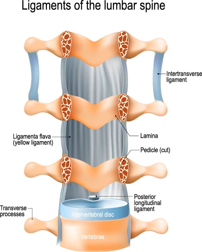
The image showcases a detailed representation of the ligaments associated with the lumbar spine, which is the lower part of the vertebral column. These ligaments are crucial for providing stability to the spine while allowing for a range of movements.
From the front to the back, we can observe the intervertebral disc, which is a fibrocartilaginous structure acting as a cushion between adjacent vertebrae. The vertebral body is not visible here, but it lies anterior to the intervertebral disc and is a major structural component of the vertebra.
The posterior longitudinal ligament runs longitudinally down the spinal column along the posterior aspect of the vertebral bodies, inside the vertebral canal. It helps to prevent hyperextension of the spine, which is excessive backward bending.
On the sides of the vertebrae, we see the transverse processes, which are small bony projections where muscles and ligaments attach. Just behind these are the laminae, part of the vertebral arch that forms the roof of the spinal canal.
The pedicle, labeled as cut in this image, is a stub of bone that connects the lamina to the vertebral body, forming the sides of the vertebral arch.
The ligamenta flava, also known as the yellow ligaments, are bands that connect the laminae of adjacent vertebrae. They are highly elastic and preserve the upright posture by helping to straighten the spine after flexing.
The intertransverse ligaments stretch between the transverse processes of adjacent vertebrae. These ligaments aid in lateral stability and prevent excessive side-to-side movements.
This diagram effectively illustrates the complexity and integration of the ligamentous structures that support the lumbar spine, highlighting their role in maintaining spinal integrity and facilitating movement.
Spinal Ligaments
The provided image offers a detailed view of the ligaments associated with the spinal column. The ligaments are crucial structures that connect vertebrae to each other and contribute to the spine’s stability, while still allowing for a range of movements.
Key ligaments illustrated in the image include:
Anterior Longitudinal Ligament: This ligament runs along the anterior surfaces of the vertebral bodies, from the pelvic surface of the sacrum to the anterior tubercle of the atlas (C1). It restricts extension movements of the spine.
Posterior Longitudinal Ligament: This one is situated within the vertebral canal along the posterior surfaces of the vertebral bodies. It is narrower and weaker than the anterior longitudinal ligament and limits flexion of the spine.
Ligamentum Flavum: Also known as the yellow ligament due to its yellowish color from elastin fibers, it connects the laminae of adjacent vertebrae. It preserves the upright posture and assists with the spine’s elastic recoil after bending forward.
Interspinous Ligaments: These are found between the spinous processes of adjacent vertebrae. They help to limit flexion of the spine.
Supraspinous Ligament: This ligament connects the tips of the spinous processes from the seventh cervical vertebra (C7) down to the sacrum, and it resists flexion of the spine.
Intertransverse Ligaments: Located between the transverse processes of adjacent vertebrae, these ligaments restrict contralateral (opposite side) lateral flexion.
Together, these ligaments form a complex system that maintains the alignment of the vertebral column, allows for controlled movements, and protects the spinal cord and nerve roots. They work in concert with the muscles and tendons surrounding the spine to perform a wide range of motions and to provide stability against various forces that the spine encounters during daily activities.
Nuchal Ligament
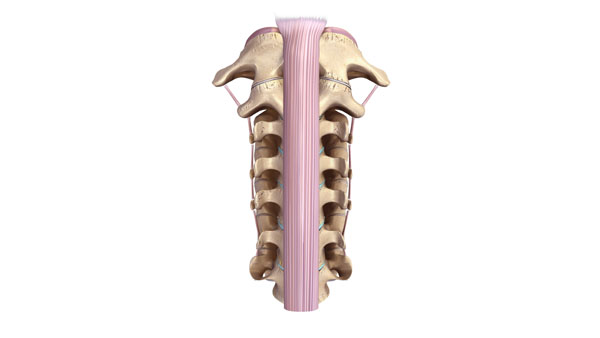
This image displays the dorsal aspect of the human spine, highlighting the nuchal ligament. The nuchal ligament is a thick, dense, and elastic connective tissue structure that extends from the external occipital protuberance and median nuchal line of the skull down to the spinous process of the seventh cervical vertebra. It is continuous with the supraspinous ligament, which extends down the length of the spine beyond the cervical region.
The nuchal ligament plays a crucial role in maintaining posture by allowing the head to return to its neutral position after flexing. It also provides an attachment site for several muscles of the neck that control the movement and positioning of the head. This ligament is particularly well-developed in species that hold their heads horizontally, such as humans, and it assists with the stabilization of the head during movement.
Stages of Disc Herniation
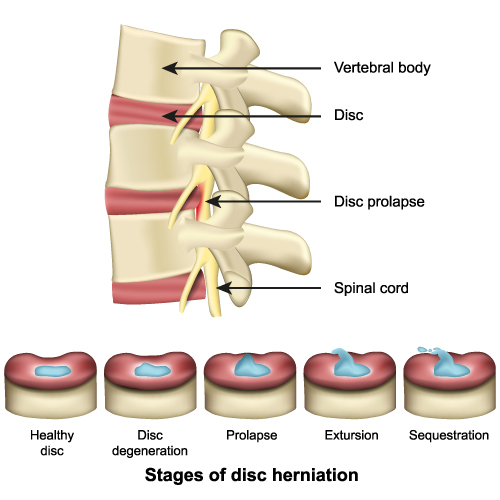
This image provides a two-part visualization related to spinal anatomy and pathology, specifically illustrating a condition known as disc herniation.
The top section of the image shows a side view of the human spinal column, with three vertebrae and the intervertebral discs between them. Each vertebra is labeled as ‘Vertebral body’. The intervertebral discs, which act as cushions between the vertebrae, are labeled as ‘Disc’. Below the disc, the ‘Spinal cord’ runs through the vertebral column. One disc is marked with ‘Disc prolapse’, where a portion of the disc material is protruding outwards, pressing against the spinal cord. This prolapse can lead to pain, numbness, or weakness due to the pressure on the spinal nerves.
The bottom section is labeled ‘Stages of disc herniation’. It shows a progression from a ‘Healthy disc’, which has an intact and hydrated blue nucleus, through stages of ‘Disc degeneration’ where the disc begins to lose hydration and height, to ‘Prolapse’, where the nucleus pushes out against the disc wall. ‘Extrusion’ is the next stage where the nucleus material breaks through the outer layer of the disc but remains within the disc’s confines. Finally, ‘Sequestration’ is depicted as a stage where fragments of the nucleus material have broken free from the disc into the spinal canal.
The sequence illustrates how disc herniation starts from a healthy disc and progresses through stages of deterioration, eventually leading to the extrusion or sequestration of disc material. The health of these discs is critical for spinal function, providing flexibility, shock absorption, and protection for the spinal cord. The progression from a healthy disc to a herniated disc can lead to significant back pain and neurological symptoms due to the impact on nearby spinal nerves.
Osteoarthritic Spine
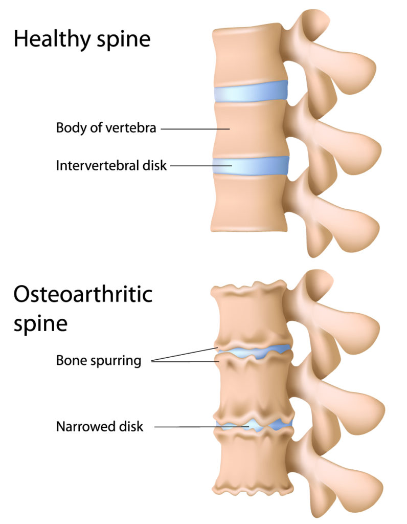
The image presents a comparison between a healthy spine and an osteoarthritic spine.
In the top half, the healthy spine is depicted with well-defined and uniformly shaped vertebral bodies. The intervertebral discs, the cushion-like pads between vertebrae, are intact and maintain a consistent height, allowing for proper spacing between the vertebral bodies. These discs are essential for absorbing shock, providing flexibility, and maintaining the integrity of the vertebral column.
In contrast, the bottom half illustrates an osteoarthritic spine. Osteoarthritis is characterized by the breakdown of cartilage and can affect any joint, including those in the spine. The vertebral bodies show signs of bone spurring, also known as osteophytes, which are bony projections that form along joint margins. These growths can occur as a natural response to the increased pressure on the degenerating cartilage and can contribute to the stiffness and pain commonly associated with osteoarthritis. Additionally, the intervertebral discs are narrowed, indicating disc degeneration. This narrowing can lead to reduced flexibility, potential nerve compression, and changes in the structure and stability of the spine.
Overall, this image serves to illustrate the structural changes that occur in the spine due to osteoarthritis, highlighting the contrast between a healthy and an affected vertebral column.
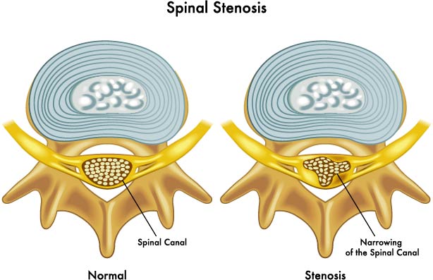
Spinal Stenosis
The illustration presents a comparison between a healthy spine and an osteoarthritic spine.
In the top half, the healthy spine is depicted with well-defined and uniformly shaped vertebral bodies. The intervertebral discs, the cushion-like pads between vertebrae, are intact and maintain a consistent height, allowing for proper spacing between the vertebral bodies. These discs are essential for absorbing shock, providing flexibility, and maintaining the integrity of the vertebral column.
In contrast, the bottom half illustrates an osteoarthritic spine. Osteoarthritis is characterized by the breakdown of cartilage and can affect any joint, including those in the spine. The vertebral bodies show signs of bone spurring, also known as osteophytes, which are bony projections that form along joint margins. These growths can occur as a natural response to the increased pressure on the degenerating cartilage and can contribute to the stiffness and pain commonly associated with osteoarthritis. Additionally, the intervertebral discs are narrowed, indicating disc degeneration. This narrowing can lead to reduced flexibility, potential nerve compression, and changes in the structure and stability of the spine.
Overall, this image serves to illustrate the structural changes that occur in the spine due to osteoarthritis, highlighting the contrast between a healthy and an affected vertebral column.
Spinal Curvature Pathology
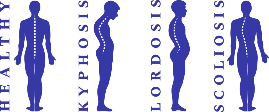
The image depicts four silhouettes representing different spinal curvatures in the sagittal and coronal planes.
The first silhouette, labeled “Healthy,” shows a normal spine with a gentle kyphotic curve in the thoracic region (upper back) and a lordotic curve in the lumbar region (lower back). This is considered the ideal spinal alignment, providing balance and allowing for efficient movement and shock absorption.
The second silhouette, labeled “Kyphosis,” demonstrates an exaggerated outward curve of the thoracic spine. This condition can lead to a hunched-back appearance and is often associated with osteoporosis or degenerative spine diseases.
The third silhouette, labeled “Lordosis,” shows an increased inward curvature of the lumbar spine. Commonly referred to as “swayback,” it can create significant lower back pain due to the added stress on the lumbar spine.
The fourth silhouette, labeled “Scoliosis,” illustrates a lateral, or sideways, curvature of the spine. Unlike the other conditions, scoliosis affects the spine in the coronal plane and can lead to an asymmetrical appearance of the back and shoulders.
Overall, these silhouettes serve as a visual reference for understanding common spinal deformities and their differences from the normal spinal curvature.
