
Table of Contents
Male Reproductive Anatomy
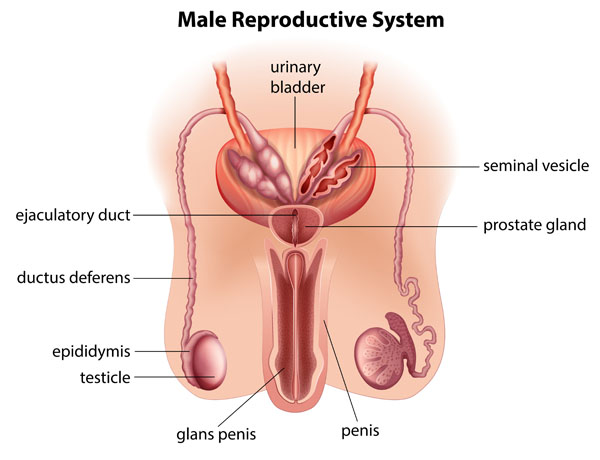
The image presents a labeled diagram of the male reproductive system, which is primarily responsible for sexual function and reproduction.
At the top of the image, we see the urinary bladder, which stores urine. It’s not part of the reproductive system, but it’s included here because of its proximity and shared structures with the reproductive organs.
Adjacent to the bladder are the seminal vesicles, which produce a significant portion of the seminal fluid. This fluid is a component of semen and provides nutrients for the sperm.
Below the bladder and seminal vesicles, the prostate gland is illustrated. The prostate secretes a fluid that nourishes and transports sperm during ejaculation. This fluid makes up about 30% of the volume of semen.
The ductus deferens, also known as the vas deferens, are long, muscular tubes that transport mature sperm to the urethra in preparation for ejaculation.
The ejaculatory ducts are formed by the fusion of the ductus deferens and the seminal vesicles. These ducts transport the seminal fluid into the urethra.
The epididymis is a long, coiled tube that sits atop the testicle and is the site of sperm maturation.
The testicles, also known as testes, are the oval organs where sperm is produced. They are also responsible for producing testosterone, the primary male sex hormone.
The image also depicts the penis, which contains the urethra through which urine and semen leave the body. The glans penis, or the head of the penis, contains a high concentration of nerve endings and is often considered the most sensitive area of the male reproductive system.
Lastly, this diagram includes an illustration of the penis in a flaccid state, emphasizing the reproductive system’s role beyond just sexual activity, highlighting its function in the excretory system as well.
Detailed View of the Male Pelvis
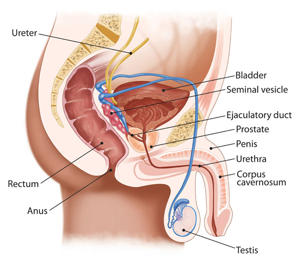
This illustration provides a detailed view of the male pelvis, highlighting components of both the urinary and reproductive systems, as well as their anatomical relationship to the digestive system.
Starting from the top, the ureter is shown. This is the tube through which urine flows from the kidney to the bladder. The bladder itself is depicted as a muscular sac-like organ that stores urine until it is excreted.
Beneath the bladder, we find the seminal vesicle, which produces a fluid that forms part of the semen. This fluid contains nutrients to nourish the sperm.
The ejaculatory duct, which is not visible externally, is the path that seminal fluid follows from the seminal vesicle and ductus deferens into the urethra.
Below the ejaculatory duct, the prostate gland is visible. It is a gland that surrounds the urethra and adds additional fluid to the semen during ejaculation.
The penis is represented extending downward, with the urethra running through its length. This is the conduit for the excretion of urine and the ejection of semen.
Within the penis, the corpus cavernosum is one of the two areas of erectile tissue. These structures fill with blood during sexual arousal, leading to an erection.
The testis is shown at the bottom of the illustration, external to the main pelvic area, housed within the scrotum. The testis produces sperm and testosterone.
Lastly, this diagram shows a section of the digestive system, namely the rectum, which is the final straight portion of the large intestine in some mammals, and the anus, which is the opening at the end of the digestive tract where the contents of the rectum are expelled.
Overall, the image integrates elements from various systems, providing a comprehensive view of their proximity and interrelated functions.
Structures of the Sperm Cell and Ovum
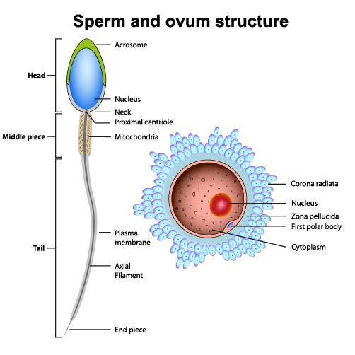
The image illustrates the structures of a sperm cell and an ovum, which are the male and female reproductive cells, respectively.
On the left side, the sperm cell is depicted with its various parts:
- The head contains the nucleus, which houses the genetic material (DNA). It’s capped with the acrosome, a vesicle that contains enzymes crucial for penetrating the outer layers of the ovum during fertilization.
- The neck is the segment immediately below the head and leads into the middle piece. It contains the proximal centriole, which plays a role in cell division after fertilization.
- The middle piece contains mitochondria, which provide the energy for the sperm’s motility, powering the tail’s movement to propel the sperm towards the ovum.
- The tail, or flagellum, exhibits a whiplike movement that propels the sperm cell forward. It’s composed of an axial filament surrounded by a plasma membrane.
- The end piece is the terminal portion of the tail and contains the axial filament.
On the right side, the ovum, or egg cell, is shown with its components:
- The nucleus of the ovum also contains genetic material and is positioned more centrally within the cell.
- The cytoplasm is the cellular material outside the nucleus, where cellular activities occur.
- Surrounding the nucleus is the zona pellucida, a glycoprotein layer that protects the ovum and is involved in the binding of the sperm cell during fertilization.
- The first polar body is a small cell that is a byproduct of the ovum’s maturation process.
- The corona radiata is a layer of cells that surrounds the ovum and provides it with necessary nutrients and support.
This image is central to understanding human reproduction, as the fusion of a sperm and an ovum, each contributing half of the genetic material, leads to the formation of a zygote, the first stage in the development of a new individual.
Anatomy of the Male Sperm Cell
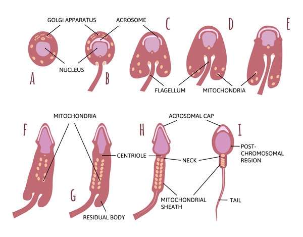
This illustration depicts the stages of sperm development and the anatomy of a mature sperm cell, known as spermatogenesis.
Starting with letter A, we see a spermatid, which is an early stage of the sperm cell. It has a prominent nucleus and is beginning to form the acrosome, a cap-like structure, from the Golgi apparatus. The acrosome is crucial for fertilization as it contains enzymes that help the sperm penetrate the egg.
In B, the acrosome has formed more distinctly over the nucleus, which contains the genetic material of the sperm. The development of the acrosome is essential for the sperm cell’s ability to fertilize an ovum.
As we progress to C, the flagellum starts to emerge. This tail-like structure is responsible for the mobility of the sperm, allowing it to swim towards the ovum.
By D, the mitochondria, which provide the energy for sperm motility, begin to gather around the portion of the flagellum that is closest to the cell body. This area is known as the midpiece.
In E, the cell is further elongated, and the residual body is left behind, which is a remnant of the cytoplasm from the spermatid.
In F, the mature sperm cell is shown with mitochondria packed in the midpiece, providing the energy necessary for the flagellum’s movement.
G shows the residual body, which is shed during sperm maturation.
In H, we see a mature sperm cell with a clearly defined acrosomal cap and centriole, which is part of the sperm cell’s microtubule organizing center, crucial for the cell’s structure.
Finally, I displays a fully mature spermatozoon with all its parts labeled, including the post-chromosomal region and tail. The mitochondrial sheath in the midpiece encircles the flagellum, and the neck connects the head of the sperm, which contains the nucleus and acrosome, to the tail.
The entire process illustrates the transformation from a simple cell to a complex structure capable of swimming, navigating the female reproductive tract, and ultimately fertilizing an egg.
Anatomical Representation of the Testicle
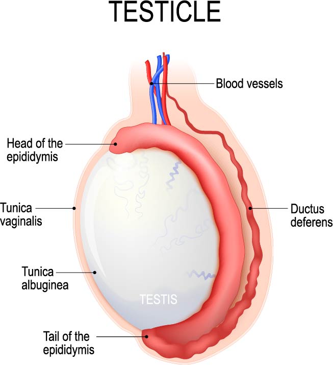
The image is an anatomical representation of a testicle, detailing its various parts and associated structures.
Central to the image is the testis itself, the oval-shaped male reproductive organ responsible for the production of sperm and the hormone testosterone. It is encapsulated by a tough, protective outer layer called the tunica albuginea. The tunica albuginea is a dense layer of connective tissue that extends inward to divide the testis into lobules.
The epididymis sits atop the testis, with its head positioned at the upper pole of the testis and its tail at the lower pole. The epididymis is a tightly coiled set of tubes that connect to the ductus deferens. It is the site where sperm mature and are stored.
The tunica vaginalis is a serous membrane that covers the front and sides of the testis. It is derived from the peritoneum during the descent of the testis into the scrotum.
The ductus deferens, also known as the vas deferens, is the tube through which mature sperm are transported from the epididymis in anticipation of ejaculation.
The image also shows the blood vessels that supply the testis, including arteries and veins, which are vital for providing oxygen, nutrients, and for the regulation of temperature, which is crucial for proper spermatogenesis.
This diagram is a useful tool for understanding the testicular anatomy and the initial path sperm take from their production site to the outside of the body.
Anatomical Structure of Complex Vascular and Nervous System
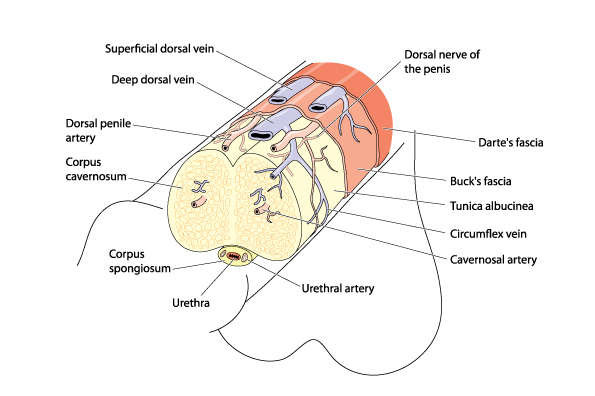
The provided image is a detailed anatomical illustration of the penis, showing the various structures that compose its complex vascular and nervous systems, as well as its protective coverings.
The penis comprises three columns of erectile tissue: two larger ones called the corpus cavernosum and a smaller one below called the corpus spongiosum, which surrounds the urethra, the tube through which urine and semen pass.
The corpus cavernosum is the primary erectile tissue that fills with blood during arousal, causing an erection. The paired structures are supplied by the cavernosal arteries.
The urethra, which runs along the ventral aspect of the penis, is surrounded by the corpus spongiosum. This helps to protect the urethra and keep it open during ejaculation. The urethral artery supplies blood to this area.
The image shows the tunica albuginea, a fibrous envelope that surrounds the corpus cavernosum and corpus spongiosum. It helps to trap blood in the erectile tissues, contributing to the rigidity of an erection.
Blood is supplied to the penis by the dorsal penile artery, and the blood is drained by veins, including the superficial and deep dorsal veins. The circulatory system is crucial for the erectile function of the penis.
Nerve supply, as indicated by the dorsal nerve of the penis, is essential for sensation and the initiation of an erection.
The penis is also enveloped in fascial layers: Dartos fascia, a layer of connective tissue that lies just beneath the skin, and Buck’s fascia, a deeper layer that encases the erectile tissues.
The circumflex vein is one of the veins responsible for draining the blood from the erectile tissues after an erection subsides.
This image provides an overview of the anatomical features that are essential for the functions of the penis, including sexual intercourse and urination.
Vasectomy Procedure Illustration
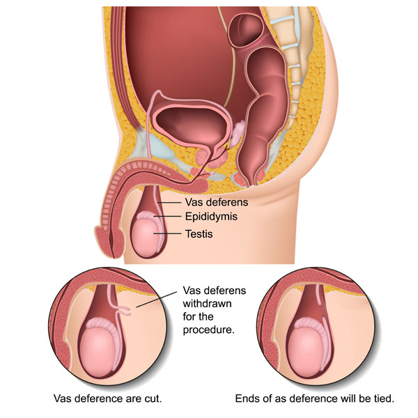
The image is a medical illustration depicting a vasectomy procedure, which is a surgical form of male sterilization used for permanent contraception. The upper part of the image provides an anatomical overview of the male reproductive organs within the pelvic region, while the lower part details the steps involved in the vasectomy process.
In the upper diagram, we see the testis, the male reproductive gland that produces sperm and testosterone. Sitting atop the testis is the epididymis, where sperm is stored and matures. The vas deferens, also known as the ductus deferens, is the tube that transports sperm from the epididymis to the ejaculatory ducts in anticipation of ejaculation.
The lower diagrams zoom in on the vas deferens to illustrate the vasectomy procedure. In the first step, a segment of the vas deferens is withdrawn from the scrotum. The second step shows the vas deferens being cut, and the final step demonstrates the ends of the vas deferens being tied or sealed. This prevents the transport of sperm from the testes to the urethra, thereby leading to sterility while not affecting the ejaculation of seminal fluid.
This procedure is typically performed as a means of permanent birth control, and while it prevents the release of sperm, it does not affect the production of male sex hormones and typically has no impact on sexual function.
Anatomical Terms and Definitions
| Term | Definition |
|---|---|
| Corpus Cavernosum | One of the two columns of erectile tissue found in the penis that fills with blood during arousal, causing an erection. |
| Corpus Spongiosum | The erectile tissue surrounding the urethra in the penis, preventing it from being squeezed shut during an erection. |
| Ductus Deferens | A muscular tube that transports mature sperm from the epididymis to the urethra in preparation for ejaculation. |
| Ejaculatory Duct | The duct formed by the fusion of the ductus deferens and the seminal vesicle, through which seminal fluid is transported into the urethra. |
| Epididymis | A long, coiled tube atop the testicle where sperm mature and are stored. |
| Glans Penis | The sensitive bulbous structure at the distal end of the penis, often considered the most sensitive area of the male reproductive system. |
| Penis | The male reproductive organ that contains the urethra, through which urine and semen are expelled from the body. |
| Prostate Gland | A gland surrounding the urethra that secretes fluid nourishing and transporting sperm during ejaculation. |
| Seminal Vesicles | Glands adjacent to the bladder that produce a significant portion of the seminal fluid, a component of semen. |
| Testicles | The male reproductive glands located in the scrotum, responsible for producing sperm and testosterone. |
| Tunica Albuginea | A fibrous envelope surrounding the corpus cavernosum and corpus spongiosum in the penis, aiding in erection by trapping blood within the erectile tissues. |
| Urethra | The tube running through the penis that carries urine from the bladder to the outside of the body and also serves as a conduit for semen during ejaculation. |
| Urinary Bladder | A hollow organ that stores urine from the kidneys before it is excreted. Though primarily part of the urinary system, it is included in reproductive diagrams due to its proximity to male reproductive organs. |
| Vasectomy | A surgical procedure for male sterilization or permanent contraception that involves cutting and sealing the vas deferens, thereby preventing sperm from entering the urethra and thus preventing fertilization. |
