
Table of Contents
Hypothalamus and Pituitary Gland Anatomy
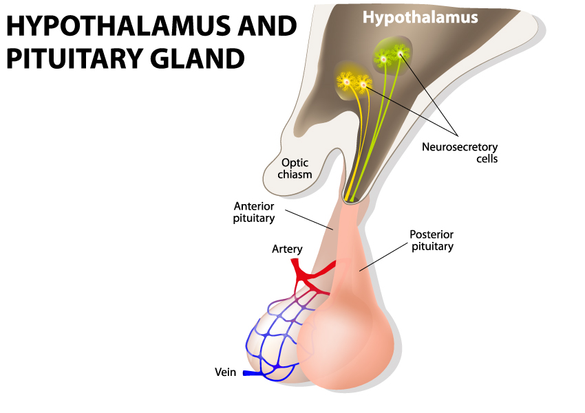
The image presents a simplified diagram of the hypothalamus and pituitary gland, two central structures within the brain that play crucial roles in endocrine function and the regulation of various bodily processes.
The hypothalamus, located at the base of the brain, is represented at the top part of the image. It is a vital brain area that controls many autonomic functions of the peripheral nervous system. It has a direct influence on the pituitary gland, which is depicted just below the hypothalamus and is connected to it by a thin stalk.
In the diagram, yellow branching structures are shown emerging from the hypothalamus. These are neurosecretory cells, which are specialized neurons that produce hormones released into the blood, affecting the pituitary gland. Neurosecretory cells send their hormonal signals directly to the pituitary gland to regulate its activity.
The optic chiasm is indicated by a horizontal formation just below the hypothalamus. This structure is where the optic nerves partially cross over, and it is located in the forebrain, directly in front of the hypothalamus. The optic chiasm is responsible for the visual coordination by relaying visual information received from the eyes to the brain.
The pituitary gland is shown in two distinct parts: the anterior pituitary and the posterior pituitary. The anterior pituitary receives releasing or inhibiting hormones from the hypothalamus via a portal blood vessel system, which is not explicitly depicted here. These hypothalamic hormones regulate the secretion of the anterior pituitary hormones.
The posterior pituitary, on the other hand, does not produce its own hormones but stores and releases hormones produced by the hypothalamus, such as oxytocin and vasopressin (antidiuretic hormone).
Vascular supply to the pituitary gland is illustrated by red and blue structures representing an artery and a vein, respectively. Arteries carry oxygenated blood to the pituitary gland, and veins carry deoxygenated blood away from it, ensuring that the gland receives the necessary nutrients and oxygen to function properly and that metabolic waste is removed.
This diagram is fundamental in understanding the anatomical and functional relationship between the hypothalamus and the pituitary gland, illustrating how these two structures work together to control a wide array of physiological processes including growth, metabolism, and reproductive functions.
Hormone Feedback Look
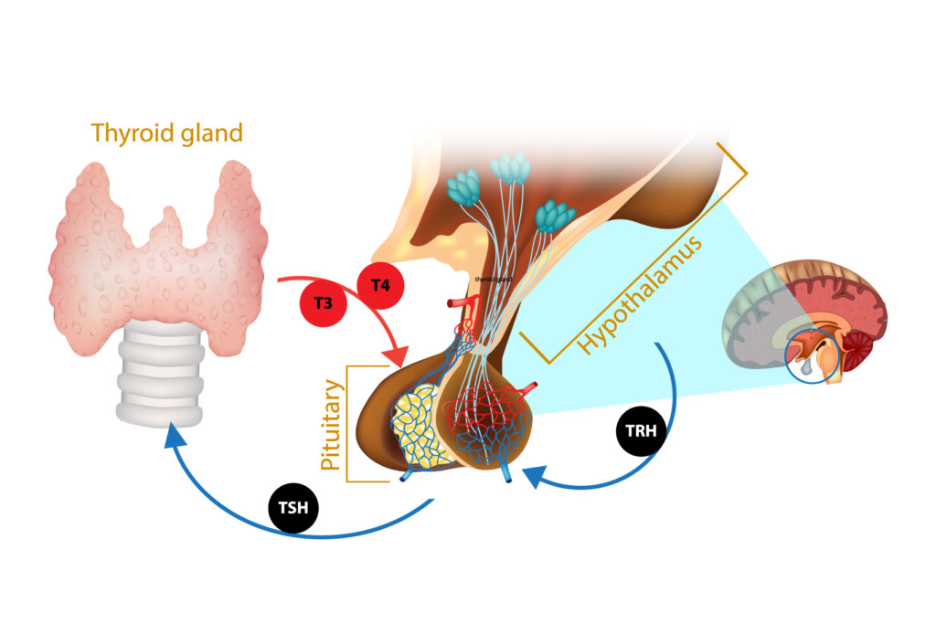
This illustration shows the feedback loop between the hypothalamus, pituitary gland, and thyroid gland, which is critical for the regulation of thyroid hormones in the body.
The hypothalamus, depicted at the top right of the image within the brain, produces thyrotropin-releasing hormone (TRH). TRH then travels to the pituitary gland, which is shown just beneath the hypothalamus. In response to TRH, the pituitary gland secretes thyroid-stimulating hormone (TSH), which circulates down to the thyroid gland, illustrated at the left side of the image. The thyroid gland, in turn, produces thyroid hormones, specifically T3 (triiodothyronine) and T4 (thyroxine).
These hormones are then released into the bloodstream and exert their effects on various tissues, helping to regulate metabolism, energy generation, and many other critical bodily functions. The levels of T3 and T4 in the blood provide feedback to both the pituitary gland and the hypothalamus to modulate the production of TSH and TRH, respectively, thereby maintaining the delicate balance of thyroid hormone levels.
Hormones Secreted by the Pituitary Gland
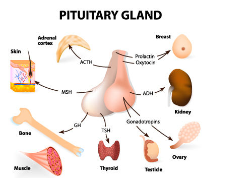
This diagram provides an overview of the various hormones secreted by the pituitary gland and their respective target organs or tissues in the human body.
The pituitary gland, often referred to as the “master gland,” is shown in the center. It is divided into two main lobes: the anterior and posterior pituitary, each responsible for releasing specific hormones.
From the anterior pituitary:
- ACTH (Adrenocorticotropic hormone) stimulates the adrenal cortex to produce cortisol and other glucocorticoids, which are vital in stress response, metabolism, and immune function.
- MSH (Melanocyte-stimulating hormone) is involved in skin pigmentation, influencing the melanocytes in the skin.
- GH (Growth hormone) affects the bone and muscle, promoting growth and development.
- TSH (Thyroid-stimulating hormone) targets the thyroid gland, encouraging it to produce thyroid hormones (T3 and T4) that regulate metabolism.
- Gonadotropins, which include LH (Luteinizing hormone) and FSH (Follicle-stimulating hormone), act on the ovaries and testicles to influence sex hormone production and gamete (egg and sperm) production.
From the posterior pituitary:
- ADH (Antidiuretic hormone or vasopressin) is directed towards the kidneys, helping the body to retain water and regulate urine concentration.
- Oxytocin targets the breasts, stimulating milk ejection, and the uterus, inducing contractions during childbirth.
Each hormone has a critical role in maintaining homeostasis and facilitating the body’s responses to various physiological demands. The diagram underscores the complexity and importance of the endocrine system’s interplay with other bodily systems.
Anatomical Terms and Definitions
| Term | Definition |
|---|---|
| ACTH (Adrenocorticotropic Hormone) | Stimulates the adrenal cortex to produce cortisol and other glucocorticoids, vital for stress response, metabolism, and immune function. |
| ADH (Antidiuretic Hormone) | Also known as vasopressin, directed towards the kidneys to help the body retain water and regulate urine concentration. |
| Anterior Pituitary | The front part of the pituitary gland that releases hormones regulated by the hypothalamus, influencing growth, metabolism, and reproductive functions. |
| FSH (Follicle-Stimulating Hormone) | A gonadotropin acting on the ovaries and testicles to influence sex hormone production and gamete production. |
| GH (Growth Hormone) | Affects bone and muscle, promoting growth and development. |
| Hypothalamus | A vital brain area located at the base of the brain, controlling many autonomic functions and influencing the pituitary gland's activity. |
| LH (Luteinizing Hormone) | A gonadotropin that acts on the ovaries and testicles, involved in the regulation of the menstrual cycle and ovulation in females, and testosterone production in males. |
| MSH (Melanocyte-Stimulating Hormone) | Involved in skin pigmentation by influencing melanocytes in the skin. |
| Neurosecretory Cells | Specialized neurons in the hypothalamus that produce hormones affecting the pituitary gland. |
| Optic Chiasm | A structure where the optic nerves partially cross over, located in front of the hypothalamus, responsible for visual coordination. |
| Oxytocin | A hormone targeting the breasts and uterus, stimulating milk ejection and inducing contractions during childbirth, released by the posterior pituitary. |
| Pituitary Gland | Known as the "master gland," located below the hypothalamus, divided into the anterior and posterior pituitary, secreting various hormones that regulate bodily functions. |
| Posterior Pituitary | The back part of the pituitary gland that stores and releases hormones produced by the hypothalamus, such as oxytocin and vasopressin. |
| T3 (Triiodothyronine) | A thyroid hormone produced by the thyroid gland, involved in regulating metabolism and energy generation. |
| T4 (Thyroxine) | Another thyroid hormone produced by the thyroid gland, crucial for metabolism and energy production. |
| TRH (Thyrotropin-Releasing Hormone) | Produced by the hypothalamus, it stimulates the pituitary gland to secrete TSH. |
| TSH (Thyroid-Stimulating Hormone) | Secreted by the pituitary gland, it stimulates the thyroid gland to produce thyroid hormones (T3 and T4). |
| Vascular Supply | The arteries and veins providing oxygenated blood to and carrying deoxygenated blood away from the pituitary gland, ensuring it receives nutrients and oxygen. |
