
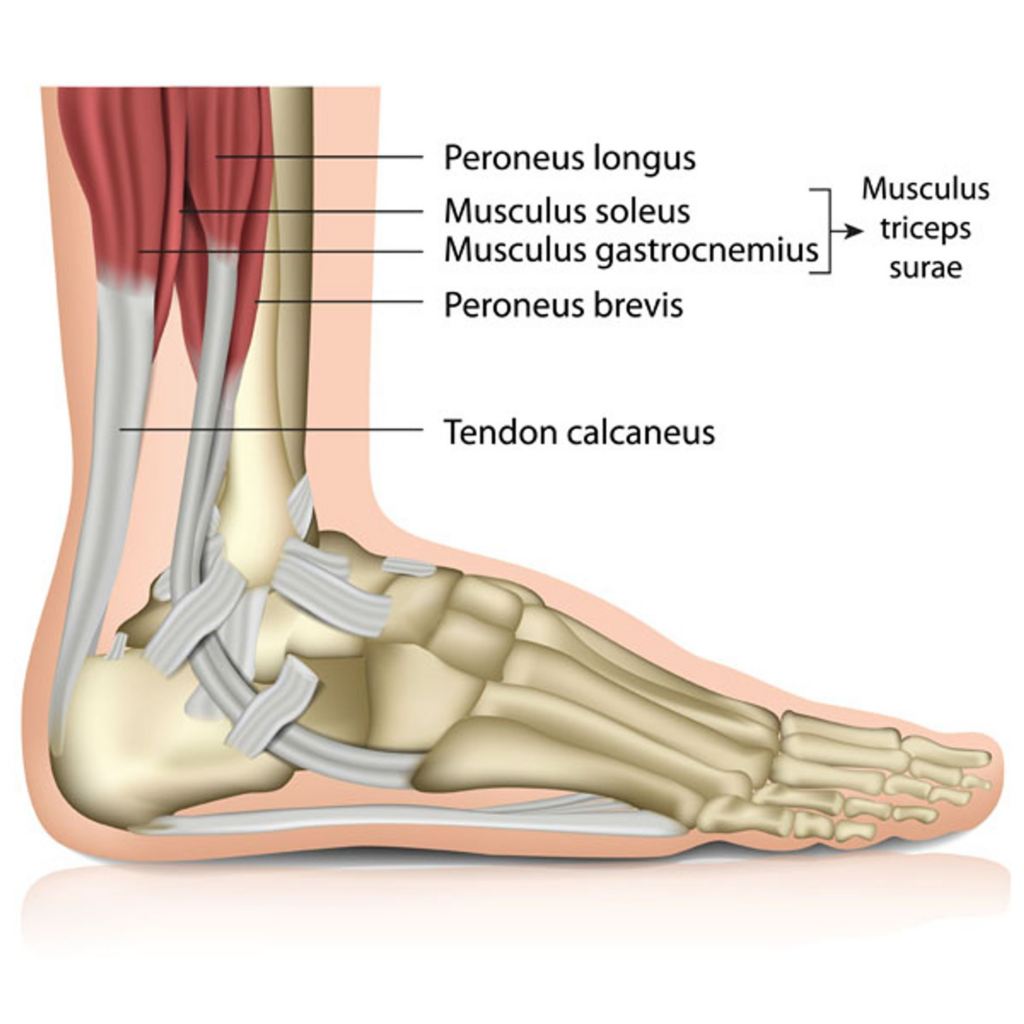
Musculus Triceps Surae
The musculus triceps surae, often referred to simply as the triceps surae, is a pair of muscles located in the posterior compartment of the lower leg. This muscle group is critical for walking, running, and jumping as it plays a major role in plantar flexion of the foot and flexion of the knee.
The triceps surae is comprised of two major muscles: the gastrocnemius and the soleus. These muscles converge into a common tendon, known as the Achilles tendon or calcaneal tendon, which attaches to the calcaneus, or heel bone.
The gastrocnemius is the larger, more superficial muscle of the triceps surae. It originates from two heads that attach to the femur, right above the knee joint. Due to its location, the gastrocnemius contributes to knee flexion as well as foot plantarflexion. It has a characteristic diamond shape and is what gives the calf its bulging, muscular appearance.
The soleus muscle, on the other hand, lies underneath the gastrocnemius. It originates from the tibia and fibula, the two bones of the lower leg, and merges with the gastrocnemius to form the Achilles tendon. Unlike the gastrocnemius, the soleus does not cross the knee joint, and thus, it only contributes to plantar flexion of the foot.
In terms of blood supply, the triceps surae receives blood from branches of the popliteal artery, specifically the sural arteries. Innervation to the triceps surae is provided by the tibial nerve, a branch of the sciatic nerve.
This muscle group is particularly prone to overuse injuries, especially in athletes or individuals who frequently engage in high-impact activities. Common pathologies include calf strains, Achilles tendinopathy, and in severe cases, rupture of the Achilles tendon. Therefore, appropriate conditioning, stretching, and strength training of these muscles are vital for preventing injury
Peroneus Longus
The Peroneus Longus, also known as the Fibularis Longus, is a muscle that plays an essential role in foot and ankle stability. It is found in the lateral compartment of the leg, running along the outer side, and is one of three peroneal (fibular) muscles, with the others being the Peroneus Brevis and Peroneus Tertius.
The Peroneus Longus originates from the head and upper part of the lateral surface of the fibula, as well as the adjacent intermuscular septa and fascia. From there, it extends downward and wraps underneath the foot. The muscle then runs obliquely across the sole of the foot to attach at the base of the first metatarsal and medial cuneiform bones.
In terms of function, the Peroneus Longus works to evert and plantarflex the foot. Eversion involves moving the sole of the foot outward, away from the midline of the body, while plantarflexion is the action of pointing the toes downward. These movements are crucial for walking, running, and balance.
The Peroneus Longus is innervated by the superficial fibular (peroneal) nerve, a branch of the common fibular (peroneal) nerve, which itself is a branch of the sciatic nerve. Blood supply to the muscle is provided by the fibular (peroneal) artery.
Like other muscles, the Peroneus Longus can be injured due to trauma or overuse, leading to conditions such as sprains or strains. In particular, this muscle is often implicated in conditions related to ankle instability and foot dysfunction, making regular strengthening and stretching exercises crucial for individuals engaged in activities with high foot and ankle demands.
Musculus Soleus
The Musculus Soleus, more commonly known as the Soleus muscle, is a powerful muscle in the posterior part of the lower leg, sometimes referred to as the calf muscle. It lies underneath the Gastrocnemius muscle and together, they form the Triceps Surae muscle group, which is crucial for standing, walking, and running.
The Soleus originates from two points: the posterior aspect of the fibula and the soleal line on the tibia. This broad and flat muscle extends downwards and combines with the Gastrocnemius muscle to form a common tendon, the Achilles tendon (Calcaneal tendon), which inserts into the posterior part of the calcaneus bone, the heel bone.
The primary function of the Soleus muscle is to perform plantarflexion of the ankle, which is the action of pointing the toes downwards. It is also involved in maintaining upright posture by counteracting the tendency to fall forward at the ankle when standing.
The Soleus is innervated by the tibial nerve, a branch of the sciatic nerve. The arterial supply is provided by branches of the posterior tibial artery and fibular (peroneal) artery.
An injury or strain to the Soleus muscle can cause pain and mobility issues in the lower leg. Common causes of Soleus muscle injuries include overuse, particularly in runners and jumpers, and inadequate warm-up before intense physical activity. Therefore, it's crucial to engage in regular stretching and strengthening exercises, which can help maintain the health and flexibility of the Soleus and other calf muscles.
Peroneus Brevis
The peroneus brevis, also known as the fibularis brevis, is one of the key muscles located within the lateral compartment of the lower leg. As part of the peroneal or fibular group of muscles, it plays a significant role in ankle and foot movement.
The muscle originates from the lower two-thirds of the lateral surface of the fibula, which is the smaller of the two long bones in the lower leg. The muscle's fibers run down the lateral side of the lower leg, in close proximity to the larger peroneus longus muscle, which lies superficial to it.
The tendon of the peroneus brevis muscle passes behind the lateral malleolus, the outer bony prominence of the ankle. It then crosses the foot diagonally to attach at the base of the fifth metatarsal, which is the long bone on the outer side of the foot that connects to the little toe. This tendon can be felt on the outside of the ankle when the foot is everted.
The main function of the peroneus brevis is to evert the foot, meaning it helps to turn the sole of the foot outward. It also assists in plantarflexion of the ankle, which is the motion of pointing the toes downwards.
The peroneus brevis muscle is innervated by the superficial fibular (peroneal) nerve, which branches from the common fibular (peroneal) nerve. This nerve allows for the sensation and control of the outer part of the lower leg and the dorsal aspect of the foot. The blood supply to the peroneus brevis muscle is primarily provided by the fibular (peroneal) artery, a branch of the posterior tibial artery.
Due to its critical role in foot movement and stability, injuries to the peroneus brevis, such as tears or sprains, can significantly impair a person's ability to walk, run, or participate in other weight-bearing activities. These injuries often result from intense physical activity, improper footwear, or ankle instability. Symptoms may include lateral foot pain, swelling, and weakness or instability of the foot and ankle. If such symptoms are present, a healthcare professional should be consulted for a thorough evaluation and appropriate treatment.
Tendon Calcaneus
The tendon calcaneus, more commonly known as the Achilles tendon, is the largest and strongest tendon in the human body. It's a crucial component of the musculoskeletal system and plays a vital role in activities such as walking, running, and jumping.
The Achilles tendon is a robust, fibrous structure that connects two important calf muscles — the gastrocnemius and the soleus — to the calcaneus, or the heel bone. The gastrocnemius muscle has two heads originating from the thigh bone (femur), while the soleus muscle originates from the tibia and fibula, the two bones of the lower leg. These muscles merge to form the Achilles tendon, which attaches to the posterior aspect of the calcaneus.
The primary function of the Achilles tendon is to transmit the force generated by the contraction of the gastrocnemius and soleus muscles to the foot, facilitating movements such as plantarflexion (pointing the foot downwards) and assisting with stability during standing and walking.
The Achilles tendon is not directly supplied by arteries; instead, it receives its blood supply from the surrounding connective tissue and the muscles to which it is attached. It has a complex vascular supply involving several arteries, including the posterior tibial artery and the peroneal artery.
However, there's a specific area of the tendon, located 2-6 cm above its attachment to the heel bone, referred to as the "watershed zone" or "critical zone," which has less direct blood flow. This area is often the site of Achilles tendon injuries due to its relative lack of blood supply, making it susceptible to degeneration and tearing.
Speaking of innervation, the Achilles tendon receives its nerve supply from the sural nerve, a sensory nerve, and the tibial nerve, a mixed sensorimotor nerve. These nerves help in sensing the position of the ankle (proprioception) and controlling muscle movement.
Injuries to the Achilles tendon, such as tendonitis (inflammation) or rupture, are common in athletes and individuals who engage in high-impact activities. Symptoms of Achilles tendon injuries can include pain, swelling, stiffness, and difficulty with foot movements. If any such symptoms are noticed, it is advisable to seek medical attention for appropriate diagnosis and treatment.
