
We are going to be writing a series of articles from our website NEHCacademy.com and our FB group over the next few weeks to share what we have found in our research of how fluid pressure plays such an important role in our Vibrational Fascia Release Technique(TM). The main purpose is to re-insert the lost variable back into the equation of why we are in pain with lack of mobility. We have plenty of real-life examples to back up our theories, and these articles are designed to provide some science behind our theories of fluid, fascia, and tuning forks.
There has been such a big focus on the solid form of fascia in the past few years as researchers and clinicians are realizing the vital role collagen fascia fibers plays in almost every aspect of our body. Previously known as connective tissue, fascia (in its many forms) has been seen as the forgotten tissue until very recently as technology and research methods have allowed us a different view of fascia in a natural environment. Jean Claude Guimberteau showed us live fascia in a video, Carla Stecco devoted her life to mapping fascia layers in an Atlas of images, and Thomas Myers determined how fascia is connected across the body in lines or distinct strips.
Bobbi Jo and I are focusing on another part of the equation that was almost impossible to measure and view in a research setting. Interstitial fluid is the plasma released from the artery walls into the extra-cellular spaces of our tissue to provide substances needed for survival or processing based on the purpose of each cell. The ability to observe fluid in its natural environment was very difficult since the act of cutting into the tissue would release the pressure and fluid from the local area. Cadaver studies and biopsies also released the fluid in the tissue prior to observation and research.
It was not until recently when researchers like Dr. Neil Theise announced the “interstitium” as the largest organ in the body. He describes his discovery of the white spaces in the slides he observes every day as a diagnostic liver pathologist, and he continued to focus on the fluid spaces in between the collagen fiber structures of our body.
Here is where we pick up the story from our point of view at New Earth Healing Center and NEHC Academy where we have found patterns to healing the body with vibration of a weighted tuning fork (128hz) in our VFRT method. We have consistently found where fluid pressure was not only pressing against pain receptors to cause pain, but there were other factors of fluid pressure producing a chain of problems that could possibly be traced to many other conditions thought to be unrelated. We invite you into our world to see things through our eyes and determine for yourself if fluid and fascia together are responsible for most chronic conditions.
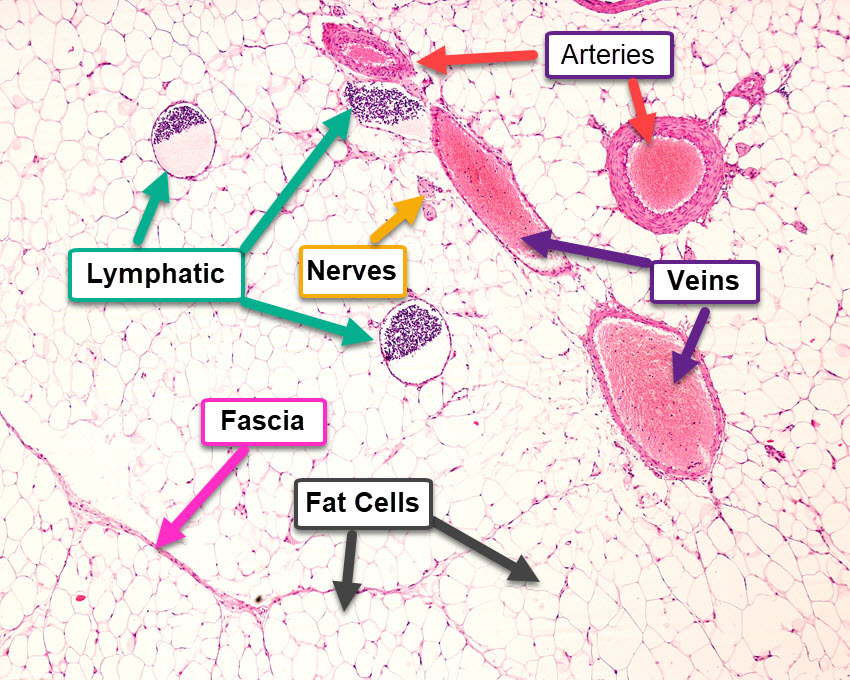
In the attached image, we see a very important and different way to see the body through a diagnostic process of immuno-staining the tissue in a live person and seeing the colors change in the tissue as staining highlights very specific parts of the body. Through high definition ultrasound, we can see the story unfolding in real time to find missing pieces to the puzzle for the first time. We can even see fluid distribution throughout the image and how damage to a certain area causes a chain of events previously un-noticed by other diagnostic techniques.
In the image, notice how the usual subjects within a superficial adipose (fat) layer are related to each other in a very compact space. We will continue to use this image and show you how to interpret the image and learn from our tissue underneath the skin as we zoom into each part for a more detailed discussion. There are a lot of things to see in this image, but this is just the beginning of our journey to learning about the body through fluid, fascia, and tuning forks.
