
Table of Contents
Thoracic Spine Anatomy Video
Thoracic Vertebrae: Lateral View
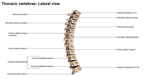
The image displays a lateral view of the thoracic vertebrae, highlighting several key anatomical features. Starting at the top, the vertebra labeled T1, or the first thoracic vertebra, shows a feature known as the spinous process. This bony projection extends posteriorly from the vertebra and can be felt through the skin as a series of bumps along the middle of the back.
Moving down, we see the superior articular process, which articulates with the inferior articular process of the vertebra above it. This articulation facilitates the movement and connection between adjacent vertebrae. Each vertebra has a transverse process extending laterally, which serves as an attachment point for muscles and ligaments. The articulatio zygapophysealis, also known as the facet joint, is where the superior and inferior articular processes meet, allowing for the articulation between vertebrae.
On the side of the vertebrae, we notice the fovea costalis processus transversi, which are the facets on the transverse processes that articulate with the tubercles of the ribs. Similarly, the fovea costalis superior and inferior are indentations on the vertebral body that articulate with the head of the ribs.
The foramen intervertebrale, or intervertebral foramen, is an opening between adjacent vertebrae through which spinal nerves exit the spinal column. Below that, the incisura vertebralis inferior and superior are notches that contribute to the formation of the intervertebral foramen when two vertebrae are aligned.
Finally, the facies articularis inferior is the inferior surface of the vertebral body that forms a joint with the vertebra below. These structures work collectively to support the thoracic cage, allow for certain movements, and protect the thoracic spinal cord.
