
Table of Contents
Basic Neuron Anatomy
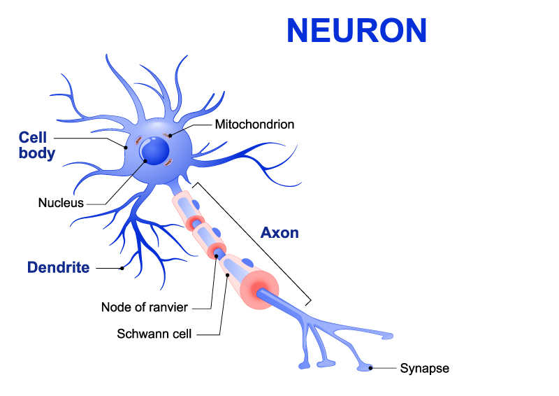
The image displays a diagrammatic representation of a neuron, which is the fundamental unit of the brain and nervous system, responsible for carrying messages throughout the body as electrical impulses. This neuron has several distinct parts:
The cell body, also known as the soma, contains the nucleus, which houses the cell’s genetic material. The cell body is the neuron’s metabolic center and is responsible for maintaining the life of the cell.
Radiating from the cell body are dendrites, which are tree-like structures that receive messages from other nerve cells and transmit these messages towards the cell body.
The axon is a long, slender projection that conducts electrical impulses away from the cell body. This part of the neuron can vary greatly in length, with some axons in the human body extending a meter or more.
Along the axon are periodic gaps in the myelin sheath known as Nodes of Ranvier. These nodes are crucial for the rapid conduction of nerve impulses along the axon, as they allow the impulse to ‘jump’ from node to node, a process known as saltatory conduction.
The axon is insulated by a fatty layer known as the myelin sheath, which is interrupted at regular intervals by the Nodes of Ranvier. The myelin sheath is produced by specialized cells; in the peripheral nervous system, these cells are called Schwann cells.
The mitochondrion within the cell body is the powerhouse of the cell, generating the energy required for the neuron to function.
At the very end of the axon are the synapses, which are the junctions through which neurons signal to each other and to non-neuronal cells such as muscles or glands. The synapse ensures the direction of the nerve impulse transmission is unidirectional and specifically targeted.
This visual encapsulates the structure of a neuron, which can be considered a marvel of biological engineering, enabling both the reception and transmission of electrical signals throughout the nervous system.
Types of Neurons
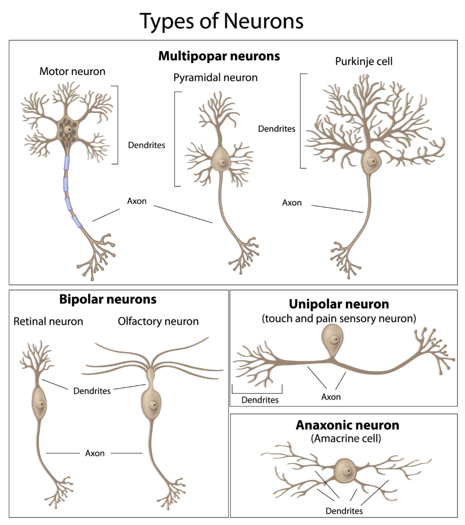
This image illustrates various types of neurons, each with distinctive structures and functions within the nervous system.
At the top left, we see a Motor neuron characterized by a long axon and multiple branching dendrites. This type of neuron transmits signals from the central nervous system to muscles, inducing movement.
Next to it, under the category of Multipolar neurons, there are two examples: a Pyramidal neuron and a Purkinje cell. Pyramidal neurons, found in the cerebral cortex, have a triangular-shaped soma and a single axon; they are involved in a variety of cognitive functions. The Purkinje cell, located in the cerebellum, has a highly complex and extensive dendritic tree, making it critical for motor coordination.
Below, in the Bipolar neurons category, there are two types: Retinal neuron and Olfactory neuron. Bipolar neurons have a simple structure with one axon and one dendritic branch. The Retinal neuron is integral to the visual system, while the Olfactory neuron is involved in the sense of smell.
The Unipolar neuron depicted is specialized for the sensory systems, particularly for touch and pain. Unlike other neurons, it has a single process that extends from the cell body and splits into two branches, functioning as both an axon and dendrite.
Finally, the Anaxonic neuron, represented here by an Amacrine cell found in the retina, has no clear axon and multiple dendrites. It is involved in the visual processing by modulating the signal between bipolar cells and ganglion cells.
Each neuron type is adapted to its specific role in processing and transmitting information, reflecting the complexity and efficiency of the nervous system.
Synapse Anatomy
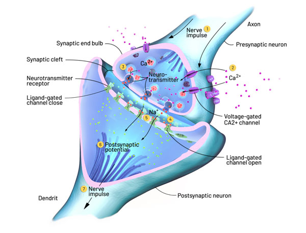
The image presents a detailed view of a synapse, which is the site of communication between neurons. This diagram breaks down the complex process of synaptic transmission:
Presynaptic Neuron: This is the neuron that sends the signal. At the end of its axon, you can see the synaptic end bulbs that contain neurotransmitters.
Axon: This is the long, thin structure of the presynaptic neuron that transmits the nerve impulse towards the synapse.
Synaptic End Bulb: This bulbous structure at the end of the axon stores neurotransmitters and is where the nerve impulse triggers their release.
Voltage-Gated Ca2+ Channel: The arrival of a nerve impulse opens these channels, allowing calcium ions (Ca2+) to enter the presynaptic neuron’s terminal. This influx of calcium is crucial for the release of neurotransmitters into the synaptic cleft.
Neurotransmitter: These are chemical messengers released by neurons to transmit signals to other neurons. They are stored in vesicles within the synaptic end bulb.
Synaptic Cleft: The small gap between the presynaptic and postsynaptic neurons through which neurotransmitters diffuse.
Neurotransmitter Receptor: These are proteins located on the postsynaptic neuron’s membrane that specifically bind to neurotransmitters.
Ligand-Gated Channel: When a neurotransmitter binds to its receptor, it can open a channel for ions like sodium (Na+) to flow into the postsynaptic neuron, initiating a postsynaptic potential.
Postsynaptic Neuron: This neuron receives the signal. The binding of neurotransmitters to receptors on its dendrite can generate a new nerve impulse.
Nerve Impulse: This is the electrical signal that travels along the neuron. In the postsynaptic neuron, the nerve impulse is initiated if enough neurotransmitters bind and cause a significant change in the membrane potential.
Postsynaptic Potential: This is the change in membrane potential of the postsynaptic neuron due to the binding of neurotransmitters and the subsequent movement of ions.
This entire process is how neurons communicate with each other, allowing for the complex signaling that underlies all nervous system functions, from reflexes to thought processes.
Schwann Cell
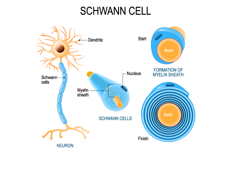
This illustration provides an educational visualization of the Schwann cell and its role in the formation of the myelin sheath around the axon of a neuron.
On the left, we see a neuron with its dendrites branching out from the cell body. The axon extends from the neuron, and along it, Schwann cells are shown. The Schwann cells are depicted as individual cells along the axon that contribute to the formation of the myelin sheath.
The myelin sheath is a fatty layer that wraps around the axon, which is crucial for insulating the axon and increasing the speed of electrical signal transmission. This is shown in the ‘Start’ phase, where a Schwann cell begins to wrap around a bare axon.
As the Schwann cell wraps around the axon, it creates multiple layers of membrane that form the myelin sheath. This process is depicted in the middle section, labeled “FORMATION OF MYELIN SHEATH,” which illustrates the progressive wrapping of the Schwann cell’s membrane around the axon.
The ‘Finish’ section of the illustration shows the axon completely enveloped by the concentric layers of the Schwann cell membrane, which is now a fully formed myelin sheath. The nucleus of the Schwann cell is seen at one end of the myelin sheath, indicating the Schwann cell’s body.
This myelination by Schwann cells is essential for the proper functioning of the nervous system, particularly in the peripheral nervous system where these cells are found. Myelination allows for the rapid and efficient transmission of nerve impulses along the axon. The gaps between adjacent Schwann cells are known as the nodes of Ranvier, which are not shown here but are important for the saltatory conduction of nerve impulses.
Myelin Sheath Formation
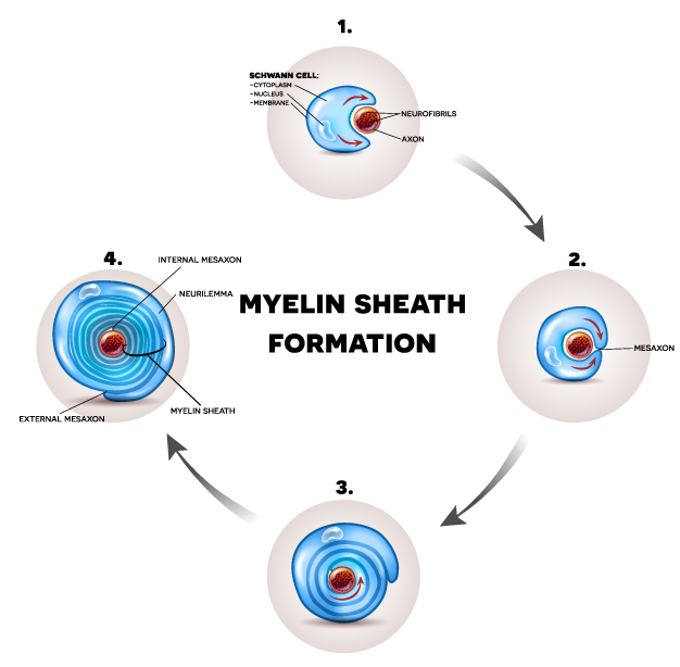
This diagram provides a step-by-step visualization of myelin sheath formation around an axon, a process crucial for rapid and efficient nerve signal transmission in the peripheral nervous system. The myelin sheath is a fatty layer produced by Schwann cells that insulates axons and facilitates the conduction of electrical signals.
Schwann Cell Attachment: The first step shows a Schwann cell with its cytoplasm and nucleus approaching an axon. The Schwann cell starts to envelop the axon, which contains neurofibrils, the structural components responsible for the transport of nutrients and organelles within the neuron.
Initiation of Wrapping: In the second step, the Schwann cell begins to wrap its membrane around the axon. A mesaxon forms, which is the point of contact where the Schwann cell membrane starts to wrap around the axon.
Progression of Wrapping: As the wrapping continues, the layers of Schwann cell membrane begin to compact around the axon. The Schwann cell’s cytoplasm is progressively squeezed out, and multiple layers of membrane accumulate.
Completion of Myelin Sheath: In the final stage, the myelin sheath is fully formed around the axon. The compacted layers of the Schwann cell membrane create the myelin sheath, and the cytoplasm is no longer visible. The neurilemma, which is the outermost layer of the Schwann cell containing the nucleus, wraps around the myelin sheath. The structure now has an internal mesaxon and an external mesaxon, which represent the inner and outer layers of the wrapped membrane, respectively.
The entire myelination process is essential for the proper functioning of neurons, particularly in the peripheral nervous system, where Schwann cells facilitate this process. The myelin sheath not only insulates the axon but also significantly increases the speed of electrical impulse conduction through saltatory conduction, where the impulse jumps from one node of Ranvier to the next.
Sensory Receptors in the Skin
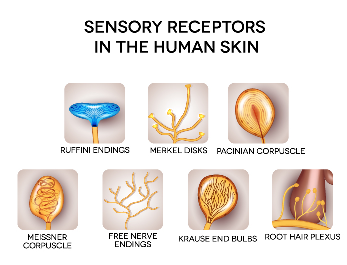
The image displays a range of sensory receptors located in human skin, each specialized for different types of sensory input:
Ruffini Endings: These are spindle-shaped receptors that respond to skin stretch and contribute to the kinesthetic sense of and control of finger position and movement.
Merkel Disks: These are slow-adapting, touch receptors that respond to light touch, and they help in detecting shapes and edges.
Pacinian Corpuscle: These are oval-shaped, fast-adapting receptors that respond to deep pressure and vibration. They are located deeper in the dermis or hypodermis.
Meissner Corpuscle: These receptors are sensitive to light touch and adapt quickly to changes in texture and slow vibrations. They are concentrated in areas like the fingertips and lips.
Free Nerve Endings: These are the most common type of nerve ending, which respond to pain, temperature, and mechanical stimuli like touch and pressure.
Krause End Bulbs: These are thermoreceptors that can perceive cold temperatures. They are also thought to detect tactile sensations.
Root Hair Plexus: These are nerve fibers that wrap around the base of hair follicles and detect hair movement, allowing for the sensation of a light touch when the hair is displaced.
Each type of receptor is adapted to its function, contributing to the rich tapestry of tactile sensations humans can perceive. These receptors work together to provide the nervous system with detailed information about the external environment as it relates to touch, pressure, temperature, and pain.
Glial Cells
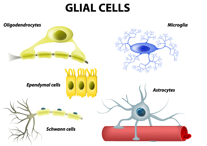
This illustration provides a visual overview of the different types of glial cells, which are non-neuronal cells that provide support and maintenance for the central nervous system (CNS) and the peripheral nervous system (PNS).
Oligodendrocytes: These cells, with their few extended arms, are found in the CNS. They produce the myelin sheath that insulates neuronal axons, enhancing the speed of electrical transmission along the axons within the CNS. Each oligodendrocyte can extend its processes to myelinate multiple axons.
Microglia: Shown with numerous branching processes, microglia are the immune cells of the CNS. They act as the first and main form of active immune defense, performing functions similar to macrophages by scavenging the CNS for plaques, damaged neurons, and infectious agents.
Ependymal Cells: These cells line the ventricles in the brain and the central canal of the spinal cord. They have a ciliated surface that helps circulate the cerebrospinal fluid, which cushions the brain and spinal cord.
Astrocytes: The star-shaped astrocytes are among the most versatile glial cells in the CNS. They maintain the blood-brain barrier, provide nutrients to nervous tissue, and play a role in repair and scarring processes following CNS injury.
Schwann Cells: These are the myelinating cells of the PNS. They wrap around the axons of peripheral nerves to form the myelin sheath, which aids in the rapid transmission of electrical signals. Unlike oligodendrocytes, each Schwann cell myelinates a single axon.
These glial cells are essential for the normal functioning of the nervous system, providing structural support, insulation, and regulation of the extracellular environment around neurons.
Alzheimer's Disease Neuron Disintegration
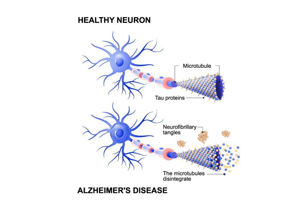
The image juxtaposes a healthy neuron with a neuron affected by Alzheimer’s disease, illustrating the cellular changes that occur in this neurodegenerative condition.
In the top half, the Healthy Neuron is shown with a well-structured network of dendrites branching out from the cell body, and an axon extending from it. Inside the axon, there are microtubules, which are part of the neuron’s cytoskeleton providing support and facilitating transport within the neuron. Tau proteins are depicted as small dots along the microtubules, stabilizing them under normal conditions.
The bottom half of the image shows a neuron affected by Alzheimer’s Disease. Here, the microtubules within the axon are disintegrating, and tau proteins are abnormally aggregated into what are known as neurofibrillary tangles. These tangles contribute to the disruption of the neuronal transport system and compromise the structural integrity of the neuron.
These pathological changes are characteristic of Alzheimer’s disease and are associated with the loss of neuron function and cell death, leading to the cognitive decline and memory loss seen in individuals affected by the disease. Neurofibrillary tangles, along with amyloid plaques (not shown in this image), are the hallmark signs of Alzheimer’s pathology.
Anatomical Terms and Definitions
| Term | Definition |
|---|---|
| Alzheimer’s Disease | A neurodegenerative disorder characterized by the accumulation of amyloid plaques and neurofibrillary tangles in the brain, leading to cognitive decline and memory loss. |
| Astrocytes | Star-shaped glial cells in the central nervous system that maintain the blood-brain barrier, provide nutrients to nervous tissue, and play a role in repair and scarring processes following CNS injury. |
| Axon | A long, slender projection of a neuron that conducts electrical impulses away from the cell body. |
| Cell Body (Soma) | The part of a neuron that contains the nucleus and genetic material; it is responsible for maintaining the life of the cell and its metabolic activities. |
| Dendrites | Branch-like structures of neurons that receive messages from other nerve cells and transmit these messages towards the cell body. |
| Ependymal Cells | Glial cells lining the ventricles of the brain and the central canal of the spinal cord, involved in the production and circulation of cerebrospinal fluid. |
| Free Nerve Endings | The most common type of nerve ending, which responds to pain, temperature, and mechanical stimuli like touch and pressure. |
| Microglia | The immune cells of the central nervous system, acting similarly to macrophages by scavenging the CNS for plaques, damaged neurons, and infectious agents. |
| Myelin Sheath | A fatty layer that insulates axons of some neurons, facilitating the rapid conduction of electrical signals. Produced by oligodendrocytes in the CNS and Schwann cells in the PNS. |
| Neurofibrillary Tangles | Abnormally aggregated tau proteins inside neurons, associated with Alzheimer’s disease, contributing to the disruption of the neuronal transport system and compromising the structural integrity of the neuron. |
| Neuron | The fundamental unit of the brain and nervous system, responsible for carrying messages throughout the body as electrical impulses. |
| Nodes of Ranvier | Gaps in the myelin sheath along the axon that allow electrical impulses to jump from node to node, facilitating rapid signal transmission. |
| Oligodendrocytes | Glial cells in the central nervous system that produce the myelin sheath insulating neuronal axons. Each oligodendrocyte can extend its processes to myelinate multiple axons. |
| Schwann Cells | Glial cells in the peripheral nervous system that wrap around axons to form the myelin sheath, aiding in the rapid transmission of electrical signals. Each Schwann cell myelinates a single axon. |
| Synapse | The junction through which neurons signal to each other and to non-neuronal cells such as muscles or glands. Ensures directional transmission of nerve impulses. |
