
Table of Contents
Interrelationship of Components Connected to Pancreas
Depicted here is an illustration showcasing the interrelation between various components of the human digestive and circulatory systems with the pancreas.
The pancreas is a glandular organ shown in yellow, which is divided into three sections: the head of the pancreas, the body of the pancreas, and the tail of the pancreas. The pancreatic duct is highlighted, running through the pancreas and allowing digestive enzymes to flow into the duodenum.
The duodenum, the first section of the small intestine, wraps around the head of the pancreas. Notably, two structures called the major duodenal papilla and the minor duodenal papilla are where the pancreatic duct and the common bile duct release their contents into the duodenum, aiding in digestion.
Above the pancreas, the gallbladder is depicted, which stores and concentrates bile produced by the liver. The common bile duct is a tube that carries bile from the gallbladder and liver to the duodenum.
To the right of the pancreas, the jejunum, which is the second part of the small intestine, is shown. It is responsible for the majority of nutrient absorption during digestion.
Behind the depicted organs are three major vessels: the inferior vena cava, which carries deoxygenated blood from the lower body to the heart; the abdominal aorta, the main artery that carries oxygenated blood from the heart to the lower body; and the portal vein, which carries nutrient-rich blood from the gastrointestinal tract to the liver.
Understanding these relationships is crucial for comprehending the digestive process and the integration of the circulatory system in nutrient absorption and distribution.
Pancreatic Islet Anatomy
The image provides an educational visual aid on the function of pancreatic beta cells within the islets of Langerhans, as they relate to insulin production and glucose uptake.
At the top, we have an anatomical depiction of the pancreas, an organ involved in both endocrine and exocrine functions. The pancreas is connected to the first part of the small intestine, the duodenum, indicating the exocrine role of the pancreas in secreting digestive enzymes.
Focusing in, we see a close-up of a pancreatic islet, also known as an islet of Langerhans. The islet is composed of a cluster of cells, each color-coded to represent different types of cells that secrete a variety of hormones. The beta cells, which are typically identified by their insulin production, are highlighted here.
At the bottom, there’s a magnified view of a single beta cell. The process illustrated shows glucose molecules being transported into the cell via glucose transporter 2 (GLUT2). Inside the beta cell, glucose is metabolized, leading to the release of insulin. Insulin, represented by purple triangles, is a hormone critical for the regulation of glucose levels in the blood. It facilitates the uptake of glucose by various tissues in the body to be used for energy or stored for future use.
This image encapsulates the critical role of pancreatic beta cells in glucose metabolism and the body’s overall energy regulation, linking the micro (cellular level) and macro (organ level) perspectives.
Type 1 Diabetes
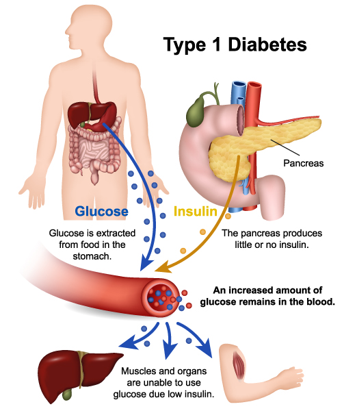
The illustration provides a visual explanation of Type 1 Diabetes and its impact on the human body. In Type 1 Diabetes, the pancreas, an organ located behind the stomach, fails to produce sufficient insulin, which is a hormone crucial for regulating blood sugar levels.
The image shows glucose, which is a primary source of energy for the body’s cells, being extracted from food in the stomach. Normally, glucose enters the bloodstream and is then used by the body’s cells for energy. However, insulin is needed for glucose to enter these cells.
In the case of Type 1 Diabetes, the pancreas produces little or no insulin. Without insulin, glucose cannot be absorbed by the muscles and other organs, which means that an increased amount of glucose remains in the blood. This high level of glucose in the blood can lead to a variety of health complications over time.
The schematic also shows the circulatory system, highlighting how the lack of insulin prevents glucose from being used by the body, which underlines the chronic nature of Type 1 Diabetes and emphasizes the necessity of insulin in the metabolism of glucose.
Type 2 Diabetes
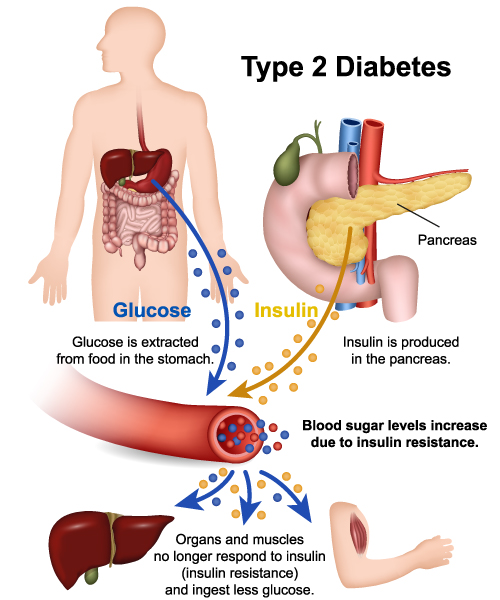
The image provides an overview of the pathophysiology of Type 2 Diabetes. In this condition, glucose is extracted from food in the stomach just as in normal metabolism, which then enters the bloodstream. The pancreas, an organ located behind the stomach, produces insulin, a hormone essential for regulating blood sugar levels by facilitating the uptake of glucose into cells for energy.
However, in Type 2 Diabetes, the body’s cells become resistant to the effects of insulin. Despite the pancreas producing insulin, the cells in the muscles and other organs do not respond effectively to it—a condition known as insulin resistance. Consequently, glucose is not adequately absorbed by these cells and remains in the bloodstream, leading to increased levels of blood sugar.
The image also shows the liver, which plays a critical role in glucose metabolism and insulin regulation. Due to insulin resistance, the liver and muscles do not take up glucose as they should, contributing to the elevated blood sugar levels that are characteristic of Type 2 Diabetes.
The condition contrasts with Type 1 Diabetes, where the pancreas produces little or no insulin, rather than an issue with the body’s response to insulin. In both types of diabetes, the failure to properly regulate blood sugar can lead to various health complications over time.
How Does Insulin Work?
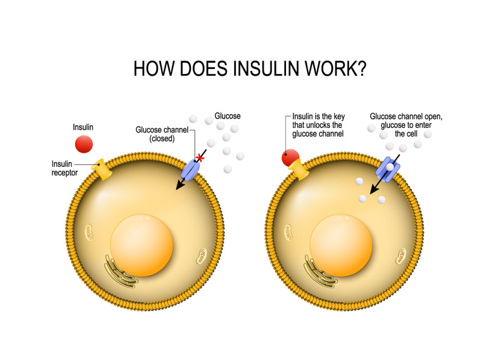
This image provides a visual explanation of the mechanism of insulin action on a cellular level. It depicts a cell with a cell membrane represented in yellow, and two states are shown side by side for comparison.
On the left side, we see a closed glucose channel with glucose molecules outside the cell unable to enter. An insulin molecule is shown hovering above an insulin receptor on the cell’s surface. This represents the state before insulin has bound to its receptor.
On the right side, the depiction changes to show the insulin molecule bound to the insulin receptor, which triggers a change in the cell membrane that allows the glucose channel to open. This illustrates how insulin acts as a key to unlock the cell, permitting glucose molecules to enter the cell. This process is vital for glucose metabolism, as it allows glucose to be taken up by cells from the bloodstream to be used for energy or stored for future use.
The comparison between the two states emphasizes the role of insulin in the regulation of blood glucose levels, and how this process is essential for maintaining homeostasis in the body. This is a critical concept in understanding how the body uses insulin to manage energy resources and the pathophysiology of conditions like diabetes where this regulatory mechanism is impaired.
Acute Pancreatitis
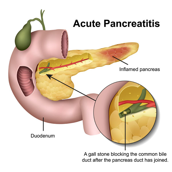
The image illustrates the condition of acute pancreatitis, which is an inflammatory process of the pancreas. The pancreas is shown with areas of redness indicating inflammation. Acute pancreatitis often presents with sudden and severe abdominal pain and can have various causes including gallstones, alcohol use, and others.
Highlighted in the image is the duodenum, the first segment of the small intestine, which closely associates with the pancreas. The pancreas secretes digestive enzymes into the duodenum through the pancreatic duct, which is depicted in green.
The inset detail shows a gallstone obstructing the common bile duct. This is significant as the common bile duct and the pancreatic duct often share a close anatomical relationship as they enter the duodenum. When a gallstone blocks the common bile duct, it can also block the pancreatic duct. This blockage can cause digestive enzymes to become trapped in the pancreas, leading to the pancreatic inflammation characteristic of acute pancreatitis.
Such a blockage is a common cause of acute pancreatitis because it prevents the normal flow of pancreatic juices and bile into the duodenum, leading to the activation of pancreatic enzymes within the pancreas itself, which starts to digest its own tissue, causing inflammation, pain, and swelling. Treatment for acute pancreatitis may involve hospitalization, pain management, fasting to rest the pancreas, and sometimes procedures to remove gallstones or correct bile duct obstructions.
Anatomical Terms and Definitions
| Term | Definition |
|---|---|
| Abdominal Aorta | The main artery that carries oxygenated blood from the heart to the lower body. |
| Acute Pancreatitis | An inflammatory process of the pancreas often caused by gallstones, alcohol use, among other causes, characterized by sudden and severe abdominal pain. |
| Adrenal Gland | An endocrine gland located above each kidney, consisting of the cortex and medulla, producing various hormones including adrenaline, noradrenaline, and cortisol. |
| Adrenaline | A hormone and neurotransmitter produced by the adrenal medulla involved in the body’s fight-or-flight response. |
| Aldosterone | A mineralocorticoid hormone produced by the zona glomerulosa of the adrenal cortex, regulating sodium and potassium balance in the body. |
| Amino Acids | Organic compounds that combine to form proteins, serving as the building blocks for protein synthesis. |
| Androgen | Sex hormones produced in the zona reticularis of the adrenal cortex, stimulating hair growth and oil production in the skin. |
| Bile | A digestive fluid produced by the liver and stored in the gallbladder, involved in the digestion and absorption of fats in the small intestine. |
| Beta Cell | A type of cell within the Islets of Langerhans in the pancreas responsible for producing insulin. |
| Blood Glucose | The main sugar found in the blood and the body’s primary source of energy. |
| Capsule | The outer protective layer of the adrenal gland, maintaining the gland's structure and protecting it from direct injury. |
| Common Bile Duct | A tube carrying bile from the gallbladder and liver to the duodenum. |
| Cortex | The outer region of the adrenal gland, divided into three zones (zona glomerulosa, zona fasciculata, zona reticularis) each producing different hormones. |
| Cortisol | A glucocorticoid hormone produced by the zona fasciculata of the adrenal cortex, involved in stress response, metabolism, and immune response. |
| Duodenum | The first section of the small intestine where the pancreatic duct and the common bile duct release their contents. |
| Estrogens | Female sex hormones produced by the zona reticularis of the adrenal cortex, influencing the female reproductive system and bone health. |
| Gallbladder | An organ that stores and concentrates bile produced by the liver. |
| Glucagon | A hormone produced by alpha cells in the Islets of Langerhans in the pancreas, raising blood glucose levels. |
| Glucose Transporter 2 (GLUT2) | A protein that facilitates the transport of glucose into cells, particularly significant in pancreatic beta cells for insulin production. |
| Glucose Uptake | The process by which cells absorb glucose from the bloodstream, facilitated by insulin. |
| Gluconeogenesis | The production of glucose from non-carbohydrate sources, stimulated by cortisol in muscles. |
| Insulin | A hormone produced by beta cells in the pancreas, regulating blood sugar levels by facilitating glucose uptake into cells. |
| Insulin Resistance | A condition where the body's cells do not respond effectively to insulin, characteristic of Type 2 Diabetes. |
| Inferior Vena Cava | A major vein carrying deoxygenated blood from the lower body to the heart. |
| Islets of Langerhans | Clusters of cells within the pancreas, containing beta cells that produce insulin and other cells producing hormones like glucagon and somatostatin. |
| Jejunum | The second part of the small intestine, responsible for the majority of nutrient absorption. |
| Medulla | The inner region of the adrenal gland producing adrenaline and noradrenaline. |
| Mineralocorticoids | Hormones produced by the zona glomerulosa of the adrenal cortex, involved in regulating the body’s salt and water balance. |
| Noradrenaline | Also known as norepinephrine, a hormone produced by the adrenal medulla causing vasoconstriction and increasing blood pressure. |
| Pancreas | A glandular organ involved in both endocrine and exocrine functions, producing digestive enzymes and hormones like insulin. |
| Pancreatic Duct | A duct running through the pancreas, allowing digestive enzymes to flow into the duodenum. |
| Pancreatic Polypeptide | A hormone produced by PP cells in the Islets of Langerhans, involved in regulating pancreatic secretions. |
| Portal Vein | A vein carrying nutrient-rich blood from the gastrointestinal tract to the liver. |
| Somatostatin | A hormone produced by delta cells in the Islets of Langerhans, regulating the secretion of other hormones. |
| Type 1 Diabetes Mellitus | An autoimmune condition where the body’s immune system destroys insulin-producing beta cells in the pancreas, leading to elevated blood glucose levels. |
| Type 2 Diabetes | A metabolic disorder characterized by insulin resistance, where the body’s cells do not respond effectively to insulin, leading to elevated blood sugar levels. |
| Ureters | Structures that transport urine from the kidneys to the bladder. |
| Zona Fasciculata | The thickest layer of the adrenal cortex, responsible for producing glucocorticoids like cortisol. |
| Zona Glomerulosa | The outermost layer of the adrenal cortex, producing mineralocorticoids like aldosterone. |
| Zona Reticularis | The innermost layer of the adrenal cortex, producing sex hormones, particularly androgens. |
