
Muscles of the Shoulder and Upper Arm
Supraspinatus
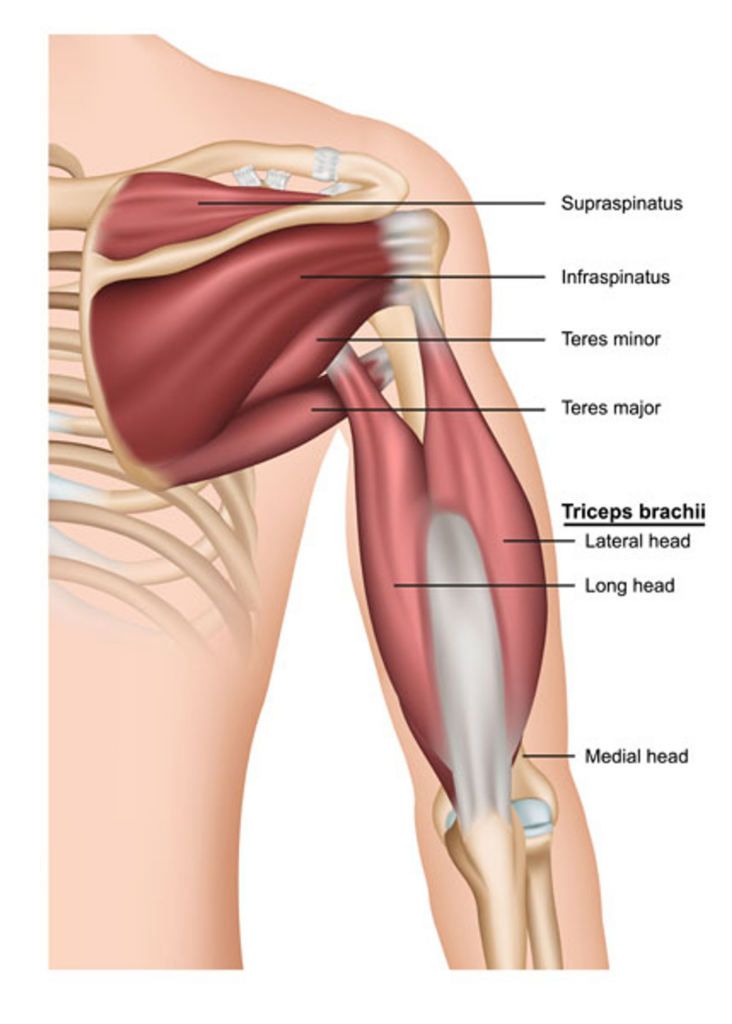
The Supraspinatus is one of the four muscles that make up the rotator cuff of the shoulder joint. It plays a vital role in stabilizing the shoulder joint and enabling shoulder movements.
The Supraspinatus muscle originates from the supraspinous fossa of the scapula, which is a depression located on the dorsal side of the scapula, above the spine. The muscle fibers travel laterally and converge into a tendon, which passes under the acromion process of the scapula, crosses the shoulder joint, and inserts onto the greater tubercle of the humerus.
This muscle is specifically responsible for the first 15 degrees of arm abduction, which is the movement of the arm away from the body in the coronal plane. After this, the deltoid muscle takes over the movement of abduction. The Supraspinatus also helps in preventing downward dislocation of the humerus, and thus plays a crucial role in maintaining shoulder joint stability.
Innervation of the Supraspinatus muscle comes from the suprascapular nerve, a branch of the upper trunk of the brachial plexus. Its blood supply is via the suprascapular artery, a branch of the thyrocervical trunk.
The Supraspinatus muscle is susceptible to injuries, often due to overuse or acute trauma. This could result in a condition commonly known as a rotator cuff tear. Symptoms may include pain, weakness, and limited range of motion. If you suspect any injury, seek medical attention promptly. To maintain the health of the Supraspinatus and other shoulder muscles, regular stretching and strengthening exercises should be incorporated into your fitness routine.
Infraspinatus
The Infraspinatus is another essential muscle within the rotator cuff of the shoulder joint, playing a key role in stabilizing this joint and facilitating certain movements of the upper arm.
The Infraspinatus muscle originates from the infraspinous fossa of the scapula, a depression located below the spine of the scapula on its posterior aspect. The muscle fibers converge to form a tendon that crosses the shoulder joint to insert on the middle facet of the greater tubercle of the humerus.
The primary function of the Infraspinatus muscle is to externally (or laterally) rotate the arm. Moreover, it assists in horizontal abduction when the arm is at 90-degree abduction. This muscle also plays a significant role in stabilizing the shoulder joint by keeping the head of the humerus firmly within the glenoid cavity of the scapula.
Like the Supraspinatus, the Infraspinatus is innervated by the suprascapular nerve, a branch of the brachial plexus. Its blood supply primarily comes from the suprascapular and circumflex scapular arteries.
Injuries to the Infraspinatus muscle, such as strains or tears, are not uncommon and often result from overuse, especially in athletes who perform repetitive shoulder movements. Symptoms may include pain, especially during external rotation of the shoulder, weakness, and reduced range of motion. In case of any such symptoms, it is crucial to seek medical attention. Regular stretching and strength-building exercises targeting the shoulder can help maintain the health and function of the Infraspinatus muscle.
Teres Minor
The Teres Minor is one of the four muscles of the rotator cuff, a group of muscles and their tendons that provide stability and facilitate movement at the shoulder joint. This small yet vital muscle plays a significant role in upper body mobility and strength.
Anatomically, the Teres Minor muscle originates from the upper two-thirds of the lateral border of the scapula, the bone that connects the humerus (bone of the upper arm) with the clavicle (collar bone). From there, the muscle fibers extend upward and laterally to insert at the lower part of the greater tubercle of the humerus.
The Teres Minor and Infraspinatus muscles work together to provide external (lateral) rotation of the arm at the shoulder joint. Additionally, the Teres Minor assists in adduction and extends the arm when the arm is raised. This muscle, therefore, plays an important role in various actions involving the arms and shoulders, such as throwing a ball, swimming, or lifting an object.
The axillary nerve, a branch of the brachial plexus, provides innervation to the Teres Minor muscle. This nerve also supplies the deltoid muscle, another key muscle in shoulder movement. The arterial blood supply to the Teres Minor muscle comes from the posterior circumflex humeral artery and the subscapular artery.
Injury to the Teres Minor muscle can result from overuse or sudden, forceful movements, leading to pain, limited range of motion, and reduced strength in the affected shoulder. If any such symptoms are noticed, it is recommended to seek professional medical help. Regular, guided exercise can help strengthen this muscle and maintain the overall health of the shoulder joint.
Teres Major
The Teres Major muscle is a thick, rounded muscle situated in the shoulder region, more specifically in the scapular region. It's commonly referred to as the "little helper" due to its assistance in the movements performed by the latissimus dorsi, a larger, more powerful muscle of the back.
Anatomically, the Teres Major originates from the posterior aspect of the scapula, specifically the lower part of the lateral border, and inserts onto the medial lip of the intertubercular sulcus of the humerus, the long bone in the upper arm. It is important to note that although the Teres Major is located near the rotator cuff muscles, it is not a part of this group.
Functionally, the Teres Major is responsible for several movements of the shoulder joint. It assists in the internal rotation (medial rotation) and adduction of the humerus, meaning it helps to rotate the arm inward and pull it toward the body. Additionally, it assists in the extension of the humerus, helping to pull the arm backward when it is raised.
The Teres Major muscle receives its innervation from the lower subscapular nerve, which is a branch of the brachial plexus, and its blood supply from the subscapular and circumflex scapular arteries.
Injuries to the Teres Major, though uncommon, can occur as a result of overuse or excessive strain, often in activities involving the repetitive or forceful use of the arm. This can result in pain and a decreased range of motion in the shoulder. Strengthening and stretching exercises, when done properly, can help maintain the health of the Teres Major and the overall stability and function of the shoulder joint.
Triceps Brachii
The triceps brachii, often referred to simply as the triceps, is a large muscle located at the back of the upper arm. Its primary function is to facilitate the extension of the elbow joint, a crucial movement in many daily activities such as pushing and throwing. The name "triceps brachii" reflects its three-part structure, with "tri-" meaning three and "ceps" meaning head.
This muscle consists of three distinct heads: the long head, the lateral head, and the medial head. Each head originates from a different location but they all come together to insert at the same point. The long head of the triceps brachii originates from the infraglenoid tubercle of the scapula, the lateral head originates from the posterior surface of the humerus above the radial groove, and the medial head originates from the posterior surface of the humerus below the radial groove. All three heads converge to insert onto the olecranon process of the ulna, which is the prominent bony projection of the elbow.
Each head of the triceps brachii muscle contributes to the overall function of the muscle, but they also have unique roles. The long head, in addition to helping extend the elbow, assists with the extension and adduction of the arm at the shoulder joint because of its origin point on the scapula. The lateral and medial heads are more involved in the extension of the forearm at the elbow joint.
Innervation to the triceps brachii muscle is provided by the radial nerve, a major peripheral nerve of the upper limb that originates from the brachial plexus. Blood supply to the muscle is provided by branches of the deep brachial artery and the superior ulnar collateral artery.
Understanding the anatomy of the triceps brachii is important for fitness and rehabilitation purposes. Strengthening the triceps can enhance the stability of the elbow joint and improve upper body strength. It's important to note that overuse or strain of the triceps can lead to injury, so proper form during exercise and adequate rest and recovery are essential.
Lateral Head
The lateral head of the triceps brachii is one of the three heads that collectively form the triceps muscle. It's located on the outer side of the upper arm, providing the triceps brachii with its distinctive three-headed appearance. Despite sharing common functionalities with the other two heads (medial and long), each head has unique characteristics and roles within the context of the muscle's overall function.
The lateral head originates from the posterior surface of the humerus, specifically the area above the radial groove. It joins the other two heads of the triceps to insert onto the olecranon process of the ulna at the elbow, forming a common tendon. The lateral head is often considered to be the most visible of the three heads when looking at the arm from an anterior view, as it gives the triceps its width.
In terms of function, the lateral head plays a crucial role in the extension of the forearm at the elbow joint. This is the primary function of the triceps brachii muscle as a whole, but the lateral head is particularly involved in providing powerful extension, especially when the arm is in a state of abduction or movement away from the center of the body.
The lateral head, like the other heads of the triceps brachii muscle, is innervated by the radial nerve. This nerve originates from the brachial plexus and is responsible for both motor and sensory innervation of the arm and forearm. Blood supply to the lateral head is provided by branches of the deep brachial artery, a major artery that serves the muscles of the arm.
In order to keep the lateral head and the entire triceps brachii muscle healthy and strong, it's important to engage in regular strength training exercises targeting this area, such as tricep pushdowns or tricep kickbacks. However, as with any physical activity, proper technique and form should be prioritized to avoid potential injuries.
Long Head
The long head of the triceps brachii, often simply referred to as the "long head", is one of the three heads of the triceps brachii muscle, situated in the upper arm. The triceps brachii's primary function is to extend the forearm at the elbow joint, but the long head has some additional functions due to its unique origin and placement within the muscle group.
The long head originates from the infraglenoid tubercle of the scapula, making it the only head of the triceps that attaches to the scapula. From its origin, it runs down the back of the arm, sharing a common tendon with the other two heads (lateral and medial) to insert onto the olecranon process of the ulna at the elbow.
The position of the long head gives it a role not only in the extension of the forearm at the elbow but also in the adduction and extension of the arm at the shoulder joint. This makes it functionally distinct within the triceps group, as the other two heads have no action at the shoulder joint.
Like the rest of the triceps brachii, the long head is innervated by the radial nerve. This nerve arises from the brachial plexus and carries signals from the brain to the muscles, allowing for voluntary movement. Blood supply to the long head is provided by the deep brachial artery and the superior ulnar collateral artery.
Exercises that specifically target the long head of the triceps include overhead triceps extensions and close-grip bench press. As with any exercise, it is important to maintain proper form and technique to prevent injury. Regular stretching can also help maintain flexibility and joint health.
Medial Head
The medial head of the triceps brachii, also referred to as the "medial head", is one of the three parts of the triceps brachii muscle, located in the upper arm. The triceps brachii's primary role is to extend the forearm at the elbow joint, but each of its heads, including the medial head, has unique characteristics due to its distinct location and structure.
The medial head of the triceps brachii originates from the posterior surface of the humerus, specifically the area inferior to the radial groove and extending to the medial intermuscular septum and lateral intermuscular septum. Its location is deeper than the long and lateral heads, sitting closer to the humerus. It converges with the other two heads of the triceps – the long and lateral – to insert onto the olecranon process of the ulna at the elbow.
Functionally, while the medial head is involved in the general action of the triceps brachii, which is the extension of the forearm at the elbow joint, it's believed to have a special role in providing fine control and low-level force for actions such as writing.
The medial head, like the other two heads, is innervated by the radial nerve, which is responsible for carrying signals from the brain to the muscle to allow for coordinated, voluntary movement. The deep brachial artery, a branch of the brachial artery, provides the blood supply to the medial head.
Exercises that work the medial head include close-grip push-ups and lying triceps extensions, also known as skull crushers. Always remember to use proper form and technique to prevent injury. Regular stretching can also aid in maintaining flexibility and joint health.