
Anatomy of Kidneys
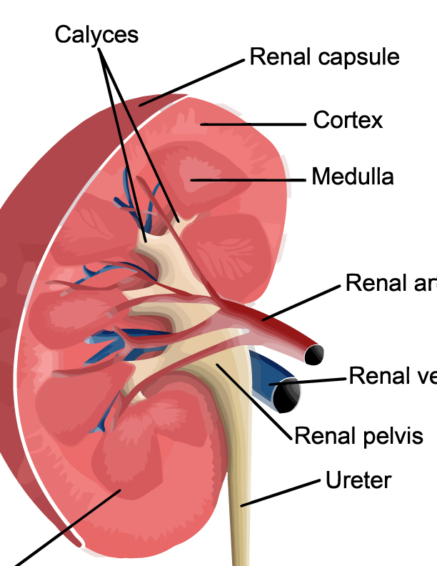
Starting from the outside, the renal capsule is a smooth, transparent sheet of dense irregular connective tissue that is continuous with the outer coat of the ureter. This capsule acts as a protective barrier against physical damage and infection, while also maintaining the kidney’s shape.
Just beneath the renal capsule is the cortex. This is the outermost layer of the kidney where blood filtration takes place. The cortex appears granulated due to the presence of numerous nephrons, the microscopic units where blood plasma is filtered. Each nephron in the cortex consists of a renal corpuscle and a renal tubule, where the blood plasma is filtered and then modified to form urine.
Deeper within the kidney is the medulla, comprised of 10 to 18 renal pyramids. These pyramids are made up of bundles of microscopic tubules and blood vessels, which create a striated appearance. The apices of these pyramids, known as renal papillae, point internally towards the calyces. The medulla’s function is to concentrate urine and conserve water, a process regulated by a complex counter-current exchange system.
The calyces are cup-shaped structures that collect urine from the renal pyramids. There are minor calyces that enclose the papillae of the pyramids, and several minor calyces converge to form a major calyx. These structures channel the urine they collect into the renal pelvis.
The renal pelvis is the central collecting region within the kidney, which then narrows to form the ureter. The walls of the renal pelvis and the ureter contain smooth muscle that contracts rhythmically to propel urine by peristalsis from the kidney to the bladder.
The renal artery, which enters the kidney at the hilum, branches off into smaller and smaller arteries until they become arterioles and finally form a capillary network around the nephrons. After the blood is filtered, the renal vein collects the cleansed blood from the capillaries and returns it to the circulation.
This image represents an intricate system where each component plays a specific role in the kidney’s function of filtering blood, maintaining fluid and electrolyte balance, and excreting waste products through urine formation. The precise architecture ensures that the kidneys can effectively carry out these vital processes.
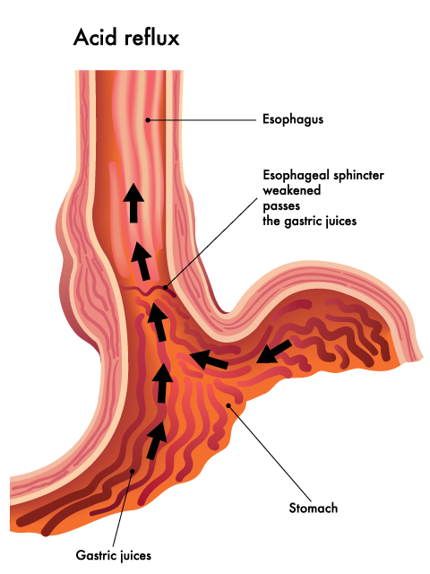
The illustration provides a visual representation of the physiological process of acid reflux, a condition where acidic gastric contents from the stomach are allowed to flow back into the esophagus. Here’s a detailed description of the components and processes depicted in the image:
- Esophagus: This is a muscular tube connecting the throat (pharynx) with the stomach. In the image, it is depicted as the vertical structure through which the black arrows are ascending, indicating the direction of the gastric juices’ movement.
- Esophageal Sphincter: Normally functioning as a valve between the esophagus and stomach, the esophageal sphincter is shown as a weakened structure in the image. It is labeled where the esophagus meets the stomach, and its compromised state is indicated, suggesting it is allowing the passage of substances that should typically be contained within the stomach.
- Stomach: Shown at the bottom of the image, the stomach is the organ that receives food from the esophagus. It secretes gastric juices, which are necessary for the digestion of food.
- Gastric Juices: These are depicted as a dark, wavy mass at the base of the esophagus, and within the stomach. The arrows emanating from this mass illustrate the reflux of these acidic juices.
- Direction of Gastric Juice Flow: The series of black arrows starting in the stomach and moving upward into the esophagus illustrates the abnormal path of the gastric juices. This reflux is caused by the weakening of the esophageal sphincter, which is failing to prevent the backward flow.
The esophagus is a fibromuscular tube, approximately 25 centimeters in length in adults, which serves as a conduit for food and liquids that have been swallowed into the mouth to reach the stomach. Its primary function is to transport what is ingested from the back of the throat, through the chest cavity, and into the stomach. It does this through coordinated, sequential muscle contractions known as peristalsis. These contractions are involuntary and move in a wave-like pattern to ensure the one-way movement of food.
The stomach, located just below the esophagus, is a muscular organ that plays a central role in the digestive process. It performs several functions, including the temporary storage of food, mechanical breakdown of food through muscle contractions, and chemical digestion of food using gastric juices. These juices are composed of hydrochloric acid and digestive enzymes, which collectively create an acidic environment designed to denature proteins and facilitate the breakdown of complex substances.
To prevent the acidic contents of the stomach from moving back up into the esophagus, a specialized ring of muscle known as the lower esophageal sphincter (LES) is located at the junction of the esophagus and stomach. The LES acts as a one-way valve, opening to allow food to pass into the stomach and closing to prevent gastric acids and food from refluxing into the esophagus.
In the case of acid reflux, the LES may not function properly. It may not close completely or may open too frequently, allowing acidic gastric contents to escape into the esophagus. This backflow of acid can irritate and damage the lining of the esophagus, causing symptoms such as a burning sensation in the chest or throat, known as heartburn. If acid reflux happens frequently, it can lead to a condition called gastroesophageal reflux disease (GERD).
Understanding these functions is critical for recognizing the importance of the LES’s role in preventing acid reflux and the potential consequences when its function is compromised.