
1914 Sutures of the Skull
Bones of the Skull:
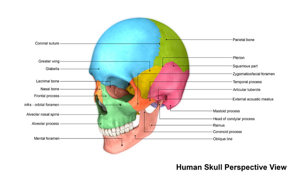
The human skull is a complex structure composed of various bones that protect the brain, support the face, and provide attachment sites for muscles. Here is a detailed description of the bones of the human skull:
Frontal Bone: The frontal bone forms the forehead and the upper part of the eye sockets. It extends backward to join the parietal bones and forms the roof of the orbits.
Parietal Bones: There are two parietal bones, one on each side of the skull. They form the sides and roof of the cranium and join at the midline through the sagittal suture.
Occipital Bone: The occipital bone is located at the posterior aspect of the skull. It forms the base of the cranium and has a large opening called the foramen magnum, through which the spinal cord passes.
Temporal Bones: There are two temporal bones, one on each side of the skull. They are situated at the lower sides of the cranium. The temporal bones house the ear structures and contribute to the sides and base of the skull.
Sphenoid Bone: The sphenoid bone is a complex bone that is located in the middle of the skull. It forms part of the base of the cranium, the sides of the skull, and the orbits. It has a central depression called the sella turcica, which houses the pituitary gland.
Ethmoid Bone: The ethmoid bone is located in the front of the skull, between the eyes. It contributes to the walls of the orbits, the nasal cavity, and the nasal septum. It contains thin, delicate plates called cribriform plates that allow the passage of olfactory nerves.
Nasal Bones: The nasal bones are two small bones that form the bridge of the nose. They articulate with the frontal bone superiorly and the maxillary bones inferiorly.
Maxilla: There are two maxillary bones, one on each side of the face. They form the upper jaw and contribute to the floor of the orbits, the roof of the mouth, and the sides of the nasal cavity.
Mandible: The mandible, commonly known as the jawbone, is the largest and strongest facial bone. It forms the lower jaw and contains the lower teeth. It articulates with the temporal bones, allowing for jaw movement.
Zygomatic Bones: The zygomatic bones, also known as the cheekbones, form the prominence of the cheeks. They articulate with the frontal bone, temporal bone, maxilla, and sphenoid bone.
Lacrimal Bones: The lacrimal bones are the smallest bones of the face. They are located within the medial walls of the orbits and contribute to the formation of tear ducts.
Palatine Bones: The palatine bones form the posterior part of the hard palate, which separates the oral and nasal cavities.
Vomer: The vomer is a thin, flat bone that forms the inferior portion of the nasal septum, dividing the nasal cavity into left and right sides.
Inferior Nasal Conchae: There are two inferior nasal conchae, one on each side of the nasal cavity. They are thin, curved bones that help create turbulence in the airflow, aiding in the warming and humidification of inhaled air.
Understanding the structure and function of the bones of the human skull is important in fields such as anatomy, anthropology, neurology, and oral and maxillofacial surgery.
Sutures:
The human skull is a complex structure comprising several bones that are connected by specialized joints called sutures. Sutures are fibrous connective tissue joints that allow for flexibility during childbirth and enable the skull bones to grow and accommodate the developing brain. Here is a detailed instruction of the major sutures of the human skull:
Coronal Suture: The coronal suture is located on the superior aspect of the skull and runs horizontally from one side to the other. It joins the frontal bone at the front with the two parietal bones on the sides.
Sagittal Suture: The sagittal suture is positioned on the midline of the skull and extends vertically from the anterior (front) to the posterior (back) direction. It connects the two parietal bones together.
Lambdoid Suture: The lambdoid suture is located at the posterior aspect of the skull, forming an inverted “V” shape. It joins the two parietal bones with the occipital bone.
Squamous Sutures: The squamous sutures, also known as the temporal sutures, are located on the lateral sides of the skull. They connect the squamous portion of the temporal bone with the parietal bone above it. There are two squamous sutures, one on each side of the skull.
Frontonasal Suture: The frontonasal suture is positioned at the midline of the skull between the frontal bone and the nasal bones. It connects the upper part of the nasal bones with the frontal bone.
Sutural Bones (Wormian Bones): Sutural bones, also called wormian bones, are small irregularly shaped bones that can sometimes be found within sutures. They are additional bones that develop in the joints between the normal skull bones and are most commonly seen in the lambdoid suture.
Understanding the location and characteristics of these sutures is important for various medical fields such as anatomy, neurosurgery, and craniofacial surgery. It is worth noting that the sutures of the human skull fuse gradually over time as an individual grows, becoming less flexible and eventually forming a solid, immovable structure.
What is the Squamous part of the skull bones?
The squamous part of the skull bones refers to the flat, scale-like portions of certain bones in the skull. Specifically, the squamous part is present in the temporal bones and the parietal bones.
In the temporal bones, the squamous part refers to the thin, flattened region that forms the lateral side of the skull. It is located just above the external ear and contributes to the sides and base of the skull. The squamous part of the temporal bone is important because it houses several structures, including the middle and inner ear structures and the temporomandibular joint.
In the parietal bones, the squamous part refers to the large, flat regions on the sides and roof of the cranium. They are positioned between the frontal bone at the front and the occipital bone at the back. The parietal bones contribute to the overall protection of the brain and also provide attachment sites for various muscles.
The term “squamous” is used to describe the flat, scale-like appearance of these portions of the bones. The squamous parts of the temporal and parietal bones play important roles in the structural integrity and functionality of the skull.
A more detailed look at sutures:
Skull sutures are developed as a part of the natural growth and development of the human skull. In early infancy and childhood, the skull bones are not fully fused, allowing for flexibility and accommodating the rapid growth of the brain. As the individual grows, the bones gradually fuse together at the sutures, creating a solid structure.
During skull development, specialized connective tissue called sutural mesenchyme fills the gaps between the developing skull bones. Over time, the sutural mesenchyme differentiates and transforms into dense fibrous connective tissue, forming the sutures. These sutures consist of fibrous tissue composed of collagen fibers, proteoglycans, and other extracellular matrix components.
Within the sutures, there are small extensions of connective tissue called Sharpey’s fibers. These fibers are collagenous strands that penetrate the bone tissue and anchor the suture firmly in place. Sharpey’s fibers are named after William Sharpey, a Scottish anatomist who first described them. These fibers enhance the stability and strength of the suture by interweaving with the bone tissue and distributing forces more evenly.
As an individual ages, the sutures gradually ossify, meaning the bones fuse together. This process, called cranial suture closure or cranial fusion, typically occurs by the late teens or early twenties. Once the sutures have fused, the skull becomes a rigid structure, protecting the brain and providing structural support.
Understanding the development and characteristics of skull sutures is important in various fields, such as craniofacial surgery, neurosurgery, and anthropology. The fusion patterns and closure timing of sutures can provide valuable information about growth and development, as well as aid in diagnosing certain craniofacial abnormalities or conditions.
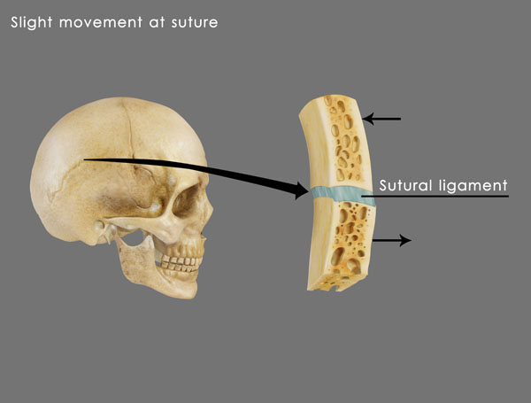
What is a sutural ligament?
A sutural ligament, also known as an interosseous ligament or an intermaxillary ligament, refers to a type of fibrous connective tissue that helps to stabilize and strengthen the joints between certain bones of the skull. These ligaments are primarily found within the sutures, which are fibrous joints that connect the individual bones of the cranium.
Sutural ligaments are composed of collagen fibers and other extracellular matrix components, similar to other ligaments in the body. They are situated within the sutures and run between the bony edges of adjacent skull bones, providing support and maintaining the alignment and stability of the bones.
The primary function of sutural ligaments is to help distribute the forces exerted on the skull during activities such as chewing, speaking, and facial expressions. They aid in absorbing and dissipating the mechanical stresses that occur at the sutures, contributing to the overall strength and integrity of the cranial vault.
It is worth noting that sutural ligaments differ from other ligaments in the body in terms of their location and structure. While most ligaments connect bones at joints, sutural ligaments are specifically found within the sutures of the skull, joining the individual cranial bones together.