
1911 Upper Arm and Shoulder Kinesiology
Muscles
The muscles of the forearm are complex and versatile, contributing to a wide range of movements. From a kinesiology standpoint, these movements are critical for various functions of the hand and wrist.
Each of these movements is crucial for the functional use of the hand and wrist. The coordination of these muscles allows for complex and precise activities, from gripping and lifting objects to performing delicate tasks requiring fine motor skills. The interplay of these muscular actions in response to nerve signals makes the forearm a fascinating area of study in kinesiology and related fields.
Here’s a detailed overview of the movements provided by the muscles of the forearm:
Flexion and Extension:
These are the primary movements of the wrist. Flexor muscles like the flexor carpi radialis, flexor carpi ulnaris, and palmaris longus are responsible for bending the wrist forward (flexion). Extensor muscles like the extensor carpi radialis longus, extensor carpi radialis brevis, and extensor carpi ulnaris straighten the wrist (extension).
Flexion and extension of the wrist are fundamental movements that involve several muscles in the forearm, each playing a specific role in controlling the motion and stability of the wrist joint. Let’s expand on these concepts:
Flexion Muscles:
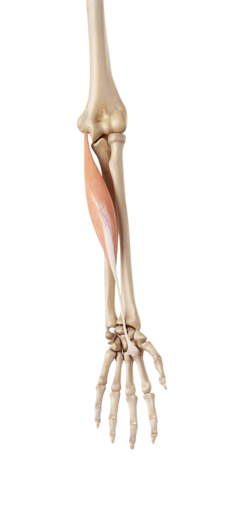
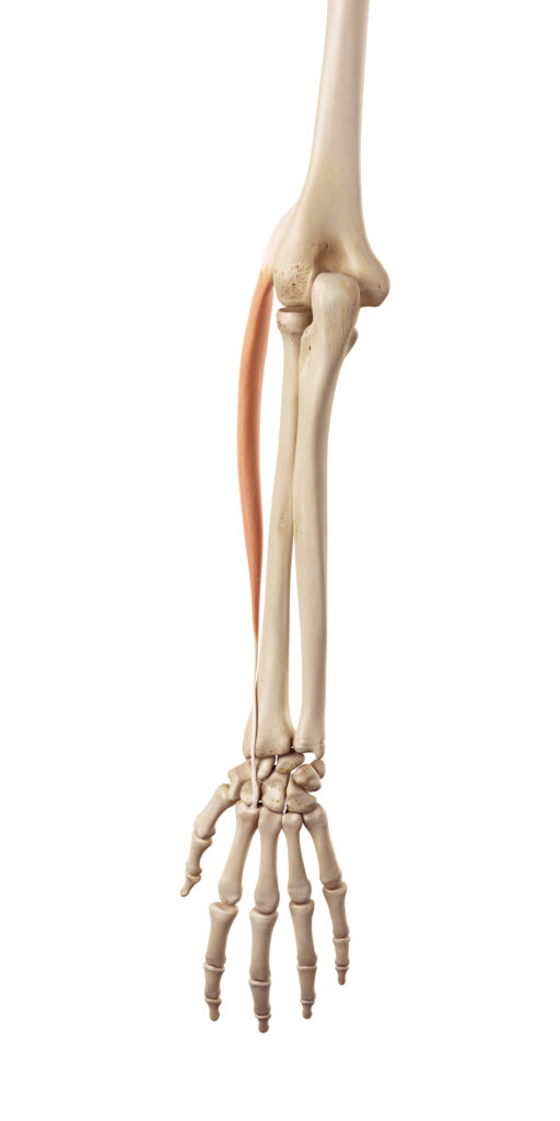
Flexor Carpi Radialis: Originating from the medial epicondyle of the humerus, this muscle runs along the forearm on the thumb side. When it contracts, it flexes and abducts the wrist, pulling it towards the thumb side.
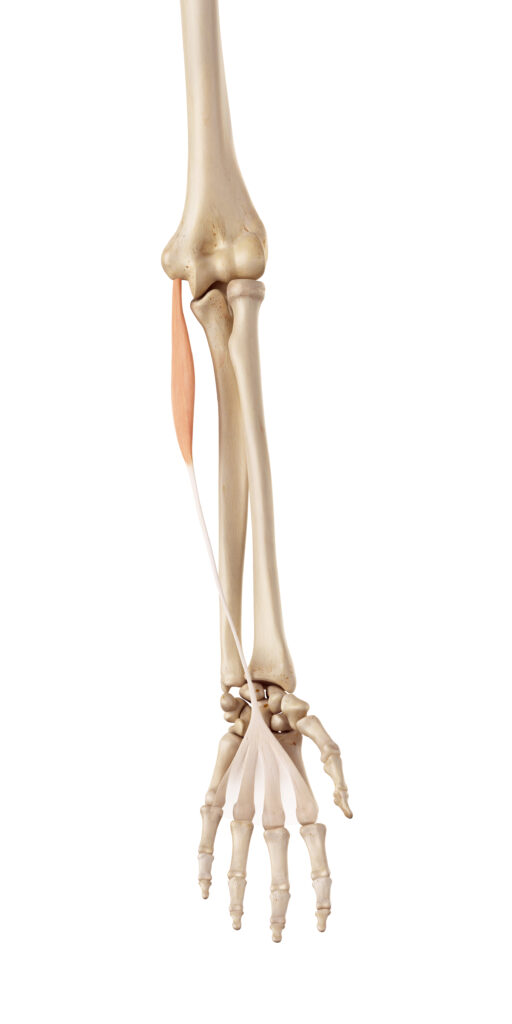
Flexor Carpi Ulnaris: This muscle also originates from the medial epicondyle but is positioned on the little finger side of the forearm. It works in concert with the flexor carpi radialis to flex the wrist but also adds a component of ulnar deviation, pulling the wrist towards the little finger side.
Palmaris Longus: Not present in all individuals, this muscle is a more superficial muscle, stretching from the medial epicondyle to the palm. It’s a weak flexor of the wrist but plays a more significant role in tensing the palmar aponeurosis, which can affect the flexor tendons in the hand.
Extension Muscles:

Extensor Carpi Radialis Longus and Brevis: These muscles originate from the lateral epicondyle of the humerus. They extend and abduct the wrist, pulling it back and towards the thumb side. The longus is more effective in wrist abduction, while the brevis is more effective in extension.
Extensor Carpi Ulnaris: It also has its origin at the lateral epicondyle and extends down the ulnar side of the forearm. This muscle extends and adducts the wrist, moving it back and towards the little finger side.
Biomechanical Aspects
- Synergistic Action: For smooth, controlled wrist movements, these muscles often work in synergy. For instance, during activities requiring precise wrist positioning, such as typing or playing a musical instrument, both flexors and extensors subtly adjust their tension to stabilize the wrist.
- Antagonistic Control: When one group of muscles contracts, the opposing group (antagonists) relaxes. This reciprocal innervation is crucial for smooth motion and prevents conflict between muscles.
- Force Distribution: The muscles are arranged to distribute force evenly across the wrist, ensuring that no single tendon bears excessive load, which could lead to injury.
- Nerve Supply: The flexors are primarily innervated by the median and ulnar nerves, while the extensors are innervated by the radial nerve. Any nerve impairment can significantly affect these movements.
Understanding the mechanics of flexion and extension is crucial in fields such as physical therapy, sports medicine, and rehabilitation, where knowledge of these actions helps in diagnosing and treating wrist injuries or conditions. For instance, repetitive strain injuries, often seen in athletes and office workers, can be better understood and managed by knowing the roles and mechanics of these muscles.
Pronation and Supination:
Pronation is the inward rotation of the forearm, which turns the palm downward or backward. Supination is the outward rotation, turning the palm upward or forward. The pronator teres and pronator quadratus are involved in pronation, while the supinator and biceps brachii assist in supination.
Pronation and supination are rotational movements of the forearm that are vital for the functional use of the hand, allowing it to adopt various orientations. These movements are facilitated by a group of muscles, each contributing to the complexity and range of motion. Let’s delve deeper into these aspects:
Pronation Muscles:
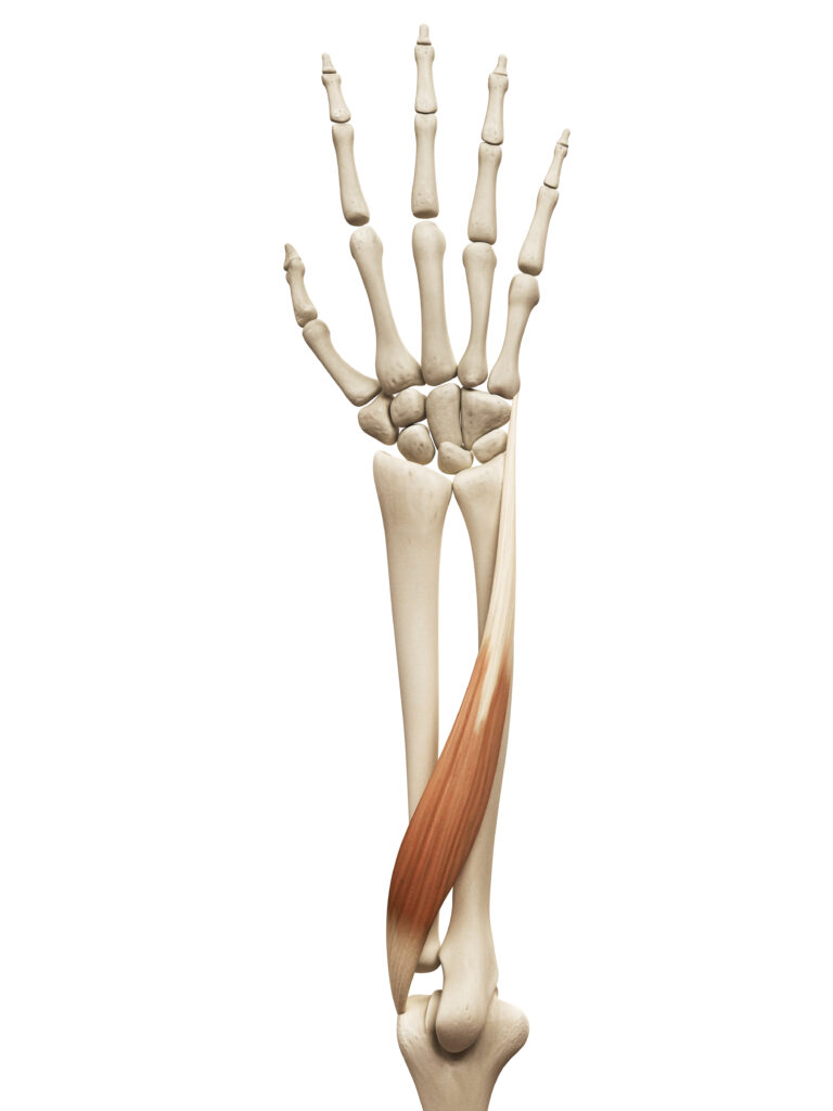
Pronator Teres: This muscle has two heads – a humeral head and an ulnar head. It originates from the medial epicondyle of the humerus and the coronoid process of the ulna, and inserts into the middle of the radius. When it contracts, it rotates the radius over the ulna, turning the palm downwards or backwards (pronation). This muscle is also active during elbow flexion.
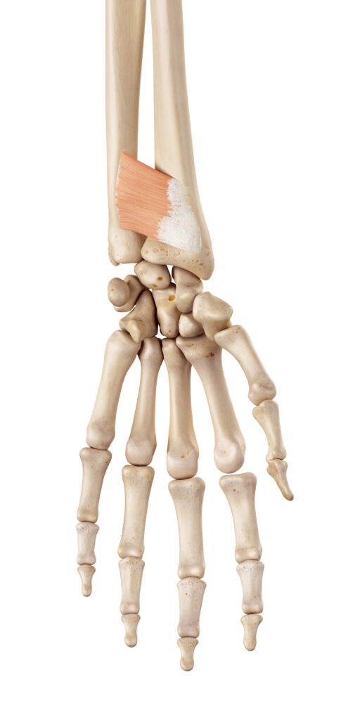
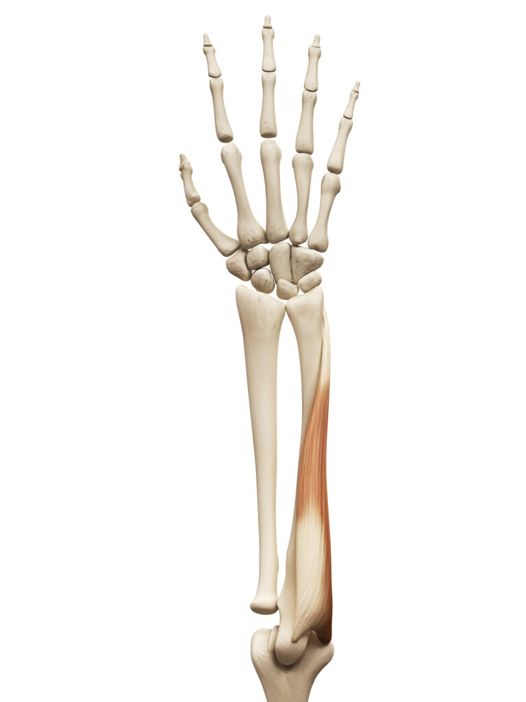
Pronator Quadratus: A deeper, square-shaped muscle, it is located near the wrist, stretching between the distal end of the ulna and the distal end of the radius. It primarily acts to pronate the forearm, especially when the elbow is extended. Its position and shape make it ideally suited for stabilizing the distal radioulnar joint during pronation.
Supination Muscles:
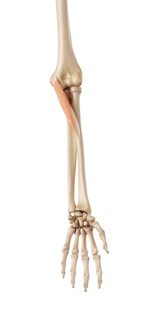
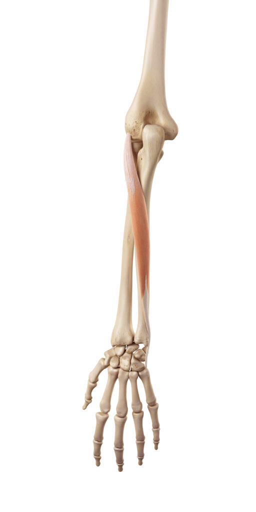
Supinator: This muscle is deep in the posterior compartment of the forearm. It wraps around the upper third of the radius, originating from the lateral epicondyle of the humerus and the ulnar collateral ligament, and inserts into the proximal radius. Its primary function is to supinate the forearm, turning the palm upwards or forwards. It’s most effective when the elbow is in an extended position.
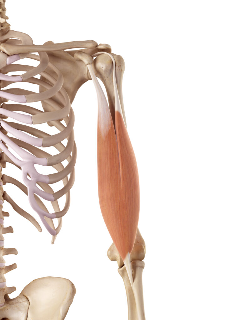
Biceps Brachii: Although primarily known for its role in flexing the elbow, the biceps brachii also plays a crucial role in supination. Its attachment to the radial tuberosity allows it to rotate the radius when it contracts, especially when the elbow is flexed. This dual role makes the biceps brachii a powerful supinator of the forearm.
Biomechanical Considerations
- Joint Involvement: The movements of pronation and supination occur primarily at the proximal and distal radioulnar joints, where the radius and ulna articulate with each other.
- Range of Motion: The average range of pronation and supination in a healthy adult is about 80°-90° in each direction. This range can vary based on individual anatomy and flexibility.
- Functional Significance: These movements are essential for numerous daily activities, such as turning a doorknob, using a screwdriver, or positioning the hand for typing or writing.
- Injury and Rehabilitation: Injuries to the muscles involved in pronation and supination, or to the nerves that innervate them, can significantly impair hand function. Conditions like biceps tendonitis, pronator teres syndrome, or radial nerve injury can disrupt these movements. Understanding the mechanics of these movements is crucial in diagnosing and treating such conditions.
In summary, pronation and supination are complex movements facilitated by a coordinated action of specific muscles. They are essential for the versatile movement of the hand and forearm, enabling a wide range of activities and contributing to the dexterity of the upper limb.
Radial and Ulnar Deviation:
Radial deviation involves moving the wrist towards the thumb side, facilitated by muscles like the extensor carpi radialis longus and brevis. Ulnar deviation is moving the wrist towards the little finger side, aided by the extensor carpi ulnaris and flexor carpi ulnaris.
Radial and ulnar deviation are movements of the wrist that involve lateral movement along the horizontal plane. These movements allow the hand to move towards the thumb side (radial deviation) or towards the little finger side (ulnar deviation), playing a critical role in the wide range of hand and wrist motions required for daily activities. Let’s delve into the specifics of these movements:
Radial Deviation Muscles:
Extensor Carpi Radialis Longus and Brevis: These muscles are located on the back of the forearm. The longus is more proximal, and the brevis is located slightly distal to it. Both originate from the lateral epicondyle of the humerus and insert into the bases of the metacarpal bones of the hand (second and third metacarpal for longus and brevis, respectively). When these muscles contract, they extend the wrist but also abduct it towards the thumb side, causing radial deviation. They play an essential role in activities like writing and gripping.

Additional Contributors: While the extensor carpi radialis longus and brevis are the primary movers, other muscles such as the abductor pollicis longus and extensor pollicis brevis also assist in radial deviation, particularly when the thumb is involved in the motion.
Ulnar Deviation Muscles:
Extensor Carpi Ulnaris: This muscle also originates from the lateral epicondyle of the humerus and inserts into the base of the fifth metacarpal. Its primary action is to extend the wrist, but it also adducts it towards the ulnar side, contributing to ulnar deviation. This muscle is particularly active during movements that require a firm grip or during actions that require applying force with the hand in an ulnarly deviated position, such as using a hammer.
Flexor Carpi Ulnaris: Originating from the medial epicondyle of the humerus and the olecranon of the ulna, this muscle inserts into the pisiform, hamate, and fifth metacarpal bones. It acts primarily to flex the wrist but also adducts it towards the ulnar side. This muscle works in conjunction with the extensor carpi ulnaris to produce ulnar deviation.
Biomechanical Aspects
- Synergistic Action: For smooth radial and ulnar deviation, these muscles often work in synergy with other forearm muscles. This coordinated action ensures balance and stability of the wrist joint during these lateral movements.
- Functional Importance: Radial and ulnar deviations are essential for tasks that require complex hand and wrist positioning, such as typing, playing musical instruments, or performing certain sports actions (like a tennis backhand).
- Range of Motion: The typical range of motion for radial deviation is about 15°, while for ulnar deviation, it is approximately 30°. This difference in range is partly due to the anatomical structure of the wrist and forearm bones.
- Role in Pathologies: Repetitive or excessive radial and ulnar deviation can lead to overuse injuries like tendinitis or carpal tunnel syndrome. Understanding these movements is crucial in occupational health, ergonomics, and in the rehabilitation of wrist injuries.
In summary, radial and ulnar deviation are essential movements for the functional versatility of the wrist and hand. They are facilitated by a group of muscles that work in concert, allowing for the complex and precise manipulation of objects and execution of various tasks involving hand and wrist movements.
Finger Movements:
The muscles in the forearm also play a crucial role in finger movements. Flexor digitorum superficialis and profundus, along with extensor digitorum and extensor indicis, are responsible for the flexion and extension of the fingers. Additionally, muscles like the flexor pollicis longus and extensor pollicis longus and brevis contribute to thumb movements.
Finger movements are fundamental for a vast array of everyday activities, from gripping objects to typing. The forearm houses several key muscles that control these movements through intricate tendons extending into the hand. Let’s delve deeper into these muscles and their roles in finger movements:
Flexion of the Fingers:
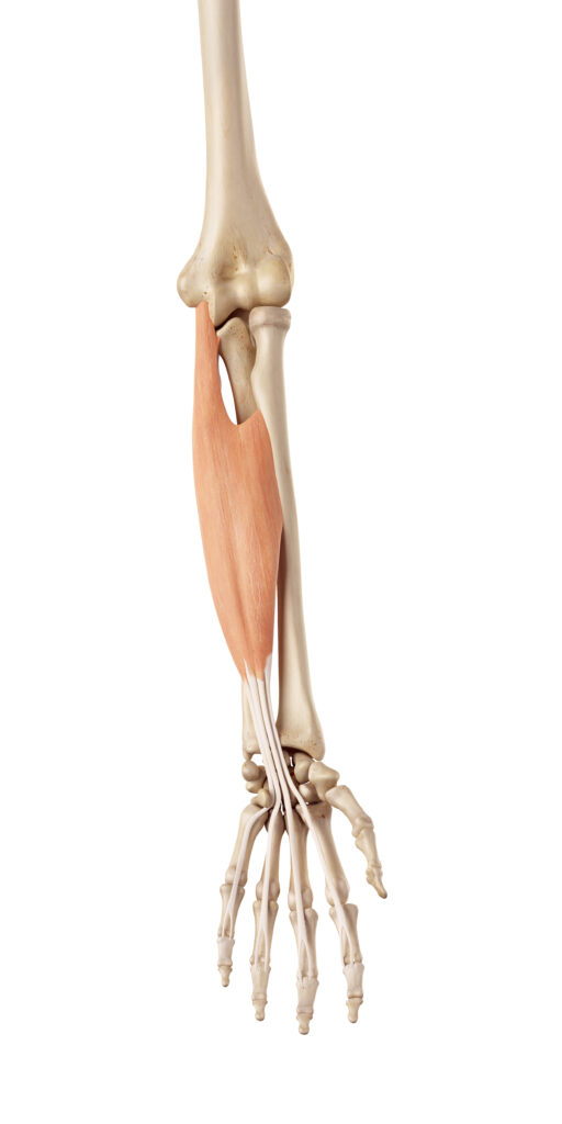
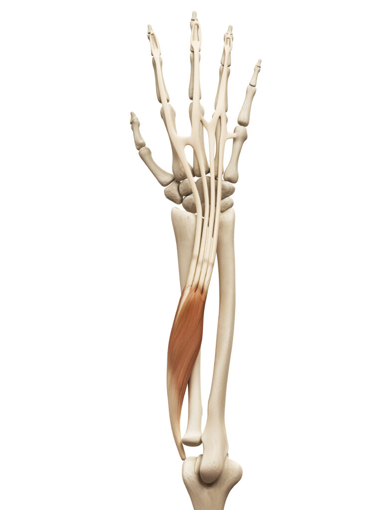
Flexor Digitorum Superficialis (FDS): This muscle is located in the anterior (front) compartment of the forearm. It originates from the medial epicondyle of the humerus, the radius, and the ulna. The FDS splits into four tendons, each attaching to the middle phalanges of the four fingers (excluding the thumb). It is primarily responsible for flexing the middle joints of the fingers (the proximal interphalangeal joints).
Flexor Digitorum Profundus (FDP): Positioned deeper than the FDS, the FDP also originates from the forearm bones and extends into the hand. Its tendons pass through the tunnels formed by the FDS and attach to the distal phalanges of the fingers. This muscle allows for the flexion of the distal interphalangeal joints and also assists in flexing the wrist.
Extension of the Fingers:
Extensor Digitorum: Located in the posterior (back) compartment of the forearm, this muscle originates from the lateral epicondyle of the humerus. It splits into four tendons, each extending to one of the four fingers. These tendons attach to the distal phalanges and are responsible for extending the fingers at all three phalangeal joints (metacarpophalangeal, proximal interphalangeal, and distal interphalangeal joints).
Extensor Indicis: This muscle is also in the posterior compartment of the forearm, adjacent to the extensor digitorum. It specifically extends the index finger and assists the extensor digitorum in this task.
Thumb Movements:
Flexor Pollicis Longus: This muscle, also located in the anterior compartment, is specifically dedicated to thumb movements. It flexes the thumb at the carpometacarpal and metacarpophalangeal joints.

Extensor Pollicis Longus and Brevis: These muscles are part of the posterior compartment. The extensor pollicis longus extends the thumb at the interphalangeal joint, while the extensor pollicis brevis is responsible for extending the thumb at the metacarpophalangeal joint. Together, they enable the full range of thumb extension and abduction.
Biomechanical Considerations
- Tendon Mechanics: The tendons of these muscles run through the carpal tunnel (for flexors) and a series of fibrous tunnels on the back of the hand (for extensors). These tunnels guide the tendons and amplify the force exerted by the muscles.
- Fine Control: The intricate coordination of these muscles allows for the fine motor skills required for tasks like writing, typing, and manipulating small objects.
- Synergistic and Antagonistic Actions: For smooth and precise finger movements, these muscles often work in synergy with others in the hand and forearm. Antagonistic muscle pairs (flexors and extensors) must be balanced in tension for effective motion.
- Clinical Relevance: Understanding the anatomy and mechanics of these muscles is crucial in treating hand injuries and conditions like tendonitis, arthritis, or carpal tunnel syndrome. Rehabilitation after hand or wrist surgery also heavily focuses on these muscles.
In summary, the muscles in the forearm play a pivotal role in controlling finger and thumb movements, enabling a wide range of functions from power grips to precise manipulations. Their complex interplay of tendons and muscle fibers is essential for the dexterous capabilities of the human hand.
Hand Movements:
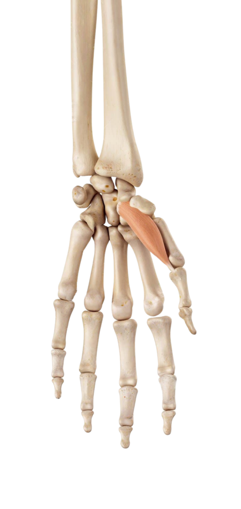
Apart from the fingers, muscles in the forearm also control various hand movements. The abductor pollicis longus and brevis enable the thumb to move away from the hand (abduction), and the adductor pollicis allows it to move towards the hand (adduction).
Hand movements, particularly those involving the thumb, are essential for the dexterous and precise manipulations that characterize human hand function. Several muscles originating in the forearm play pivotal roles in these movements. Let’s explore these muscles and their contributions to hand movements in more detail:
Abduction of the Thumb
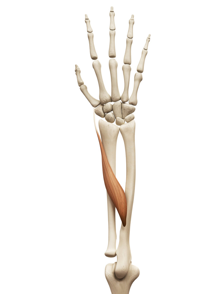
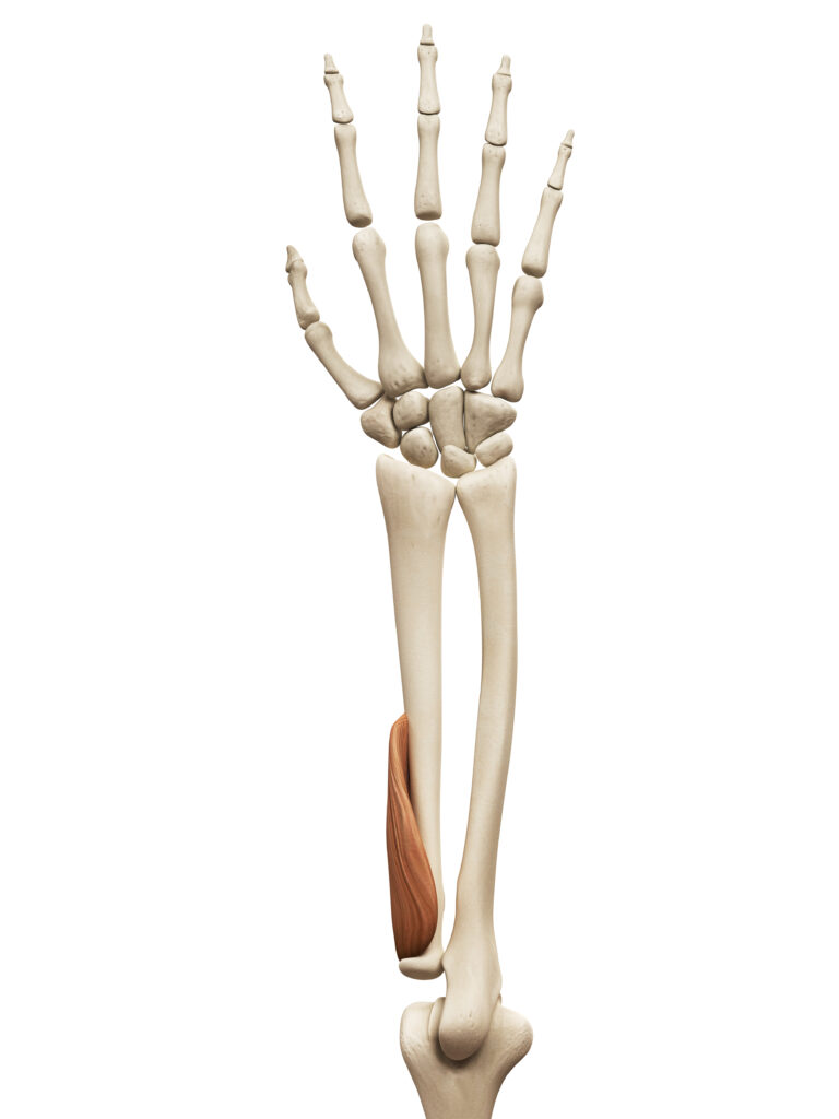
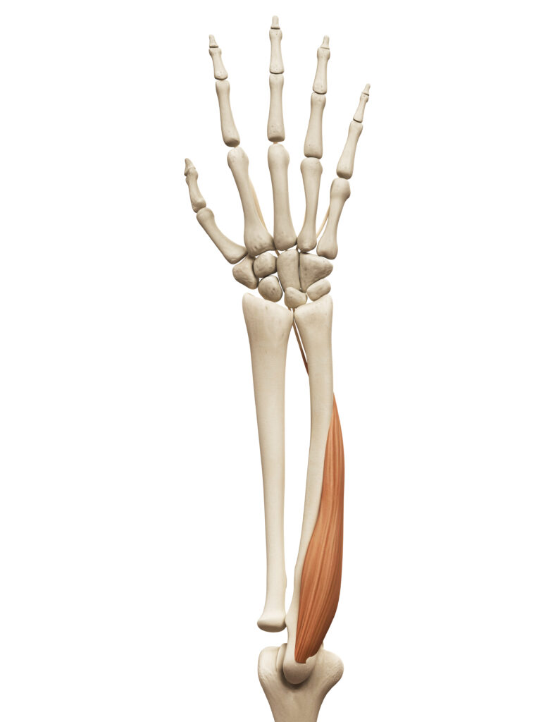
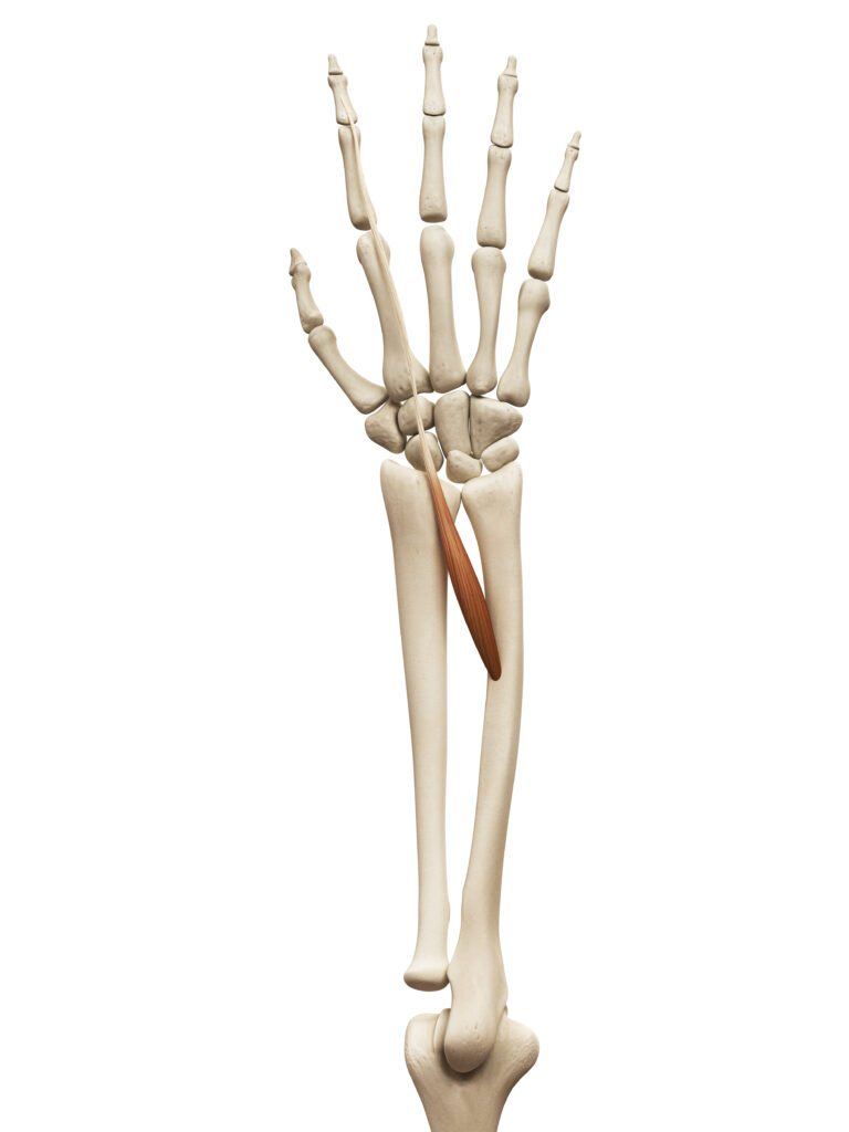
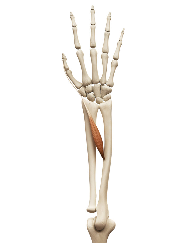
Abductor Pollicis Longus (APL): This muscle is part of the posterior compartment of the forearm. It originates from the radius and ulna and inserts into the base of the first metacarpal bone of the thumb. When the APL contracts, it abducts the thumb, moving it away from the palm of the hand. This movement is crucial for grasping and manipulating objects, allowing the thumb to oppose the fingers.
Abductor Pollicis Brevis (APB): Although not originating in the forearm, the APB is a significant muscle in the thumb’s abduction. It’s part of the thenar eminence (the muscular bulge at the base of the thumb) and plays a vital role in the fine control of thumb movements.
Adduction of the Thumb
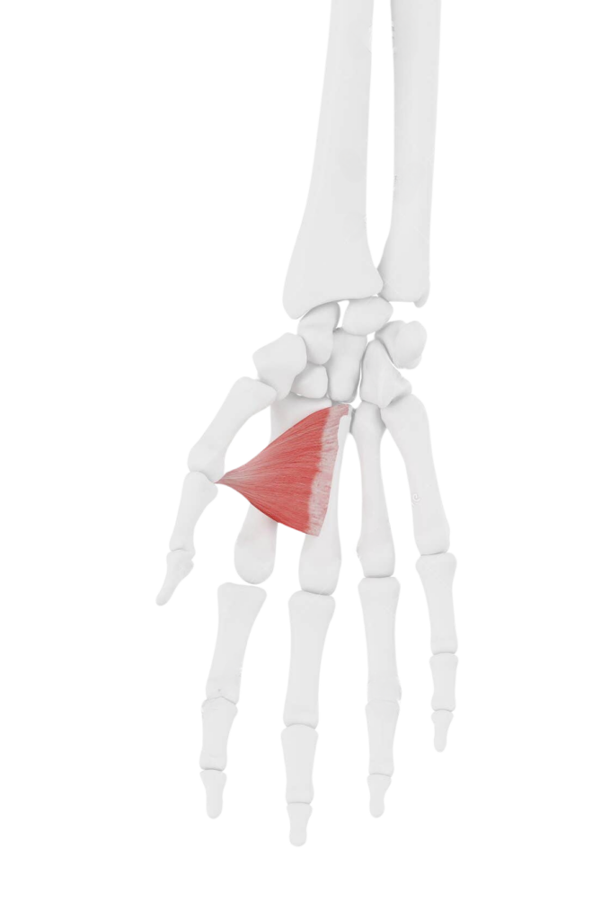
Adductor Pollicis: This muscle is located in the hand rather than the forearm but is an essential component of thumb movements. It has two heads – the oblique and transverse heads – and it originates from the third metacarpal and the capitate bone, respectively. It inserts into the base of the proximal phalanx of the thumb. The primary action of the adductor pollicis is to draw the thumb towards the palm, effectively opposing the abductors.
Other Hand Movements
- Opposition and Reposition: Muscles like the opponens pollicis (also in the thenar eminence) work to rotate the thumb’s metacarpal, bringing the tip of the thumb in contact with the fingertips (opposition). This action is countered by muscles like the abductor pollicis longus and brevis to return the thumb to its neutral position (reposition).
- Lateral Pinch and Grip: The coordinated action of these muscles allows for the lateral pinch (between the thumb and side of the index finger) and various grip forms, from precision grips (like holding a pencil) to power grips (like holding a hammer).
Biomechanical Considerations
- Synergistic Action: For functional hand movements, the forearm and hand muscles often work in synergy. This coordination is crucial for complex movements like grasping, where multiple muscles activate simultaneously.
- Tendon Pathways: The tendons of the forearm muscles travel through specific tunnels and sheaths in the wrist and hand, ensuring smooth and guided movement.
- Nerve Supply: The radial nerve innervates the abductor pollicis longus, while the median nerve typically innervates the abductor pollicis brevis and opponens pollicis. The ulnar nerve innervates the adductor pollicis. The integrity of these nerves is crucial for proper muscle function.
- Clinical Relevance: Understanding these muscles and their actions is important in treating hand dysfunctions and injuries. Conditions like carpal tunnel syndrome, De Quervain’s tenosynovitis, and thumb arthritis directly affect these muscle functions.
In summary, while some key muscles involved in thumb and hand movements originate in the forearm, others are located in the hand itself. Their coordinated actions enable a wide range of complex and precise movements, underlying the hand’s extraordinary functionality in daily life and specialized tasks.
Wrist Stabilization:
Some muscles in the forearm, while not directly causing visible movements, play a critical role in stabilizing the wrist during hand and finger movements. This stabilization is essential for the fine control needed for tasks such as writing or manipulating small objects.
Wrist stabilization is a crucial aspect of hand and forearm function, particularly during tasks that require precision and control. Several muscles in the forearm, while not primarily responsible for the visible movements of the wrist, play a vital role in maintaining wrist stability. This function is essential to provide a steady base from which the fine motor skills of the hand can operate. Let’s delve deeper into the concepts and muscles involved in wrist stabilization:
Muscles Involved in Wrist Stabilization:
Extensor Carpi Radialis Longus and Brevis: These muscles, while primarily involved in wrist extension and radial deviation, also contribute to wrist stabilization. When the fingers are flexed, these muscles contract to prevent excessive wrist flexion, thereby maintaining a stable and extended position of the wrist.
Flexor Carpi Radialis and Ulnaris: Similarly, these muscles, known for their role in wrist flexion and deviation, help stabilize the wrist in extension movements. They counterbalance the extensors, maintaining an equilibrium that stabilizes the wrist during tasks like lifting objects.
Extensor Carpi Ulnaris: This muscle is particularly important in stabilizing the ulnar side of the wrist. It plays a role in preventing excessive ulnar deviation, especially during activities that require a strong grip or during radial deviation movements.
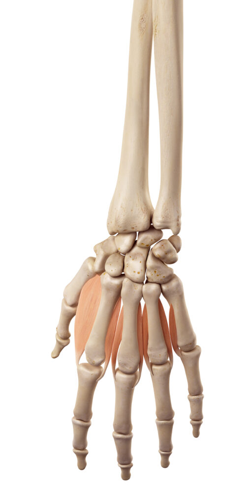
Deep Forearm Muscles: The deeper muscles of the forearm, such as the interossei and lumbricals, also contribute to wrist stability by maintaining the balance of forces across the wrist joint.
Mechanisms of Wrist Stabilization
- Co-contraction: This is when antagonist muscle groups (flexors and extensors) contract simultaneously to stabilize a joint. In the case of the wrist, co-contraction of the wrist flexors and extensors provides a steady position for precise finger movements.
- Dynamic Stabilization: During movements of the hand and fingers, the wrist stabilizers dynamically adjust their level of contraction to adapt to changing forces and positions. This adaptability is essential for tasks that require complex hand movements, such as playing musical instruments or performing surgical procedures.
- Proprioception and Neuromuscular Control: Proprioceptive feedback and neuromuscular control are key elements in wrist stabilization. Sensory information from the wrist joint and surrounding tissues helps coordinate muscle activity for precise movement and stability.
Clinical and Functional Significance
- Importance in Fine Motor Tasks: For activities requiring fine motor skills, like writing, sewing, or assembling small parts, a stable wrist provides the necessary support for precise finger movements.
- Rehabilitation and Strength Training: After wrist injuries, such as fractures or sprains, or in conditions like carpal tunnel syndrome, exercises targeting wrist stabilizers are crucial for regaining function and preventing future injuries.
- Role in Sports and Occupational Health: Athletes and workers engaged in repetitive wrist and hand movements benefit from strong wrist stabilizers to prevent overuse injuries and enhance performance.
In summary, wrist stabilization is a complex function involving various muscles in the forearm. These muscles work collectively to maintain a balanced and stable wrist position, which is fundamental for the execution of fine motor tasks and the overall functionality of the hand and upper limb. Understanding and maintaining the strength and coordination of these muscles is important for injury prevention, rehabilitation, and optimal hand function.