
TF:LVL2_M4 Nervous System
Nervous System:
Definition in Wikipedia states that a nervous system “coordinates its actions by transmitting signals to and from different parts of its body. The nervous system detects environmental changes that impact the body, then works in tandem with the endocrine system to respond to such events.” We can see by these two statements that as a system, there are transmission-based functions which responds to certain events with help from the endocrine system. It is our endocrine system that is responsible for carrying out actions using hormones as signaling chemicals sent through our circulatory system to the proper location for action.
There are two components of the nervous system. The Central Nervous System (CNC) consists of the brain and spinal cord, and the Peripheral Nervous System (PNS) continues the transmission system from the spinal cord into other parts of the body through long bundles of axon fibers.
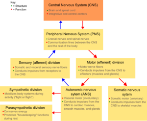
By Fuzzform at English Wikipedia. [CC-BY-SA-3.0 (http://creativecommons.org/licenses/by-sa/3.0/)], via Wikimedia Commons
The direction of travel determines the type and purpose of the nerves. Motor nerves are used to transmit information from the brain to the body to it can take action based on information from the sensory nerves supplying the brain with information about the condition of the body. There are also mixed nerves around the spinal column that coordinates motor, sensory, and autonomic (automatic) signals outwards into the body. These mixed spinal nerves are considered part of the peripheral nervous system.
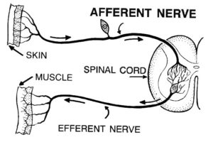
By Pearson Scott Foresman [Public domain], via Wikimedia Commons
The peripheral nervous system can be broken down into somatic nerves for voluntary movement, autonomic nerves to coordinate the “fight or flight” responses, and enteric nerves for the gastrointestinal system. We consider the autonomic and enteric nervous systems to operate involuntary without constant conscious thought.
When it comes to transmission, nerve cells called neurons are specially designed to send electrochemical signals along the axon fibers. These neuron cells are able to rapidly communicate cell-to-cell across synapse gaps which convert electrical signals into chemicals and then back to electrical signals on the other side.
Another important cell in the nervous system are the glial cells which support non-neural activity like providing nutrients, hold the neurons in place, create a protective myelin electrical insulator for the neuron cells, and provide help guide the route of neurons through axon fibers. The rapid speed required for neuron transmission is accomplished by this myelin wrapper surrounding the axons which allows for a more efficient electrical pathway.
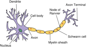
Contributed under CC license: https://commons.wikimedia.org/wiki/File:Neuron.svg
Protection of the nervous system:
Our central nervous system is protected by physical barriers in the brain and spinal cord by layers of meningeal membranes in addition to the bone structures of the skull and spinal vertebrae. There is also a chemical blood-brain barrier protecting the brain from certain chemicals in the bloodstream. Even though there are layers of physical protection for the CNS, any damage occurring to this system can have extreme and serious consequences.
In the peripheral nervous system, some nerves are protected deep inside the body while others near the surface are susceptible to damage and resulting conditions ranging from pain to loss of sensation and muscle control. Damage to nerves are usually associated with the inflammatory response of surrounding tissue which creates pressure that can pinch or close off communication along the axon. The nerves themselves can become inflamed or experience conditions associated with various diseases like diabetes and become vitamin deficient, infected, or filled with toxic substances. Nerves can become temporarily numb or stiff and lose normal function due to mechanical pressure, low temperatures, or certain chemical interactions.
The Autonomic Nervous System (ANS):
Our overall nervous system is controlled by a combination of conscious thought like moving your hand to pick up a pencil (voluntary) or unconscious thought like breathing or the beating of your heart (involuntary). These automated, involuntary, and regulatory actions are under control of the autonomic nervous system to provide an adaptive process for constantly changing conditions.
The ANS consists of three distinct nervous systems called sympathetic, parasympathetic, and enteric (gastrointestinal). The sympathetic and parasympathetic nervous systems work together to produce opposite actions requiring to the body to prepare for a “fight or flight” or return to a more relaxed condition required for rest, digestion, and healing “rest and digest” or “feed and breed”. We will focus on these two systems for our discussion of the ANS since they both impact how our body deals with chronic inflammation and resulting pain.
Autonomic (vegetative ) Nervous System:
- Regulated by hypothalamus
- Unconscious regulation of:Heart rate, Digestion, Respiratory Rate, Pupillary Response, Urination, Sexual Arousal
- The ANS is connected to the motor side of the nervous system, and most functions are involuntary with a few somatic voluntary controls as well.
Location:
Sympathetic nerves come from the spinal cord in the thoracic and lumbar regions to connect with various organs while the parasympathetic nerves come primarily form the cranial and sacral regions. The splanchnic nerves are usually associate with the sympathetic nervous system and the vagus nerve is well known in association with the parasympathetic nervous system.
The organs of the body receiving information from the autonomic nervous system receive a connection from both the sympathetic and the parasympathetic to modulate their functions.
In the image of the sympathetic innervation, we can see the route of the network of nerve fibers as they branch outwards to service the various organs involved in the sympathetic nervous system process.
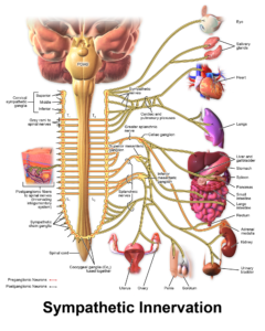
Blausen.com staff (2014). “Medical gallery of Blausen Medical 2014”. WikiJournal of Medicine 1 (2). DOI:10.15347/wjm/2014.010. ISSN 2002-4436. [CC BY 3.0 (https://creativecommons.org/licenses/by/3.0)], from Wikimedia Commons
Sympathetic:
- Diversion of blood flow (vasoconstriction) away from the GI Tract and skin while the blood flow is increased to the skeletal muscles and lungs. Heart rate and blood pressure are also increased through vasoconstriction to speed up flow to the muscles and lungs.
- Greater oxygen exchange by releasing epinephrine to the lungs to increase oxygen exchange
- Vision enhancement through pupil dilation to let in more light.
- Digestive processes are slowed down through intestinal constrictions.
Parasympathetic:
- Blood flow returns to the GI Tract and air to the lung are returned to a more efficient process.
- The vagus nerve return the heart rate and pressure back to normal.
- Pupils constrict to bring the vision back to a normal distance.
- Digestion is returned to normal through stimulation of the salivary glands.
In the image of the parasympathetic innervation image, we can see the nerve fibers branching outwards to signal the various organs to disengage from the “fight or flight” process and return to a more relaxed and sustainable condition. Notice how the vagus nerve is involved in both the sympathetic and parasympathetic processes.
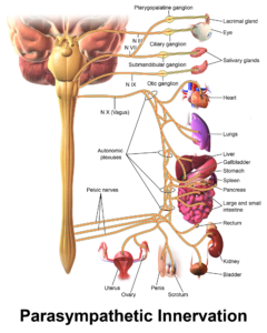
Blausen.com staff (2014). “Medical gallery of Blausen Medical 2014”. WikiJournal of Medicine 1 (2). DOI:10.15347/wjm/2014.010. ISSN 2002-4436. [CC BY 3.0 https://creativecommons.org/licenses/by/3.0)], from Wikimedia Commons