
TF:LVL2_M3 Lymphatic System
Lymphatic System Flow:
We will focus on an understanding of the lymphatic system as it applies to healing the body with weighted tuning forks. As with most body systems the lymphatic system has many layers, processes, and functions, but we are only going to discuss those aspects necessary for helping the body heal with vibration.
Wikipedia describes the lymphatic system as being a part of the vascular system which is another name for the cardiovascular system which circulates blood throughout the body. As we filter through the myriad of systems in the description, the main point to understand is that a vascular system carries blood and several other substances through a tube-like network using the pressure created by the heart.
Another way of separating these systems into schools of thought is the unique nature in which both the cardiovascular system has its own complete cycle of circulating blood outwards to feed oxygen and nutrients to the body and returning the blood to be recycled. Since the cardiovascular system is a complete circulatory loop, then where does the lymphatic system come into play?
Let’s take a closer look at how the circulatory system cycles the blood through the entire body. It helps to define the role of the blood vessels to better understand how they circulate through the body and interact with the lymphatic vessels.
Arteries transport oxygenated blood and nutrients away from the heart to the cells of tissue throughout the body. Pulmonary arteries are slightly different since they carry de-oxygenated blood to the lungs to re-oxygenate the blood supply for a return trip through the veins. In our discussion, we will focus primarily on those “systemic” arteries and further into the “peripheral” arteries circulating oxygenated blood throughout the body.
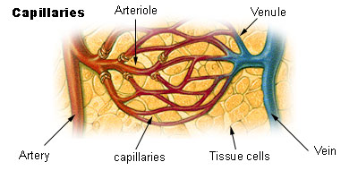
As the arteries branch out into smaller diameter vessels, the blood moves through arterioles into the capillaries for transfer into the surround cells. These tiny capillary vessels are significant to our blood and lymphatic circulation discussion because this is the part where red blood cells, gases, and nutrients pass through the capillary walls and into the interstitial tissue space. The diameter of a capillary is less than red blood cells, so the blood cells must distort and pass through these very thin capillary walls. In the image below, we can see the arteries moving through the arteriole and into a network of capillaries surrounded by a sea of tissue cells and interstitial fluid. Notice how the return trip of the blood circulation consists of venules which bring the returning fluids from the capillary drain field back into the larger veins.
Zeerka-HD [CC BY-SA 4.0 (https://creativecommons.org/licenses/by-sa/4.0)], from Wikimedia Commons
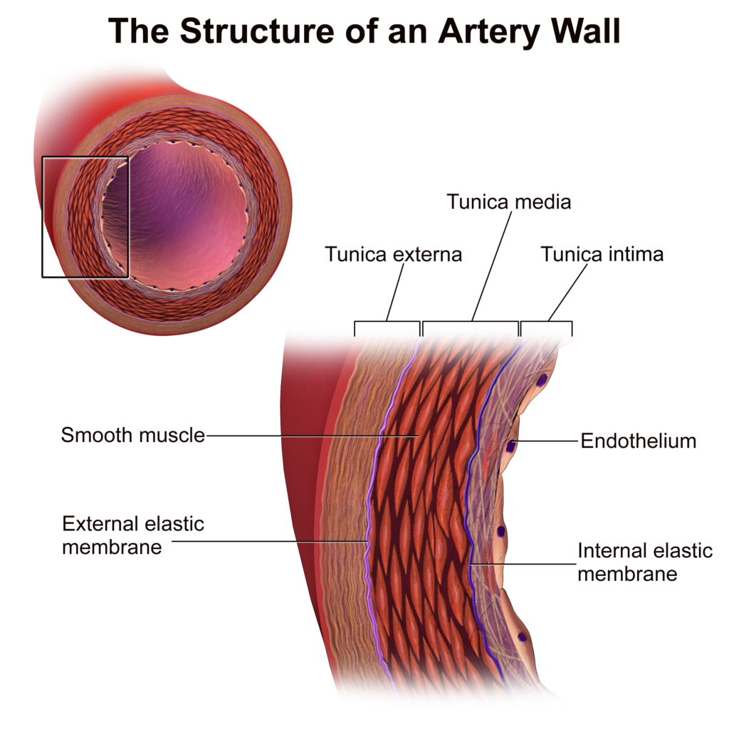
Before we move away from the artery functions, it’s important to point out the micro-anatomy with an outer wall consisting of collagen “connective tissue” that we know as fascia. The next layer is made of smooth muscle tissue and more flexible fascia fibers called elastin. The inner-most layer is lined by a single layer of endothelium cells which acts as a protective barrier and perform various immune functions.
Blausen.com staff (2014). “Medical gallery of Blausen Medical 2014”. WikiJournal of Medicine 1 (2). DOI:10.15347/wjm/2014.010. ISSN 2002-4436. [CC BY 3.0 Wikimedia Commons]
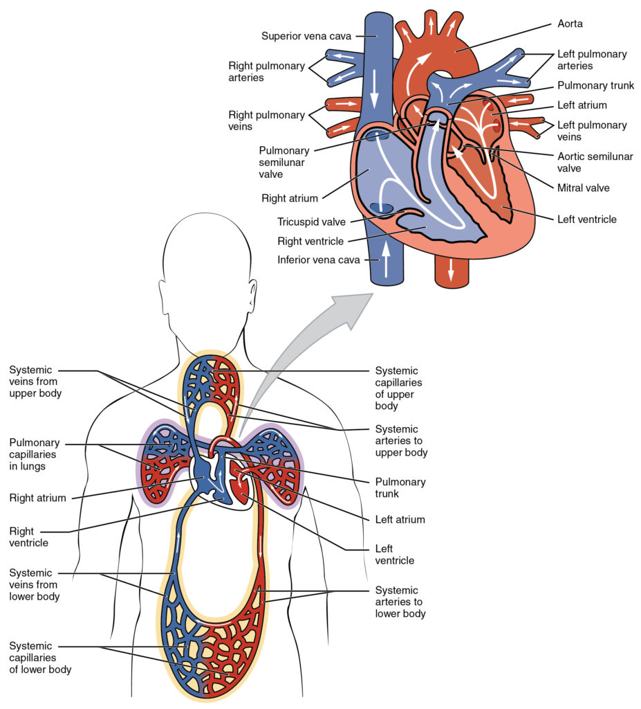
In the next image, we see the entire cycle of the blood flowing from the heart through the arteries, the capillary fields, and back to the heart through system veins. Notice there are two separate system loops with one above the heart and one below the heart. This information will be important when we discuss the lymphatic system since the lymph vessels connect to the same capillary fields and system loops above and below the heart. On a separate note, also notice there is a separate pulmonary loop specific to the circulation of blood through the lungs.
By OpenStax College [CC BY 3.0 (https://creativecommons.org/licenses/by/3.0)], via Wikimedia Commons
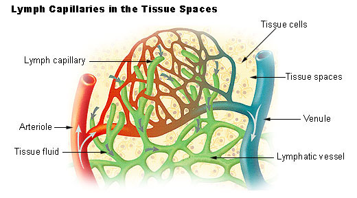
Now that we have discussed the entire flow of the blood circulatory system as a closed loop system, we can focus on how the lymphatic system connects to collect the materials in the interstitial spaces of the capillary field. In the next image, we can see the lymphatic vessels wrapped and intertwined with the arteries and capillaries within the tissue cells. Also notice how all of the systems in this image are at a very microscopic size to communicate and interact at a cellular level with the surrounding tissue.
The lymphatic vessels start out in this central meeting space as very tiny lymph capillaries and combine into much larger vessels to process and carry the collected fluids back to the area near the heart for a return to the blood circulation.
By Arcadian, 14 Oct 2006, CC 3.0, https://commons.wikimedia.org/wiki/File:Illu_lymph_capillary.jpg
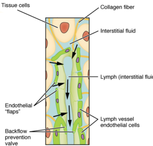
When we take a closure look at the lymph capillaries in the next image, we can see the vessel in made of a single layer of those same epithelial cells as the inner lining of the artery walls. The lymph vessel cells are designed with “flaps” to bring in the interstitial fluid floating in between the tissue cells. According to Wikipedia, the interstitial fluid collected by lymph capillaries consists of various combinations of sugars, salts, fatty acids, amino acids, coenzymes, hormones, neurotransmitters, white blood cells, and cell waste-products. The composition of fluid materials depends on the location and type of tissues.
Returning our focus back to the image of a lymph capillary, notice the unique nature of the lymph vessel endothelial cells with “backflow prevention valves”. The entire lymphatic system does not have a pumping system like the heart in the cardiovascular system. Therefore, the fluid (called lymph) is circulated through the vessels primarily through pressure created by the surrounding tissue and various movements of the body. The backflow valves prevent lymph fluid from flowing backwards due to gravity.
Before we move out of the microscopic view, notice the lymph vessels are intertwined with collagen fibers in addition to the arteries and veins of the cardiovascular system. These collagen fibers are part of the fascia “extracellular matrix” (ECM) network and a very important component of a much larger, interrelated system where we will focus on three parts throughout our training. The relationship between the interstitial fluid floating inside of the fascia matrix and the lymph vessels responsible for removing and recycling the interstitial fluid.
By CFCF [CC BY-SA 4.0 (https://creativecommons.org/licenses/by-sa/4.0)], from Wikimedia Commons
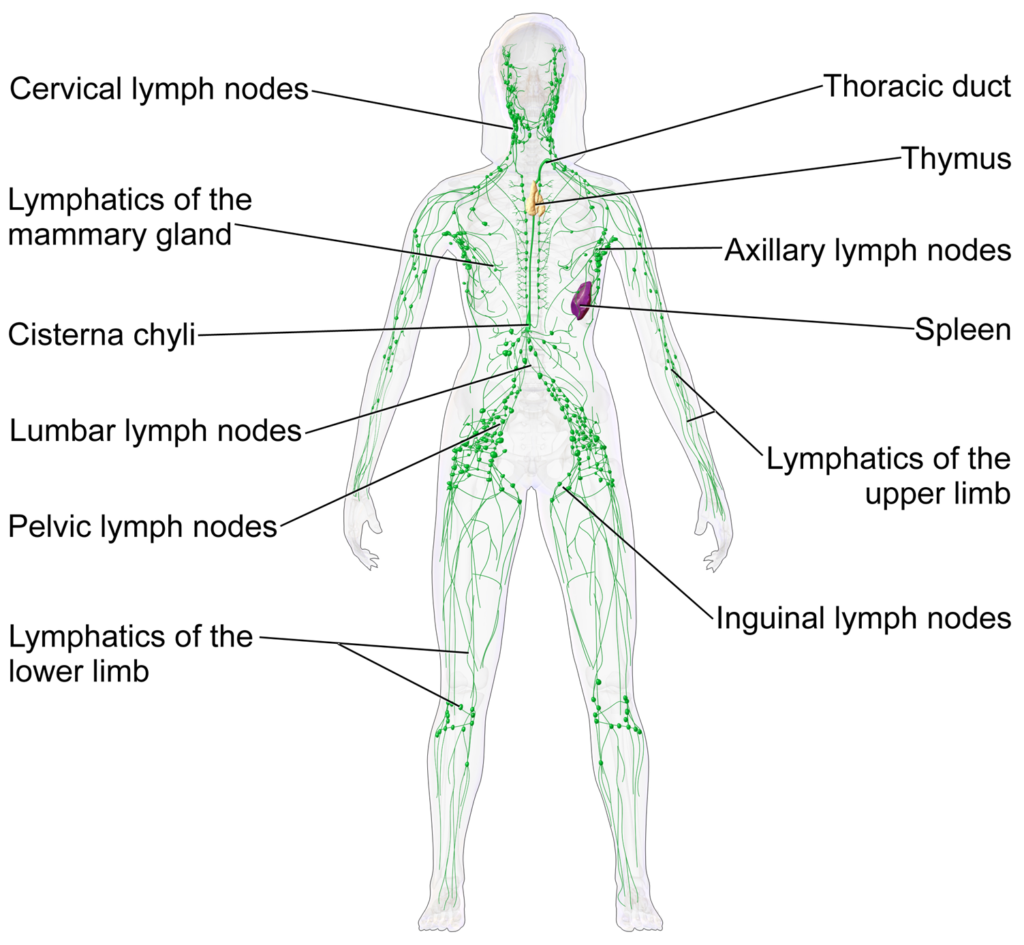
In the next image, we can see the complexity of lymphatic vessels branching and passing through various parts of the body both inside and outside of the rib cage. The lymphatic system also consists of the thymus, spleen, and tonsils to support immune and digestive system functions. In our discussion of the lymphatic system, we will focus primarily on the way the lymph fluids flow through the various vessels and nodes as a function of removing interstitial fluids from the tissues.
Since the lymph nodes and vessels are created with fascia collagen fibers and surrounded by the fascia network, we are able to affect the condition of the lymphatic system and hopefully clear blocked areas and return the system to normal operations.
Blausen.com staff (2014). “Medical gallery of Blausen Medical 2014”. WikiJournal of Medicine 1 (2). DOI:10.15347/wjm/2014.010. ISSN 2002-4436.
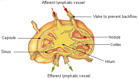
Throughout the lymphatic system, there are several hundred lymph nodes grouped in various locations of the body where injury might occur like the groin, knees, neck, and armpits. These nodes tend to swell when the lymphatic flow is blocked either up or downstream. A group of swollen nodes can be responsible for blocking entire sections of the body from a proper removal and processing of interstitial fluid.
An example is the lymph nodes behind the knees which create a choke point for communication of several other systems like nervous and cardiovascular along with the flow of lymph below the knees. We found that draining the lymph nodes behind the knee is part of the process and technique for helping with edema of the legs.
By SEER [Public domain], via Wikimedia Commons
Based on our research on the lymphatic system, we learned to move lymph through the lymphatic system towards the heart. Lymph fluids above the heart should be moved south towards the heart. Anything below the heart should be moved north towards the heart.
What we have found with working on the lymphatic system and draining nodes with the weighted tuning forks is that you have to clear out the nodes closest to the heart first before working on the furthest nodes. For example, start at the base of the neck and clear that area of lymph fluid and work up the neck and into the face in that order. Along the way, you are loosening up the blockages preventing the lymph from flowing. Inflammation causes a lot of blockages as it creates pressure and pinches off flow to several systems in addition to the lymphatic system.
If you are able to assist the flow of lymph by pushing it along the lymphatic system, it is important to know that you should push the fluids along the same direction of flow. The lymphatic system has a series of valves that only allow fluid to flow in one direction during its normal operation. However, if you are “clearing the way” by putting your tuning forks on the lower nodes first, this will relax the tissues and help everything above drain easier. Sometimes just draining one node at the bottom will automatically cause everything above it to drain immediately without any further action. These are two different actions of pushing the fluid or just clearing out the lower nodes first to assist in draining.
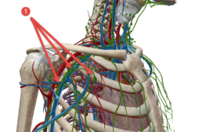
The lymph field (1) underneath the collar bone is where the lymph exchanges clean blood with the circulatory system (1) so this is technically the end of the line where “above and below the heart” connect. Clearing these areas of excess interstitial fluid build up and tension from the fascia fibers can help return lymph flow.