
Supporting Content
We have successfully connected the dots at New Earth Healing Center and NEHC Academy to explain the science supporting our clinical approach to rapid pain relief, reduction in mobility restrictions, and resolution of tissue and organ dysfunction. Our flagship unique technique of Vibrational Fascia Release Technique (VFRT) focuses mainly on rapid pain relief and mobility restrictions for a single joint. This technique is part of a larger Tuning Fork Vibration Therapy program still in development for training and certification that widens the focus on any dysfunction needing ongoing therapy. Our clinical case studies have shown a very rapid response beyond what many hand, tool, and electrical device approaches and therapies can reach in a single session.
These are big claims of clinical results, so it was necessary to determine the physiology of why a weighted tuning fork can provide such rapid results to support our theories and concepts. We believe that we have those answers.
Interstitial Fluid Pressure
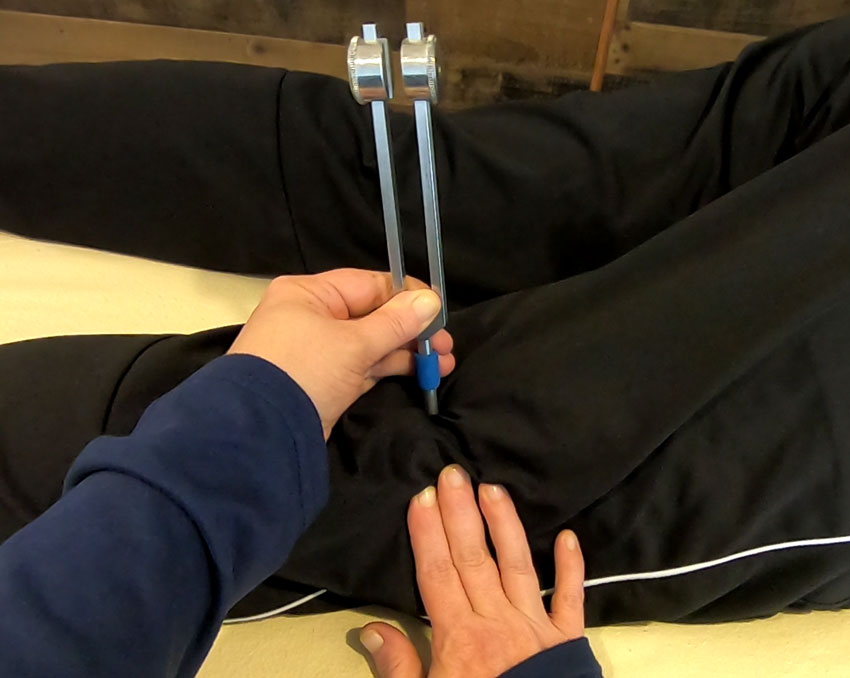
Most of our work revolves around the concept of interstitial fluid pressure building up in edema pockets throughout most of our tissue, organs, and systems. It is our theory that a 128hz weighed tuning fork placed in the right spot with the correct amount of pressure can cause the pressurized fluid to diffuse through the membrane or layer to release the pressure. It is ultimately the pressure release that is responsible for a reduction in pain and restoration of joint mobility in as little as a single tuning fork placement.
We needed a better understanding of why the fluid can transfer through a layer or membrane only when introduced to compression and vibration from a weighted tuning fork versus just hand pressure, tools, or percussion-based powered tools. Why were we feeling the tissue rapidly deflate under our hands during a single placement? It turns out that the answer had been in front of us this whole time. The reason the dots were so difficult to connect is due to the existing terminology used in the sound healing community and traditional thinking when it comes to tuning forks in general.
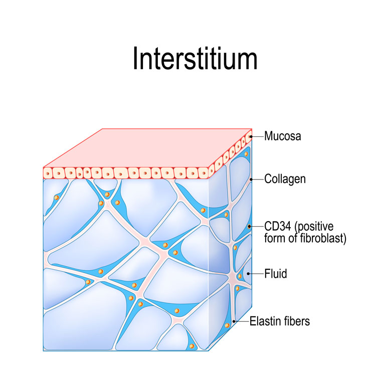
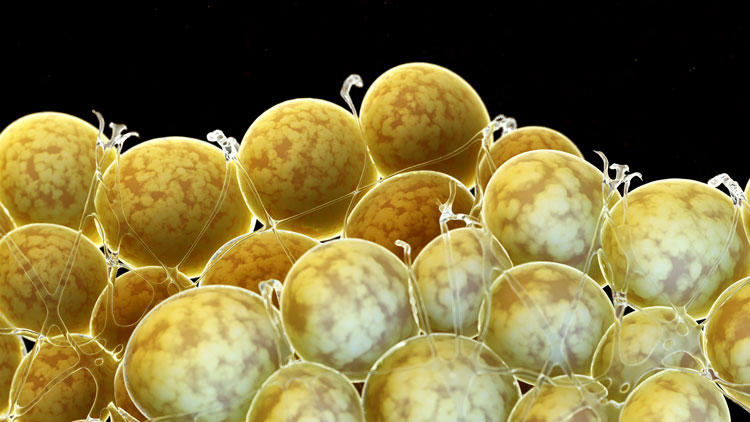
In a 2018 paper published in Scientific Reports, the authors discovered the presence of a reticular pattern of spaces using new technology from a Probe-based Confocal Laser Endomicrosopy (pCLE) where fluid-filled spaces can be identified and studied in live tissue. Until this point, pathologists were relying on methods of immunostaining to identify different tissue in samples processed and preserved with chemicals and drained of any remaining interstitial fluid. Pathologists returned to their tissue samples with a different focus and found the tell-tale “cracks” along with another discovery of fibroblast-like cells linked together and lining one side of nearby collagen bundles. These pockets of fluid are often referenced as third spacing (1), rivulets (4), or small free fluid vesicles (4).
Quarantine Process
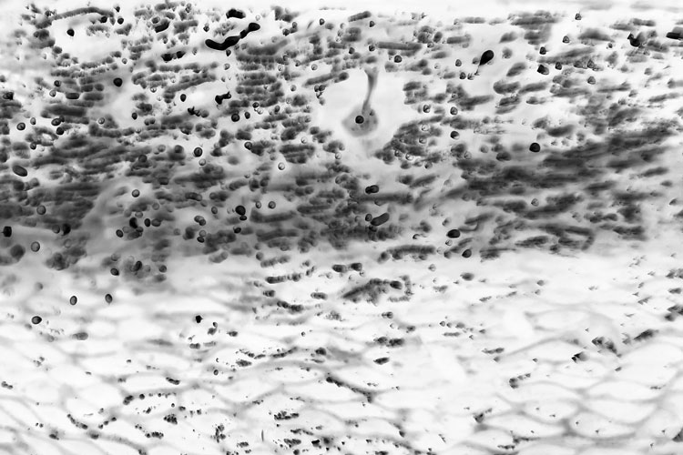
It is our theory at NEHC that our interstitial spaces are surrounding foreign matter and toxins with a membrane to quarantine the dangerous material from damaging surrounding tissue. In a Live video for Pathcast.com, Dr. Neil Theise, Pathologist and one of the authors for the previously mentioned article on the interstitium discussed in detail the discoveries he was finding through electron microscopy. He shows pathology images of cells adhering to the collagen fibers with the other side unlined which is directly exposed to the interstitial fluid space. These are not technically endothelium cells due to their lack of a basement membrane, and Dr. Theise believes these membranes are part of a pre-lymphatic system created in response to the presence of foreign material.
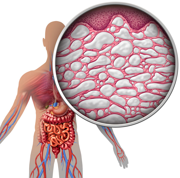
Both the video and the 2018 paper show images of injected pigment particles surrounded by membranes where the authors believe this discovery shows a mechanistically important structure of fluid-fill edema spaces with implications for tissue dysfunction. Their paper summarizes the existence of larger macroscopic and visible spaces which have a serious impact on fluid flow, compression dynamics, and resolution of tissue dysfunction. They further implicate these fluid “sinuses” as necessitating disordered fluid dynamics in organs and tissue throughout the body with a connection to disease, fibrosis, and cancer metastasis. The authors recommended sampling interstitial fluid within these spaces as part of a diagnostic process which furthers our theory on quarantined spaces.
Lack of Lymphatic Drainage
When the conditions within the interstitial space is dysfunctional with free fluid providing pressures beyond the ability to force fluid into the lumen of the lymphatic intake vessels but rather a more passive “filtration” essentially slowing down the removal of fluid. (6)
In medical literature, we find several anatomical and physiological references to the impact interstitial fluid pressure has on the initial lymphatic capillaries responsible for intake of fluid from the extracellular space. These thin endothelial cells of the lymphatic intake valves are designed to expand and open wider in the presence of increased fluid pressure because they are anchored to the surrounding collagen fibers. When the fibers spread apart to accommodate for the change in pressure, the lymphatic intake flow responds as well. However, this emergency back up process only works until the pressure of edema overwhelms the local area causing the intake valves to close or fail to provide proper suction required to bring fluid into the initial lymphatics. (7, 8)
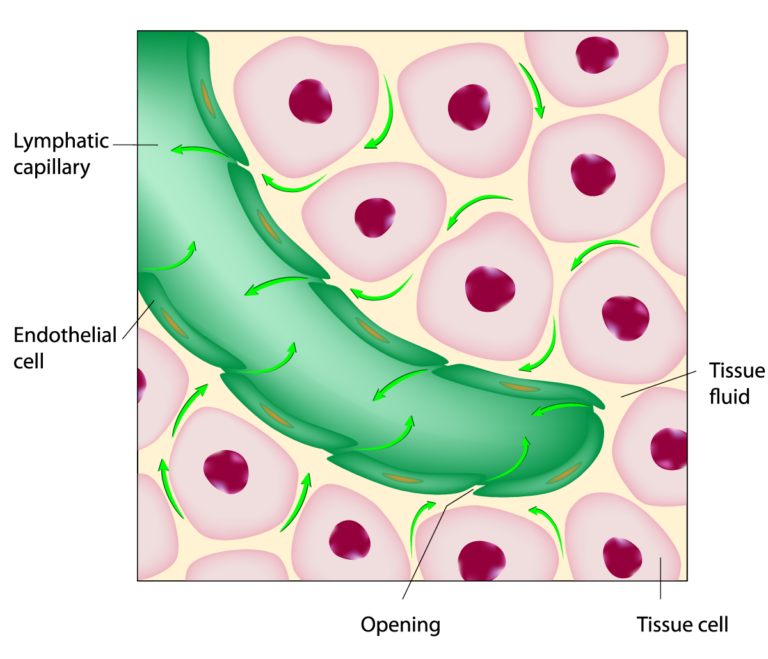
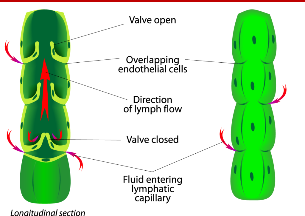
Since negative pressure is needed for suction of fluid into the intake valves, a small amount of fluid pressure in the interstitial space still protects against edema and promotes fluid flow provided it does not exceed the lymphatic capillary capacity. (9)
When the superficial adipose layer develops progressive interstitial fibrosis or adhesions, 30-50% of people experience higher levels of edema with a typical clinical diagnosis of lymphedema. In this condition, interstitial fluid is more static, plasma proteins are forced out of the arterial capillaries, and chronic inflammation damages the surrounding collagen structure of the extracellular matrix and ultimately destroys the lymphatic intake function. The increase in fibrosis, size of the adipose cells, and creation of new adipose cells leads to high resistance to manual compressive therapy due to the inability to move edema fluid pockets through the interstitial spaces. (10)
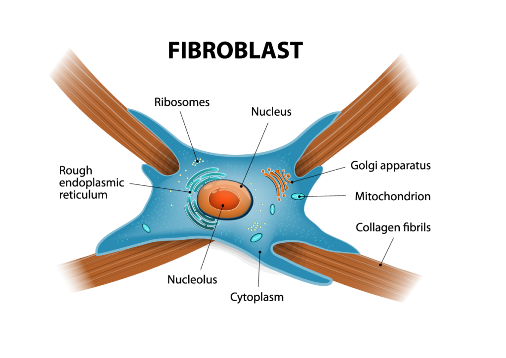
There is also a physiological function of the venous capillaries to reabsorb interstitial fluid based on losses from the arterial leakage and fluid pressure. Both hydrostatic and osmotic pressure are tightly controlled to prevent edema in the surrounding tissue in what we usually consider as “swelling.” (13) The speed or velocity of interstitial fluid is restricted due to the structure of the collagen (fascia fibers) and proteoglycan gel. The contents of the interstitial fluid is a combination of substances in different amounts with water as the base liquid and larger substances like proteins driving basic physiological movement of fluid. Fibroblast generated proteoglycan proteins are an important part of the extracellular space because their bottle brush shape provides a mechanism to slow the flow to fluid and protect the vessels and cells within the extracellular space from pressure.
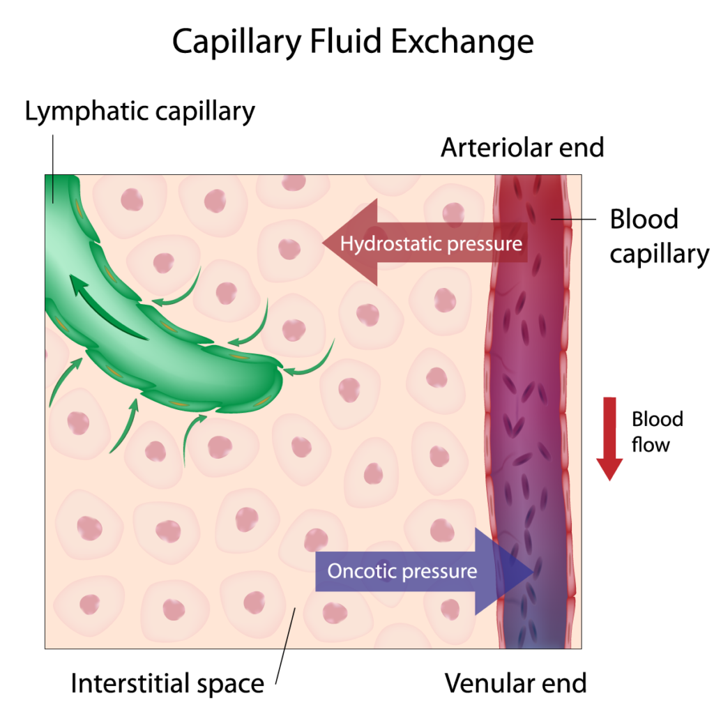
There is also a physiological function of the venous capillaries to reabsorb interstitial fluid based on losses from the arterial leakage and fluid pressure. Both hydrostatic and osmotic pressure are tightly controlled to prevent edema in the surrounding tissue in what we usually consider as “swelling.” (13) The speed or velocity of interstitial fluid is restricted due to the structure of the collagen (fascia fibers) and proteoglycan gel. The contents of the interstitial fluid is a combination of substances in different amounts with water as the base liquid and larger substances like proteins driving basic physiological movement of fluid. Fibroblast generated proteoglycan proteins are an important part of the extracellular space because their bottle brush shape provides a mechanism to slow the flow to fluid and protect the vessels and cells within the extracellular space from pressure.
Connection to Ultrasound Non-thermal Therapies
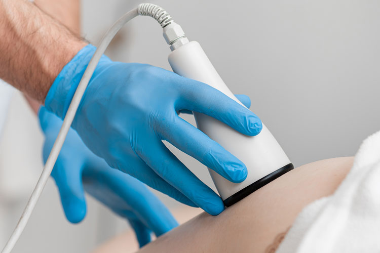
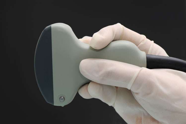
We looked deeper into an already existing therapeutic field of Ultrasound Therapy where a medical ultrasound device is placed on the person with the settings arranged to deliver either thermal (heat) therapy or non-thermal therapy. The non-thermal therapy still produces some heat due to the rapid friction and shearing between cells, but the equipment turns on and off (cycles) to allow time for heat dissipation without damaging the cells. It is in this area that we find our answers to the question of how fluids can propagate through membranes and layers with a well-known phenomenon in ultrasound physics known as acoustic cavitation. The bubbles that form within cell walls and other membranes provide a porous environment to change the physiology of the membrane to allow changes not only to the components inside, but also the materials which pass through the membrane using channels or through porous holes created by the cavitation effect. (2)
Cavitation and Micro-streaming
The concept and physics of cavitation is well known in the clinical ultrasound imaging community since the presence of bubbles within a medium where sound vibration is propagating (moving through) will cause the mechanical vibrations to scatter rather than returning to receiver. Ultrasound imaging relies on high frequency mechanical vibration moving through a tissue from the paddle or transducer and eventually bouncing off a layer of tissue where the densities greatly vary. The receiving function of the transducer calculates the travel time from and to the paddle for a visual representation of contrast between two neighboring tissue. Frequency, signal strength, and several other settings are controlled to optimize for certain tissue locations and contrast without putting too much vibration into the tissue to scatter the signal or losses from heat as the cells rapidly rub against each other. Heat generated in a sound vibration environment is normally considered absorption because the initial decibel level of the transmission is reduced when mechanical vibration is converted into a different for of energy since heat abides by electromagnetic physics.
The non-thermal effect of cavitation is primarily in the reduction of the membrane potential, changes to the lipid structure, an increase to the membrane permeability, and ionic conduction. (2) These physiological components are all important to therapeutic methods focused on relieving fluid pressure for pain relief and restoration of musculo-skeletal dysfunction. The physics of cavitation includes both the formation of the bubbles and the incredible effects from their collapse which brings on a completely new physiological phenomenon with a release of energy and bioluminescence. It is our purpose in this article to focus mainly on the physical effects of cavitation as applied to affecting fluid permeability and flow across a membrane at both the cellular level and macroscopically at the tissue level.
Cavitation can have two main effects when mechanical vibration passes through a liquid to separate air bubbles from the fluid. High intensity and low frequency ultrasound vibration can develop a stable, small bubbles, and higher ultrasound frequencies can create a more unstable condition where cavitation bubbles grow larger and collapse abruptly. (2) The difference between stable and unstable cavitation conditions are likely to explain the difference in resulting membrane permeability versus a complete destruction of membranes observed in certain cancer research using ultrasound therapies.
A subset component of cavitation is microstreaming where small vortices or eddies are developed on a very small level within cell cytoplasm and extracellular within the interstitial fluid space. Microstreaming is observed to change the metabolic activity within fibroblast cells resulting in a change to bloodstream and interstitial fluid flow. (2)
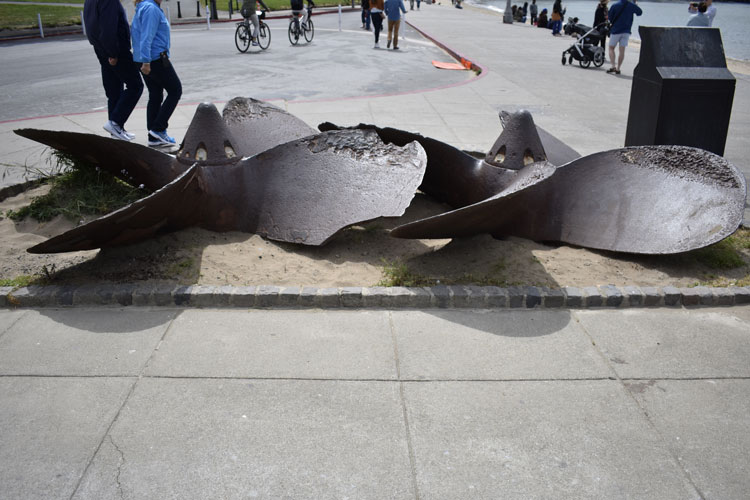
The study of cavitation physics has been documented long before a biological application with ultrasound application. Marine engineers studied the effects of cavitation on propellers after it was discovered that the tiny bubbles produced pitting within the metal blades leading to decay and destruction of the entire propeller. This is just one of the areas where my 22 years of experience in underwater physics in the United States Navy crosses with this current work using tuning forks for vibrational therapy. Cavitation from submarine and surface ship propellers were some of the loudest sounds to propagate through the water for eventual reception by a listening device. https://www.youtube.com/watch?v=MyZzhwYMytc In the 1970s, propeller manufactures like Toshiba altered designs of the entire propeller system to allow for more efficient thrust power while masking the effects of cavitation bubbles on giving away their stealth capabilities. https://www.youtube.com/watch?v=4YOyy6Jfz8E The physics of cavitation was well known in my community because the effect of tiny collapsing bubbles produced so much power that the resulting shock wave could travel for miles through the water. Imagine what cavitation and microcurrents could do when they explode within a membrane or tissue layer.

Lipid Drainage Using Tuning Forks
NEHC has experienced through our clinical results that lipids were draining through the membranes of adipose cells because of a large volume reduction and subsequent elimination of what we believed to be lipid related in a typical “fat burning” treatment. Our clients would lose anywhere from 1 to 7 inches in diameter in only 45 minutes during a treatment. We believe some of the fluid was edema-based, but that didn’t account for the difference in the tissue consistency and elimination properties experienced directly following a treatment.
We find our answers again in the studies and clinical application of ultrasound to reduce fat by releasing lipid in the adipose tissue. In the book The Mechanical Vibration: Therapeutic Effects and Application, many of the things just discussed are documented with corresponding research including this ultrasonic fat reduction treatment.
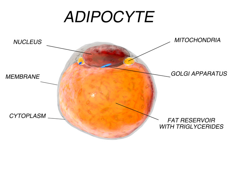
The authors found a cavitation effect in adipose (fat) tissue causing both fragmentation and leakage of the lipids into the interstitial fluid space. They have determined that ultrasound frequencies cause the cavitation phenomena at the fat/water interface which leads to adipocyte rupture and triglyceride release. When ultrasound is concentrated in a “defined subcutaneous area to produce selective lipolysis and clinically relevant reduction of the volume of subcutaneous fat pad while avoiding any damage to surrounding tissues.” (2)
Examples of Vibration Destroying Cancer Cells
We have heard of some of these reports of Fabian Maman, Anthony Holland, and the international team of scientists in Israel who successfully used low-frequency ultrasound to cause tumor cells to explode. Although this example is not the intended result of our vibrational therapy, the concept of cavitation or micro-bubbles used to affect cell membranes is now well known throughout the world. Another example is HistoSonics who has built a histotripsy platform to provide focused ultrasound to destroy targeted tissue without damaging surrounding tissue. Christine Gibbons shows an animation of the cavitation effect in the tissue during a Ted Talk in 2016. (See Time mark 4:02) https://www.youtube.com/watch?v=GKh2XFmsUx4 Please keep in mind that we are not destroying cells in vibrational therapy. These examples are showing extreme results from high powered sound. Our tuning forks only provide low frequency, low amplitude vibration to the cells and membrane to still allow for fluid propagation rather than destroying the membrane.
Weighted tuning fork produce ultrasound frequencies
In a study published in the American Journal of Physics in 1992, researchers studied an unweighted 384hz tuning fork and measured a wide range of frequencies produced by the unbalanced nature of the design. (3) Results from this research found multiple frequencies produced by several different modals or bending of the tuning fork in different directions. A tuning fork creates a myriad of frequencies well beyond what is stamped on the side. The report also discussed the results of adding mass (weight) to the inner and outer tines like a weighted tuning fork. A simple self-test can determine whether a weighted tuning fork can produce ultrasonic vibrations at the base. A person only needs to strike an unweighted tuning fork and hold the base up to their ears to observe no auditory sound received from the vibration of the base. This indicates the frequencies propagating from the base are above 20, 000hz which is considered the ultrasonic range or mechanical vibration outside of the human hearing range.
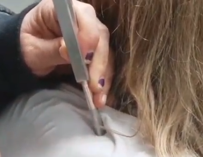
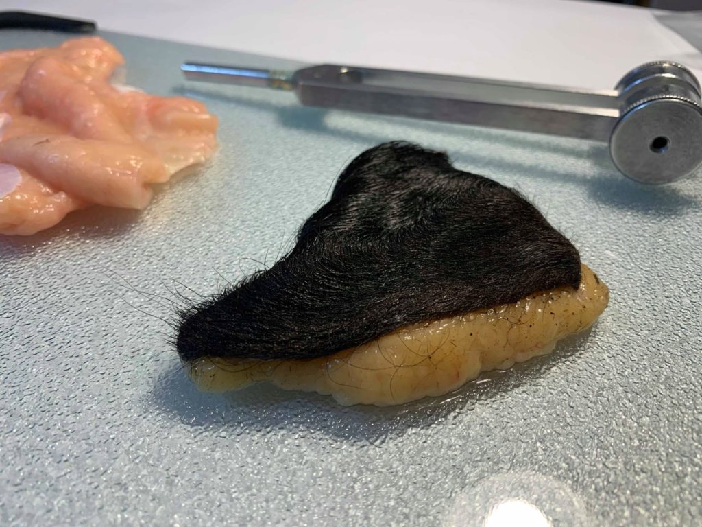
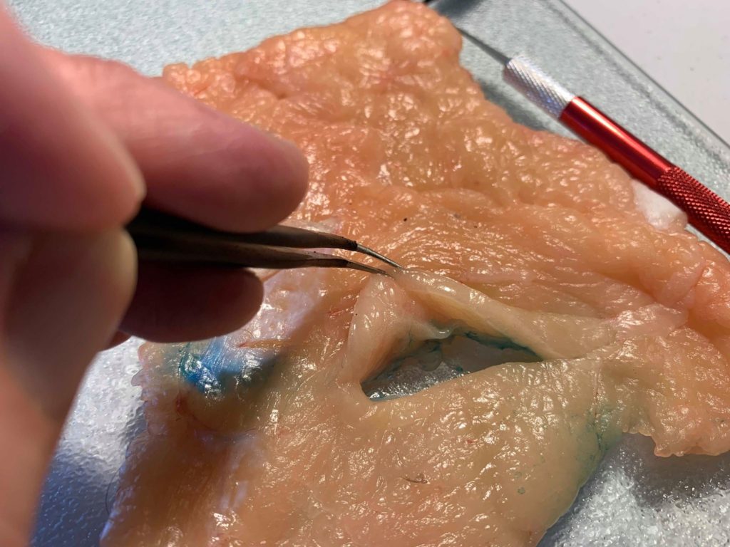
We conducted our own research using bovine tissue to study the effects of a 128hz weighted tuning fork using a 4000x zoom endoscope, and we have video of the fluid escaping from the interstitial fluid spaces with bubbles appearing at the base of the tuning fork indicating the presence of cavitation. We can observe fluid volume reducing from the brown adipose layer only while the tuning fork was vibrating. It appears to us that the weighted tuning fork appear to produce the necessary environment for the well-known acoustic cavitation phenomenon to occur within tissue, membranes, and layers.
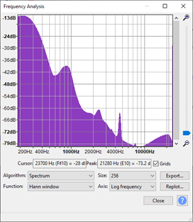
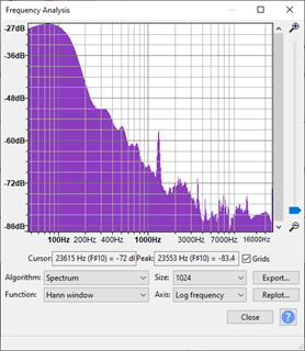
We also recorded audio from our weighted 128hz tuning fork using a Zoom H6 Digital Recorder and a shotgun microphone to process the frequency ranges. Audio was captured at the longitudinal axis near the tines (Image A) and directly from the base pointed directly at the shotgun microphone (Image B). Both audio files remained in an uncompressed .WAV format for frequency analysis in Audacity software. We observed multiple frequencies across the hearing range as previously mentioned in the 1992 American Journey of Physics study. We observed a clear ramp up of loudness (dB) in the range around 20, 000hz which is above and beyond the ultrasound range required for generating cavitation is human tissue. In normal frequency spectrum analysis, decibel levels tend to fall of in strength over higher ranges since high frequencies cannot travel as far due to conversation of mechanical vibration into heat.
An increase in strength at the ultrasound range could mean the diameter of the tuning fork allows for short wavelengths of ultrasound frequencies to travel in a sound channel within the confines of the metal handle without experiencing propagation losses. I have observed this same phenomenon in my work in the Navy by running thousands of computer simulations in the underwater environment to discover “hot spots” of channels throughout the vertical columns of water where certain narrowband frequencies can travel much further without experiencing heavy transmission losses. It is clear to us that the 128hz tuning fork generates narrowband frequencies in the ultrasound range and also transmits those frequencies through the fork length and out of the base.
References:
1. Benias, P.C., Wells, R.G., Sackey-Aboagye, B. et al. Structure and Distribution of an Unrecognized Interstitium in Human Tissues. Sci Rep 8, 4947 (2018). https://doi.org/10.1038/s41598-018-23062-6
2. Saggini, Raoul & Carmignano, Simona & Saggini, Andrea & Giuliani, Elisabetta & Ancona, Emilio & Corsetti, Enrico & Giuliani, Livio & D’Agostino, Cristina & Cascian, Michele & Lucarelli, Manuela. (2017). The Mechanical Vibration: Therapeutic Effects and Applications. 10.2174/97816810850811170101.
3. Rossing, Thomas & Russell, Daniel & Brown, David. (1992). On the acoustics of tuning forks. American Journal of Physics. 60. 620-626. 10.1119/1.17116. https://www.researchgate.net/publication/259017541_On_the_acoustics_of_tuning_forks
4. Khonsary SA. Guyton and Hall: Textbook of Medical Physiology. Surg Neurol Chapt. 16, 189-201 Int. 2017;8:275. Published 2017 Nov 9. doi:10.4103/sni.sni_327_17
5. ISTVÁN RUSZNYÁK, MIHÁLY FÖLDI, GYÖRGY SZABÓ, Lymphatics and Lymph Circulation (Second Edition), Pergamon, 1967, ISBN 9780080120225, https://doi.org/10.1016/B978-0-08-012022-5.50001-3. (http://www.sciencedirect.com/science/article/pii/B9780080120225500013)
6. ISTVÁN RUSZNYÁK, MIHÁLY FÖLDI, GYÖRGY SZABÓ, CHAPTER IX – FILTRATION AND ABSORPTION THROUGH SEROUS MEMBRANES, Editor(s): ISTVÁN RUSZNYÁK, MIHÁLY FÖLDI, GYÖRGY SZABÓ, Lymphatics and Lymph Circulation (Second Edition), Pergamon, 1967, Pages 475-510, ISBN 9780080120225, https://doi.org/10.1016/B978-0-08-012022-5.50013-X. (http://www.sciencedirect.com/science/article/pii/B978008012022550013X)
7. Ikomi, F, Hiruma, S. Relationship between shape of peripheral initial lymphatics and efficiency of mechanical stimulation–induced lymph formation. Microcirculation. 2020; 27:e12606. https://doi.org/10.1111/micc.12606
8. Byung-Boong Lee, Stanley Rockson, John Bergan; Principles and Practice of Lymphedema Surgery, Chapter 4: Anatomy and Structural Physiology of the Lymphatic System
9. Scallan J, Huxley VH, Korthuis RJ. Capillary Fluid Exchange: Regulation, Functions, and Pathology. San Rafael (CA): Morgan & Claypool Life Sciences; 2010. Chapter 3, The Lymphatic Vasculature. Available from: https://www.ncbi.nlm.nih.gov/books/NBK53448/
10. Sidawy, A., Perler, B. and Rutherford, R., 2019. Rutherford’s Vascular Surgery And Endovascular Therapy. 9th ed. Chapt 10. United States: Philadelphia, PA : Elsevier, [2019].
11. http://justinablack.com/tuning_fork_freqs/
12.https://sites.google.com/site/lucidanalysis/examples/tuningfork
13. Biofluid Mechanics-An Introduction to Fluid Mechanics Macrocirculation, and Microcirculation, David Rubenstein, Wei Yin, Mary Frame, Academic Press, 2012, ISBN: 978-0-12-381383-1