
Table of Contents
Basic Sacrum Anatomy
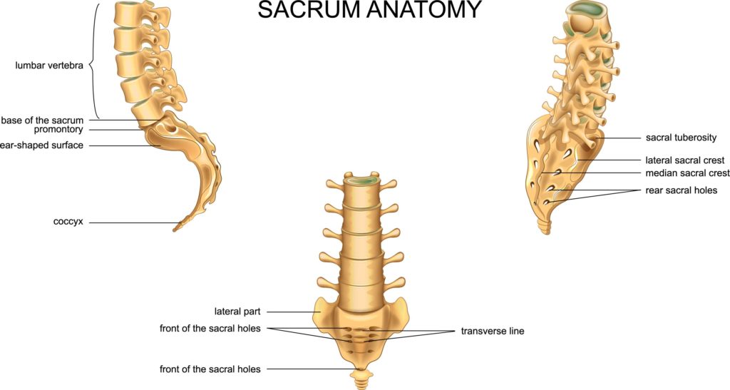
In this illustration, we’re looking at the anatomy of the sacrum, a crucial component of the human skeletal system. The sacrum is a large, triangular bone at the base of the spine and is key to forming the pelvis.
To the left, the sacrum is shown in relation to the lumbar vertebrae above it. You can observe how the base of the sacrum, which has a pronounced promontory, articulates with the last lumbar vertebra. The anterior aspect of the sacrum presents an ear-shaped surface, which is part of the sacroiliac joint, connecting the sacrum to the ilium of the pelvis.
Below the sacrum, the coccyx, or tailbone, is depicted. The coccyx is the terminal segment of the vertebral column and is composed of several fused vertebrae.
In the center, the sacrum is shown from a posterior view. Notice the medial sacral crest running down the midline, which represents the fused spinous processes of the sacral vertebrae. Flanking the crest are the posterior sacral foramina, which are openings that allow for the passage of nerves and blood vessels.
On the right, the lateral view of the sacrum is displayed. Here you can see the lateral sacral crest, which corresponds to the fused transverse processes of the sacral vertebrae, and the sacral tuberosity, which serves as a point of attachment for ligaments. The anterior sacral foramina are also visible; they align with the foramina on the posterior side and serve the same purpose.
Finally, the anterior view at the bottom center shows the sacrum head-on, highlighting the front of the sacral holes and the transverse line, which indicates the fusion points of the sacral vertebrae.
Understanding the anatomy of the sacrum is essential, particularly in fields such as orthopedics, neurology, and physiotherapy, as it plays a significant role in supporting the weight of the upper body and providing stability to the pelvis.
Spinal Conditions in Lumbar and Sacrum Region
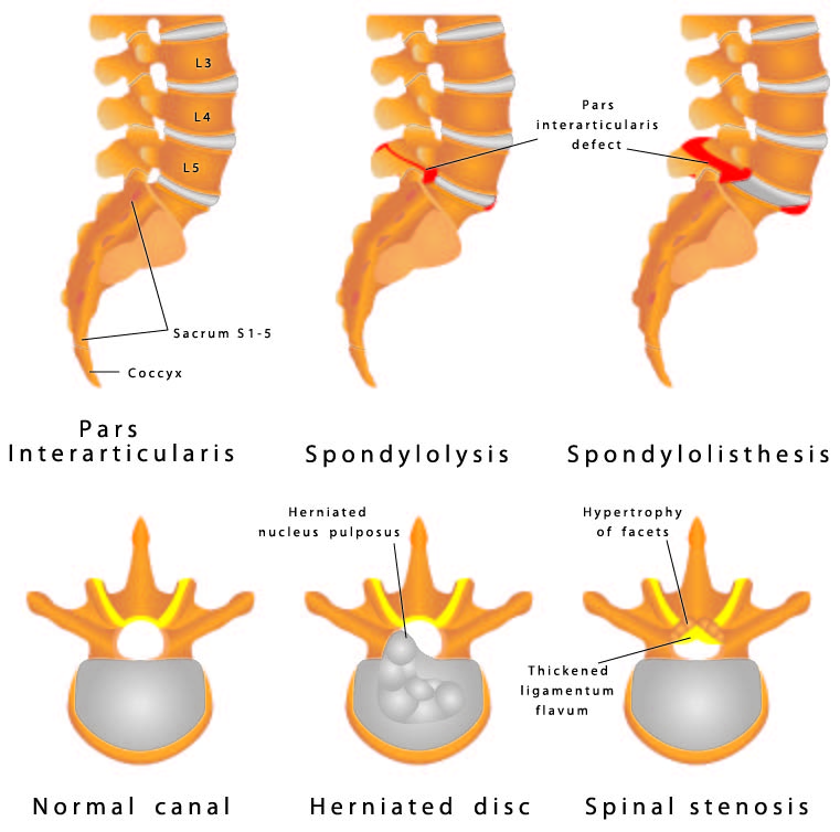
This image presents various spinal conditions along with a depiction of the lumbar spine and sacral region. The top left corner shows a normal alignment of the lumbar vertebrae labeled L3, L4, and L5, leading down to the sacrum, labeled S1-5, and ending at the coccyx, also known as the tailbone.
On the top right, we observe a condition known as spondylolysis. This is characterized by a defect or fracture in the pars interarticularis, which is the part of the vertebra located between the superior and inferior articular processes. This defect is clearly marked and is a common cause of lower back pain, especially in athletes.
Below are three axial views of the lumbar spine illustrating different conditions.
The first condition, shown on the bottom left, is normal anatomy with the spinal cord passing through the vertebral canal without any obstruction.
The middle image represents a herniated disc, where the nucleus pulposus, the soft center of the spinal disc, protrudes out of its normal boundary and impinges on the spinal canal. This can cause pain, numbness, or weakness along the path of the affected nerve.
The final image on the bottom right illustrates spinal stenosis. This condition is characterized by the narrowing of spaces in the spine, which can put pressure on the spinal cord and nerves. The image points out hypertrophy of the facets, which are the joints that allow for movement between spinal vertebrae, and thickening of the ligamentum flavum, a ligament that connects the laminae of the vertebrae. Both of these changes can contribute to the narrowing of the spinal canal.
These conditions are significant as they can lead to various symptoms and may require different treatments ranging from physiotherapy to surgical interventions. Understanding the anatomy and potential pathological changes of the spine is vital for diagnosis and management of back pain and spinal disorders.
Sacrum and Spinal Cord Relationship
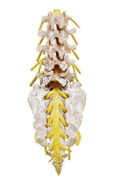
The image displays a posterior view of the human spinal column, highlighting the intricate relationship between the vertebrae and the spinal cord.
At the core, you can see the spinal cord, represented in yellow, extending from the base of the skull down through the canal formed by the successive alignment of the vertebrae. The spinal cord is a vital structure in the central nervous system, responsible for transmitting messages between the brain and the rest of the body.
Surrounding the spinal cord are the vertebrae, which are individual bones that form the vertebral column. These bones are categorized into different regions based on their location along the spine: cervical (neck), thoracic (mid-back), lumbar (lower back), sacral (pelvis), and coccygeal (tailbone).
Each vertebra consists of a vertebral body, which is the thick oval segment in front of the spinal cord, and a vertebral arch behind the spinal cord. The arch is formed by pedicles, laminae, and processes that serve as attachment points for muscles and ligaments.
Between the vertebrae are intervertebral foramina, which are openings that allow spinal nerves to exit the spinal column and branch out to the body. These nerves are critical for sensory and motor functions.
The spinal column provides protection to the spinal cord, supports the body’s weight, and allows for a flexible, controlled range of motion. It is a central structure in human anatomy, essential for both movement and sensation.
Spinal Cord Anatomy in Lumbar and Sacrum Region
The image provided is an anterior view of a portion of the human spinal column, specifically focusing on the lumbar region and the sacrum. The lumbar vertebrae are the largest segments of the movable part of the vertebral column and are characterized by their thick, robust bodies designed to bear significant weight and stress.
The anterior (frontal) perspective shows the vertebral bodies, which are the large, cylindrical parts of the vertebrae that stack on top of each other to form the front part of the spinal column. Between these bodies are the intervertebral discs, which are not visible in this model. These discs provide cushioning and allow for flexibility and movement of the spine.
Projecting posteriorly from the vertebral bodies are the spinal processes, which in this image appear as the yellow structures behind the vertebrae. These bony projections provide attachment points for muscles and ligaments.
Descending within the bony canal formed by the vertebral arches is the spinal cord, also depicted in yellow. The spinal cord is a crucial conduit for information traveling between the brain and the peripheral nervous system.
Below the lumbar vertebrae, the sacrum is visible. The sacrum is a large, triangular bone at the base of the spine, forming the back of the pelvis. It consists of five fused vertebrae and articulates with the hip bones to form the sacroiliac joints.
This anatomical representation is essential in the study of human biomechanics, orthopedics, and neurology, as it provides a clear view of the structures that support our body’s framework and protect the neural elements that control our body’s movements and functions.
Sacrum Vertebra: Lateral View
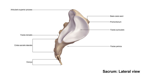
This image depicts the lateral view of the sacrum, which is the triangular bone situated at the lower end of the spine and is wedged between the two hip bones in the pelvis.
At the top of the sacrum, we have the base of the sacral bone, known as the basis ossis sacri, which is the broad, upper part of the sacrum that articulates with the last lumbar vertebra. Just below the base is the promontory, a prominent bulge that marks the anterior edge of the base and is an important anatomical landmark in obstetrics.
Moving anteriorly from the promontory, there is the pelvic surface, labeled here as facies pelvica, which is slightly concave and faces the pelvic cavity. It contributes to the formation of the pelvic brim and supports the weight of the abdominal organs.
On the dorsal side, which is the back surface facing away from the pelvic cavity, you have the dorsal surface, labeled as facies dorsalis. This roughened area provides attachment points for ligaments and muscles of the back.
Along the edge of the sacrum, we can see the lateral sacral crest, crista sacralis lateralis, which is a continuation of the transverse processes of the sacral vertebrae. This crest serves as an attachment for the sacroiliac ligaments and also forms the lateral border of the posterior sacral foramina.
The auricular surface, termed here as facies auricularis, is located on the side of the sacrum and articulates with the ilium to form the sacroiliac joint, which is essential for transmitting weight from the upper body to the pelvis and legs.
Finally, at the very bottom of the sacrum is the coccyx, also known as the tailbone. It is a small, triangular bone composed of several fused segments that form the terminus of the vertebral column.
This lateral view of the sacrum is crucial for understanding the anatomy of the pelvis and the spine, as well as for the study of body mechanics, particularly in how weight is transferred and distributed across the lower part of the skeleton.
