
Hand and Wrist Extrinsic Muscles
Muscles: The Powerhouse of the Hand
The muscles of the hand are the dynamic engines that drive movement, allowing us to perform tasks ranging from powerful gripping to delicate manipulation. Technically, there are no muscles that originate solely within the wrist itself. The wrist’s movement and stability are controlled by muscles that originate in the forearm and extend through the wrist via tendons. Here’s a closer look at these two main groups:
Extrinsic Muscles: Strength and Gross Movement
The extrinsic muscles of the hand are powerful and versatile, enabling a wide range of large movements. Here’s a more detailed look at their essential functions:
Flexors and Extensors: The Dynamic Duo
The extrinsic muscles are categorized into flexors and extensors, each with specific functions:
- Flexors: These muscles are responsible for bending the fingers and wrist. They enable actions like making a fist, gripping objects, or performing pull-ups. Flexor tendons run through the carpal tunnel, a narrow passageway in the wrist.
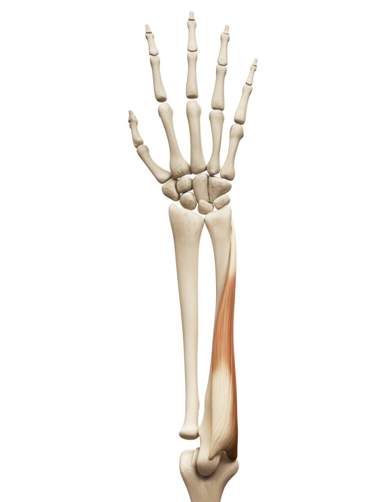
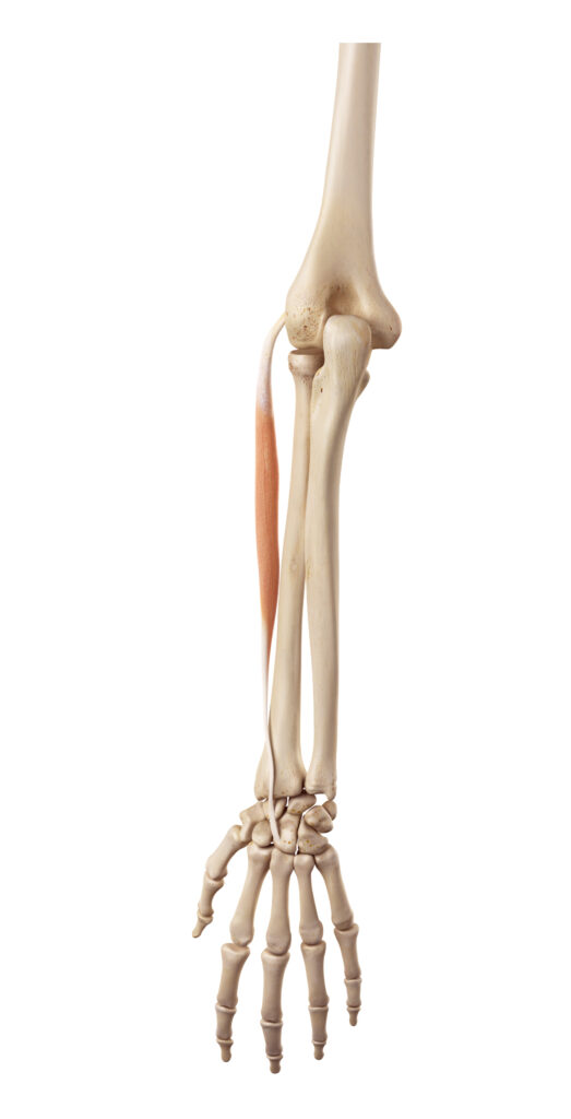
Flexor Carpi Radialis:
Helps in flexing the wrist and assists in radial abduction (moving the wrist towards the thumb).
Flexor Carpi Radialis (FCR): Anatomy and Function The Flexor Carpi Radialis (FCR) is one of the key muscles in the forearm that plays a crucial role in wrist movement. Here’s an in-depth look at its characteristics, functions, and clinical relevance:
Anatomy
- Origin: The FCR originates from the medial epicondyle of the humerus, a bony prominence on the inside part of the upper arm.
- Insertion: It inserts into the base of the second metacarpal bone in the hand.
- Innervation: It is innervated by the median nerve, which carries signals from the brain to control the muscle’s contraction.
Function
- Wrist Flexion: The primary action of the FCR is to flex the wrist, meaning it bends the wrist forward, bringing the palm closer to the forearm.
- Radial Abduction: It also assists in radial abduction, which is the movement of the wrist towards the thumb side (radial side). This action helps in actions like opening a jar or turning a key.
Synergistic Action
- Working with Other Muscles: The FCR works in synergy with other flexor muscles, such as the Flexor Carpi Ulnaris, to provide controlled flexion of the wrist. It also works with extensor muscles to stabilize the wrist during various activities.
Clinical Relevance
- Repetitive Strain Injury (RSI): Overuse of the FCR, due to repetitive actions like typing or manual labor, can lead to inflammation and pain.
- Carpal Tunnel Syndrome: The FCR tendon runs through the carpal tunnel, and swelling of this tendon can contribute to compression of the median nerve.
- Palpation and Treatment: Therapists may palpate the FCR to identify tenderness or swelling. Targeted treatments like tuning fork therapy or heat application may be used to alleviate discomfort and restore normal function. The Flexor Carpi Radialis is more than just a wrist flexor; it plays a multifaceted role in the dynamic function of the wrist and hand. Its ability to both flex the wrist and abduct it radially allows for a wide range of movements that are integral to many daily activities. Understanding the FCR’s anatomy and function is essential for therapists working with wrist-related issues, as targeted assessment and treatment can address dysfunctions related to this key muscle. Whether dealing with chronic overuse injuries or acute pain, a nuanced approach to the FCR can lead to effective healing and a return to normal wrist function.
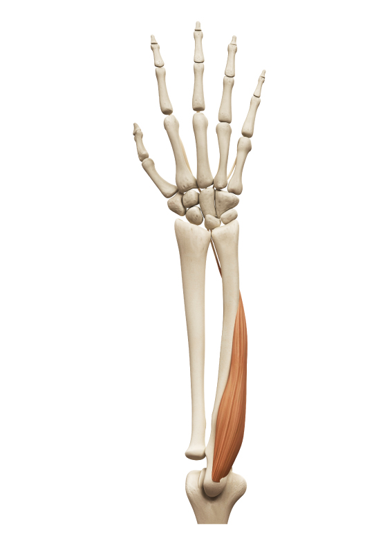
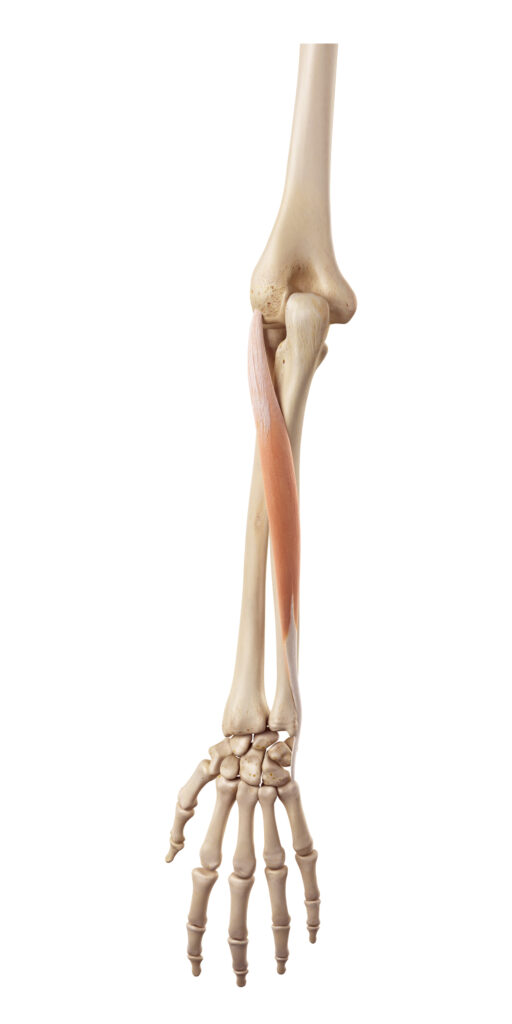
Flexor Carpi Ulnaris:
Anatomy
- Origin: The FCU has two heads of origin: the humeral head originates from the medial epicondyle of the humerus, and the ulnar head originates from the olecranon and posterior border of the ulna.
- Insertion: It inserts into the pisiform, hook of the hamate, and the base of the fifth metacarpal bone.
- Innervation: The FCU is innervated by the ulnar nerve.
Function
- Wrist Flexion: Similar to the Flexor Carpi Radialis, the FCU aids in flexing the wrist, bending it forward.
- Ulnar Abduction (Adduction): The FCU’s unique function is ulnar abduction (also known as adduction), moving the wrist towards the little finger side (ulnar side). This movement is essential in actions like gripping or holding objects with the palm.
Synergistic and Antagonistic Action
- Synergy with Other Flexors: The FCU works with other flexor muscles, such as the Flexor Carpi Radialis, to provide controlled and balanced wrist flexion.
- Antagonistic to Extensors: The FCU acts antagonistically to the extensor muscles, providing a counterbalance during wrist movements.
Clinical Relevance
- Tendinitis and Strain: Overuse or strain of the FCU can lead to inflammation and tenderness, commonly seen in activities that require repetitive gripping or wrist flexion.
- Cubital Tunnel Syndrome: Since the FCU is innervated by the ulnar nerve, issues with this nerve (such as cubital tunnel syndrome) can affect the FCU’s function.
- Palpation and Treatment: Therapists might palpate the FCU to detect abnormalities and may employ treatments like tuning fork therapy, stretching, or heat application to alleviate symptoms and restore function.
The Flexor Carpi Ulnaris is an essential player in wrist and hand movement, contributing to both flexion and ulnar abduction. Its dual actions enable a variety of complex hand functions, from simple gripping to more intricate tasks. Understanding the FCU’s unique anatomy, biomechanics, and common pathologies provides insights for therapists working with wrist-related issues. Whether addressing chronic conditions like tendinitis or acute injuries, a targeted approach to the FCU can offer relief and facilitate recovery, enhancing the overall function and well-being of the wrist and hand
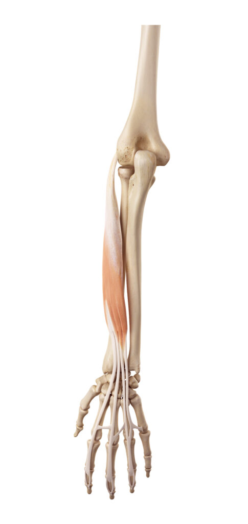
Flexor Digitorum Superficialis (FDS):
The Flexor Digitorum Superficialis (FDS) is a unique muscle in the forearm with specific functions related to the fingers. Here’s an in-depth look at the Flexor Digitorum Superficialis:
Anatomy
- Origin: The FDS has two heads of origin: the humeroulnar head, which originates from the medial epicondyle of the humerus, and the radial head, which originates from the radius.
- Insertion: The tendons of the FDS insert into the middle phalanges of the four fingers (index through little finger).
- Innervation: The FDS is innervated by the median nerve.
Function
- Flexion of Middle Phalanges: The primary function of the FDS is to flex the middle phalanges of the fingers. This action is essential in activities like making a fist, gripping objects, or playing musical instruments.
- Assists in Wrist Flexion: Though its primary role is in finger flexion, the FDS also contributes to wrist flexion.
Synergistic and Antagonistic Action
- Synergy with Other Flexors: The FDS works in coordination with other flexor muscles of the forearm, including the Flexor Digitorum Profundus, to provide controlled flexion of the fingers.
- Antagonistic to Extensors: It acts antagonistically to the extensor muscles of the fingers, balancing the forces during finger movements.
Clinical Relevance
- Repetitive Strain Injury (RSI): Overuse of the FDS, particularly in activities requiring repetitive finger flexion, can lead to inflammation and pain.
- Trigger Finger: Swelling or constriction of the FDS tendon sheath can cause a condition known as trigger finger, where the finger becomes locked in a bent position.
- Palpation and Treatment: Therapists may palpate the FDS to identify areas of tenderness or dysfunction and may use treatments like tuning fork therapy or stretching to alleviate symptoms and restore normal function.
The Flexor Digitorum Superficialis plays a vital and specific role in the movement of the fingers. Its ability to flex the middle phalanges enables many daily tasks and specialized activities, from holding a cup to playing a piano. Understanding the FDS’s anatomy, function, and common pathologies is essential for therapists working with finger-related issues. A targeted and nuanced approach to the FDS can offer effective treatment for conditions like repetitive strain injury or trigger finger, enhancing finger function and overall hand health.
Flexor Digitorum Profundus (FDP):
The Flexor Digitorum Profundus (FDP) is another critical muscle in the forearm, with specific functions related to the distal phalanges of the fingers. Let’s explore the Flexor Digitorum Profundus in detail:
Anatomy
- Origin: The FDP originates from the upper 3/4 of the anterior and medial surfaces of the ulna, interosseous membrane, and deep fascia of the forearm.
- Insertion: The tendons of the FDP insert into the bases of the distal phalanges of the four fingers (index through little finger).
- Innervation: The FDP is unusual in that it has dual innervation. The medial half (serving the little and ring fingers) is innervated by the ulnar nerve, while the lateral half (serving the index and middle fingers) is innervated by the median nerve.
Function
- Flexion of Distal Phalanges: The primary function of the FDP is to flex the distal phalanges of the fingers. This action allows intricate movements like pinching, typing, or creating intricate art.
- Assists in Wrist and Other Finger Joints Flexion: Besides flexing the distal phalanges, the FDP contributes to flexion at the wrist and other finger joints.
Synergistic and Antagonistic Action
- Synergy with Other Flexors: The FDP works in harmony with other flexor muscles of the fingers, such as the Flexor Digitorum Superficialis, to provide coordinated flexion of the fingers.
- Antagonistic to Extensors: It acts antagonistically to the extensor muscles of the fingers, balancing the forces during finger extension.
Clinical Relevance
- Repetitive Strain Injury (RSI): Similar to other flexors, overuse of the FDP can lead to inflammation and pain, particularly in activities requiring repetitive finger movements.
- Tendon Injuries: Trauma or strain to the FDP tendons can result in decreased ability to flex the distal phalanges.
- Palpation and Treatment: Therapists may palpate the FDP to find areas of tenderness or dysfunction, utilizing treatments like tuning fork therapy, stretching, or strengthening exercises to restore normal function.
The Flexor Digitorum Profundus is essential for the fine motor control and strength of the fingers, particularly in flexing the distal phalanges. Its unique anatomy, dual innervation, and specific function enable a wide array of complex hand movements. Understanding the FDP’s role is vital for therapists working with finger-related conditions, as targeted assessment and treatment can address dysfunctions related to this key muscle. Whether dealing with chronic overuse injuries or acute trauma, a nuanced approach to the FDP can lead to effective healing and a return to normal finger function.
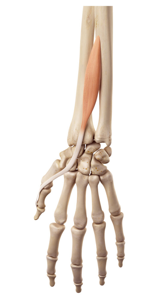
Palmaris Longus (PL):
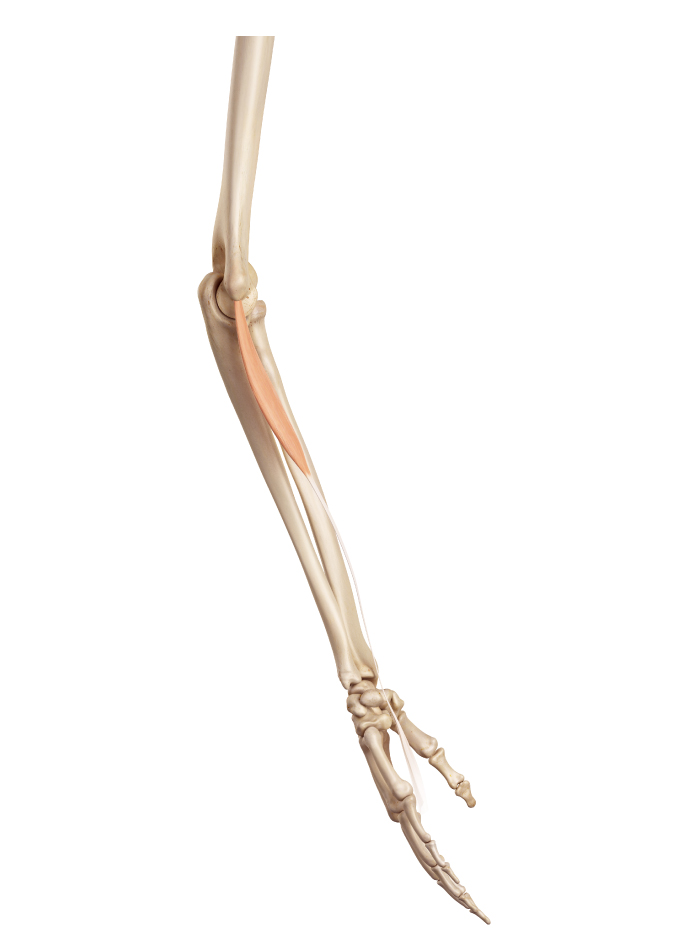
Anatomy
- Origin: The Palmaris Longus has its origin point at the medial epicondyle of the humerus. This bony prominence serves as a common origin for several other flexor muscles of the forearm, adding complexity to the functional anatomy of this region.
- Insertion: This muscle extends to insert into the palmar aponeurosis, a broad and thin fascial layer situated in the palm of the hand. This specialized connective tissue plays a key role in the hand’s grip strength and overall functionality.
- Innervation: Neural control for the Palmaris Longus is provided by the median nerve, which is responsible for both sensory and motor functions in the forearm and hand. The median nerve’s involvement ensures that the muscle operates with precision and coordination.
- Variability in Presence: One of the most striking features of the Palmaris Longus is its variable presence. This muscle is absent in approximately 14% of individuals. This absence can manifest in a unilateral or bilateral manner and is known to vary among different ethnic groups.
Function
- Assists in Wrist Flexion: Although not a primary mover, the Palmaris Longus assists in flexing the wrist. It works in synergy with other flexor muscles to provide a controlled and balanced movement, especially during tasks that require a strong grip or precise manipulation of objects.
- Tensioning the Palmar Aponeurosis: The PL also plays a role in tensing the palmar aponeurosis, which may help in grip functions.
Clinical Relevance
- Identification: The PL can often be seen or felt by opposing the thumb and little finger while flexing the wrist, though it’s absent in some individuals.
- Graft Material: Due to its minimal functional importance, the PL tendon is commonly used as a graft material in surgical procedures, such as tendon repair in other parts of the body.
- Repetitive Strain Injury (RSI): Like other flexor muscles, overuse of the PL can potentially lead to strain or inflammation.
- Palpation and Treatment: Therapists may palpate the PL to check for tenderness or dysfunction and may employ treatments like tuning fork therapy or stretching if needed. The Palmaris Longus is a unique and somewhat enigmatic muscle, given its absence in a significant portion of the population and its minimal contribution to wrist function.
Despite its relatively minor role, it serves as a fascinating example of human anatomical variation. Its presence or absence is typically of little functional consequence, but understanding its anatomy and potential clinical applications (such as its use in grafting) can be valuable for healthcare professionals. Its role in wrist flexion and potential relevance in certain therapeutic contexts adds to the complex tapestry of the forearm’s musculature. Whether palpating the forearm in a clinical setting or considering surgical interventions, the Palmaris Longus offers insights into the diversity and adaptability of the human body.
Coordinated Function of Forearm Flexor Muscles
The combined action of the muscles we’ve discussed—Flexor Carpi Radialis, Flexor Carpi Ulnaris, Flexor Digitorum Superficialis, Flexor Digitorum Profundus, and Palmaris Longus—provides the intricate and powerful movements required for various hand functions. Let’s explore how these muscles work together:
Making a Fist
- Flexor Digitorum Superficialis and Profundus: These muscles flex the middle and distal phalanges of the fingers, curling them into a fist.
- Flexor Carpi Radialis and Ulnaris: Flexing the wrist adds stability and strength to the fist, especially in actions like punching or gripping tightly.
Gripping Objects
- Combination of Flexors: Gripping requires coordinated action among all the flexor muscles, allowing the fingers to wrap around objects of various shapes and sizes.
- Palmaris Longus: Though its role is minor, the Palmaris Longus may assist in tensing the palmar aponeurosis, enhancing the grip.
Holding Onto Things (e.g., Handlebar, Pen)
- Flexor Carpi Radialis and Ulnaris: Flexing and stabilizing the wrist provide a strong base for holding objects.
- Flexor Digitorum Superficialis and Profundus: Flexing the fingers allows them to conform to the shape of the object, whether it’s the slender shaft of a pen or the thicker grip of a handlebar.
- Synergistic Action: Holding onto things requires constant adjustments and fine control, achieved through the synergistic action of these muscles.
Clinical and Therapeutic Considerations
- Assessment: Therapists may assess the coordinated function of these muscles in actions like gripping or holding, looking for weaknesses or imbalances.
- Tuning Fork Therapy: Targeted vibration therapy may be applied to specific muscles or tendons to alleviate discomfort, improve fluid movement, and enhance overall hand function.
- Rehabilitation: Following injuries or surgeries, rehabilitation may focus on restoring the coordinated action of these muscles, enabling the return to daily activities and specific tasks. The intricate dance of the forearm’s flexor muscles allows us to perform a wide array of daily tasks, from simple actions like holding a cup to specialized skills like playing a musical instrument. Understanding how these muscles work together offers insights into the biomechanics of the hand and provides a foundation for therapeutic interventions. Whether addressing chronic conditions, acute injuries, or simply appreciating the marvel of human movement, the coordinated function of these muscles underscores the complexity and adaptability of the human body.
Extensors: Straightening and Lifting
Extensor Carpi Radialis Longus and Brevis (ECRL and ECRB):
The Extensor Carpi Radialis Longus (ECRL) and Extensor Carpi Radialis Brevis (ECRB) are vital muscles in the forearm that contribute to wrist extension and radial abduction. Let’s explore these muscles in detail:
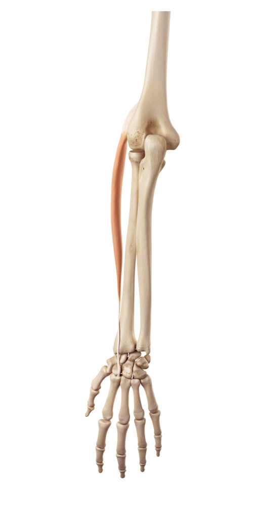
Extensor Carpi Radialis Longus (ECRL)
- Origin: Lateral supracondylar ridge of the humerus.
- Insertion: Base of the second metacarpal bone.
- Innervation: Radial nerve.
The ECRL originates from the lateral supracondylar ridge of the humerus, a prominent bony feature found on the upper arm bone. This muscle extends downward to insert itself at the base of the second metacarpal bone in the hand. The strategic positioning of its insertion point allows the ECRL to exert a powerful influence on wrist extension and radial deviation. When it comes to neural control, the ECRL is innervated by the radial nerve, a major nerve originating from the brachial plexus that provides motor and sensory functions to the arm’s posterior and lateral aspects. The radial nerve’s involvement ensures that the ECRL performs its movements with precision and coordination.
Extensor Carpi Radialis Brevis (ECRB)
- Origin: Lateral epicondyle of the humerus.
- Insertion: Base of the third metacarpal bone.
- Innervation: Deep branch of the radial nerve.
In the realm of anatomical study, the arm presents a complex interplay of muscles, each serving specific functions that contribute to the overall movement and stability of the arm and hand. Among these muscles, two key players stand out for their role in wrist extension and radial deviation: the Extensor Carpi Radialis Longus (ECRL) and the Extensor Carpi Radialis Brevis (ECRB).
While closely related to the ECRL, the ECRB has distinct anatomical features that make it unique. Its origin point is situated at the lateral epicondyle of the humerus, a bony prominence located near the elbow joint. From here, the muscle extends to insert at the base of the third metacarpal bone, providing it with the leverage to contribute to wrist extension and radial deviation, albeit in a slightly different manner than its longer counterpart. Neural control of the ECRB is provided by the deep branch of the radial nerve, which ensures that the muscle operates in synergy with other extensor muscles of the forearm.
Function
- Wrist Extension: Both the ECRL and ECRB are responsible for extending the wrist, meaning they straighten the wrist and lift the back of the hand.
- Radial Abduction: These muscles also aid in radial abduction, moving the wrist toward the thumb side (radial side). This movement is crucial in actions like spreading the fingers or positioning the hand to grasp objects.
Synergistic and Antagonistic Action
- Synergy with Other Extensors: The ECRL and ECRB work in coordination with other extensor muscles of the wrist, such as the Extensor Carpi Ulnaris, to provide balanced and controlled wrist extension.
- Antagonistic to Flexors: They act antagonistically to the flexor muscles of the wrist, ensuring smooth transitions between flexion and extension movements.
Clinical Relevance
- Lateral Epicondylitis (Tennis Elbow): Overuse or strain of the ECRB can contribute to lateral epicondylitis, a painful condition affecting the outer elbow.
- Wrist Injuries: Dysfunction in these muscles may contribute to wrist pain or instability, particularly in activities requiring repetitive wrist extension.
- Palpation and Treatment: Therapists may palpate the ECRL and ECRB to identify areas of tenderness or dysfunction. Treatment modalities like tuning fork therapy, stretching, or strengthening exercises may be employed to restore normal function.
The Extensor Carpi Radialis Longus and Brevis play a vital role in the dynamic function of the wrist, enabling extension and radial abduction. These movements are foundational to many daily tasks and specialized skills, from lifting objects to playing sports like tennis. Understanding the anatomy, function, and common issues associated with the ECRL and ECRB provides insights for therapists and medical professionals working with wrist-related conditions. A targeted approach to these muscles, whether through assessment, treatment, or rehabilitation, can lead to effective healing and a return to normal wrist function.
Extensor Carpi Ulnaris (ECU):
The Extensor Carpi Ulnaris (ECU) is a significant muscle in the forearm that plays a key role in extending the wrist and assisting in ulnar abduction. Let’s delve into the details of this muscle:
Anatomy
- Origin: The ECU originates from both the lateral epicondyle of the humerus (common extensor origin) and the dorsal surface of the ulna.
- Insertion: It inserts into the base of the fifth metacarpal bone.
- Innervation: The ECU is innervated by the posterior interosseous nerve, a branch of the radial nerve.
In the study of human anatomy, particularly concerning the musculature of the forearm and hand, the Extensor Carpi Ulnaris (ECU) emerges as a muscle of significant functional importance. Situated on the ulnar side of the forearm, the ECU serves vital roles in wrist extension and ulnar deviation, contributing to a wide range of movements that extend from simple grasping to complex manipulative tasks.
Extensor Carpi Ulnaris (ECU)
The origin of the ECU is dual-natured, emanating from two distinct anatomical landmarks. The first point of origin is the lateral epicondyle of the humerus, commonly referred to as the common extensor origin. This bony prominence near the elbow serves as the starting point for several extensor muscles of the forearm. The second origin point lies on the dorsal surface of the ulna, the bone on the medial side of the forearm. This dual origin provides the ECU with a robust anchoring structure, enabling it to exert force efficiently across the wrist joint.
Progressing distally from its points of origin, the ECU extends to insert itself into the base of the fifth metacarpal bone. This specific point of insertion enables the muscle to exert its influence on the pinky side of the hand, thus playing a critical role in wrist extension and ulnar deviation.
In terms of neural control, the ECU is innervated by the posterior interosseous nerve, a specialized branch of the radial nerve. This nerve branch is crucial for providing the ECU with the neural impulses required for precise and coordinated movements. The posterior interosseous nerve ensures that the muscle’s actions are well-synchronized with the other muscles of the forearm, contributing to the fluidity and accuracy of wrist and hand movements.
In summary, the Extensor Carpi Ulnaris serves as a key player in the anatomical and functional landscape of the forearm and hand. Its unique origin points, specific insertion, and specialized neural innervation all contribute to its role in wrist extension and ulnar deviation. Understanding the anatomical details of the ECU provides us with a more comprehensive view of the complexity involved in even the most mundane movements, illuminating the intricate design and functional sophistication of the human musculoskeletal system.
Function
- Wrist Extension: The primary function of the ECU is to extend the wrist, allowing the hand to move away from the palm side.
- Ulnar Abduction (Adduction): The ECU also assists in ulnar abduction (also known as adduction), moving the wrist towards the little finger side (ulnar side). This action helps in activities like swinging a hammer or stabilizing the wrist during gripping.
Function of the Extensor Carpi Ulnaris (ECU)
Wrist Extension
The ECU’s foremost role is in the extension of the wrist, a movement that facilitates the hand moving away from the palm side. This action is fundamental to numerous tasks, including but not limited to lifting objects, typing, and even simple gestures like waving. Wrist extension is indispensable in activities that demand an extended range of motion and the ability to apply force in an upward direction. The ECU’s specific anatomical configuration—originating from the lateral epicondyle of the humerus and the dorsal surface of the ulna, and inserting into the base of the fifth metacarpal—optimally positions it to execute this function effectively.
Ulnar Abduction (Adduction)
In addition to wrist extension, the ECU plays a critical role in ulnar abduction, also commonly referred to as adduction. This function involves moving the wrist towards the little finger side, or the ulnar side. Such a movement is especially useful in activities that require a lateral force, such as swinging a hammer. It also stabilizes the wrist during gripping tasks, providing a counterbalance to the forces applied by the thumb and the radial side of the hand.
This action of ulnar abduction underscores the ECU’s versatility and its importance in functional activities. Whether you are gripping a steering wheel, swinging a tennis racket, or merely picking up a glass, the ECU contributes to the stability and agility of the wrist. Its role in ulnar abduction complements its primary function in wrist extension, making it a key player in the intricate ballet of muscles that orchestrate the movements of the forearm and wrist.
In conclusion, the Extensor Carpi Ulnaris is not just an anatomical structure but a dynamic entity with roles that are crucial to our everyday function. Its dual capabilities in wrist extension and ulnar abduction allow for a broad spectrum of movements, contributing to both the versatility and the specialization of the human hand and forearm. Understanding these functions in detail provides a deeper insight into the mechanical genius of human physiology, highlighting the intricacies that allow us to perform a wide array of tasks with both power and precision.
Synergistic and Antagonistic Action
- Synergy with Other Extensors: The ECU works in coordination with other extensor muscles of the wrist, such as the Extensor Carpi Radialis Longus and Brevis, to provide controlled wrist extension.
- Antagonistic to Flexors: The ECU acts antagonistically to the flexor muscles, particularly the Flexor Carpi Ulnaris, balancing the forces during wrist movements.
In the complex choreography of muscular action that governs human movement, the Extensor Carpi Ulnaris (ECU) is far from a solo performer. Its role in the musculoskeletal system is nuanced by its relationships with other muscles, functioning both in synergy and in antagonism to facilitate a broad range of motions and ensure the stability of the wrist. Below, we delve into these dynamic interactions to better understand the ECU’s place within the broader context of muscular function.
Synergistic and Antagonistic Action of the Extensor Carpi Ulnaris (ECU)
Synergy with Other Extensors
The ECU does not work in isolation when executing wrist extension; it acts in concert with other extensor muscles, particularly the Extensor Carpi Radialis Longus (ECRL) and Extensor Carpi Radialis Brevis (ECRB). This coordinated effort allows for a controlled and balanced extension of the wrist. By sharing the workload among these muscles, the body can achieve more refined, stable, and powerful movements. For instance, the combined action of the ECU with the ECRL and ECRB is integral to activities that require both strength and precision, such as lifting a heavy object or manipulating a tool.
Antagonistic to Flexors
While the ECU collaborates with its fellow extensors, it acts in opposition to the flexor muscles, particularly the Flexor Carpi Ulnaris (FCU). This antagonistic relationship is crucial for balancing the forces exerted on the wrist during movement. When the ECU contracts to extend the wrist, the FCU simultaneously relaxes to allow this movement, and vice versa. This interplay ensures that neither extension nor flexion becomes excessive, thereby preventing potential injury and enabling a full range of controlled motion.
The Extensor Carpi Ulnaris serves as both a collaborator and a counterbalance within the muscular network of the forearm and wrist. Its synergistic action with other extensors allows for controlled and powerful wrist extension, while its antagonistic relationship with flexor muscles ensures a balanced and stable range of motion. These dynamic interactions amplify the ECU’s functional significance, revealing the muscle as a key component in the intricate system that underpins our physical capabilities. Understanding the synergistic and antagonistic actions of the ECU offers a richer, more nuanced view of the biomechanics involved in even the simplest wrist movements, thereby illuminating the marvel of the human musculoskeletal system.
Clinical Relevance
- Repetitive Strain Injury (RSI): Overuse or strain of the ECU, particularly in activities requiring repetitive wrist extension or ulnar abduction, can lead to inflammation and pain.
- Subluxation: The ECU tendon can subluxate or dislocate from its groove in certain wrist positions or following injury, causing pain and snapping sensation.
- Palpation and Treatment: Therapists may palpate the ECU to identify areas of tenderness or dysfunction and may utilize treatments like tuning fork therapy, splinting, or targeted exercises to restore normal function.
The Extensor Carpi Ulnaris is vital for both the extension and ulnar abduction of the wrist, enabling a wide range of movements that are essential for daily tasks and specialized activities. Understanding the ECU’s anatomy, biomechanics, and common pathologies is crucial for therapists and medical professionals working with wrist-related issues. Whether addressing chronic conditions like repetitive strain injury or acute issues like tendon subluxation, a targeted approach to the ECU can lead to effective treatment and restoration of wrist function. The ECU’s role in stabilizing and moving the wrist underscores the complexity and adaptability of the forearm’s musculature.
In a clinical context, the Extensor Carpi Ulnaris (ECU) holds particular relevance due to its susceptibility to various conditions and injuries. Understanding the anatomical details and functional roles of this muscle can aid healthcare providers in the diagnosis and treatment of a range of issues affecting the forearm and wrist. Below we explore the clinical implications associated with the ECU, including repetitive strain injuries, tendon subluxation, and the approaches to palpation and treatment.
Clinical Relevance of the Extensor Carpi Ulnaris (ECU)
Repetitive Strain Injury (RSI)
One of the most common issues associated with the ECU is Repetitive Strain Injury (RSI). Activities that require repetitive wrist extension or ulnar abduction can overuse or strain this muscle, leading to inflammation and pain. Occupations or hobbies that involve continuous wrist movement, such as typing, painting, or certain sports, can make individuals more susceptible to developing RSI in the ECU. Prompt diagnosis and intervention are essential to prevent the condition from worsening and affecting the individual’s quality of life.
Subluxation
Another clinical concern related to the ECU is tendon subluxation or dislocation. The ECU tendon can become dislodged from its groove in specific wrist positions or following an injury. This condition can cause a range of symptoms, including pain and a snapping sensation during wrist movement. Subluxation often necessitates immediate medical attention to prevent further complications, such as chronic instability or tendon degeneration.
Palpation and Treatment
For diagnosis and treatment planning, therapists may use palpation techniques to identify areas of tenderness or dysfunction in the ECU. By applying pressure along the muscle and its tendon, healthcare providers can isolate points of concern that may require further investigation or targeted treatment. Various therapeutic approaches may be employed to restore normal function to the ECU, ranging from tuning fork therapy to address tissue imbalances, to splinting for joint stabilization, and targeted exercises to improve strength and flexibility.
The Extensor Carpi Ulnaris is not only of anatomical and functional importance but also holds significant clinical relevance. Understanding its roles and potential vulnerabilities allows for more accurate diagnosis and effective treatment of conditions affecting this muscle. The range of clinical issues that can involve the ECU underscores the need for comprehensive knowledge about its anatomy, function, and synergistic and antagonistic relationships with other muscles. Such understanding is crucial for healthcare providers who aim to offer the most effective treatments for disorders affecting the forearm and wrist.
Extensor Digitorum (ED):
The Extensor Digitorum (ED) is an essential muscle in the forearm, playing a vital role in extending the fingers and the wrist. Let’s explore the Extensor Digitorum in detail:
Anatomy
- Origin: The Extensor Digitorum originates from the lateral epicondyle of the humerus, part of the common extensor origin.
- Insertion: The tendons of the ED insert into the dorsal surface of the middle and distal phalanges of the four fingers (index through little finger).
- Innervation: The ED is innervated by the posterior interosseous nerve, a branch of the radial nerve.
Function
- Finger Extension: The primary function of the ED is to extend the fingers, meaning it straightens the fingers from a flexed position. This action is vital in activities like opening the hand, spreading the fingers, or releasing a grip.
- Wrist Extension: The ED also contributes to wrist extension, allowing the hand to move away from the palm side.
Synergistic and Antagonistic Action
- Synergy with Other Extensors: The ED works in coordination with other extensor muscles of the wrist and fingers to provide controlled and balanced extension.
- Antagonistic to Flexors: It acts antagonistically to the flexor muscles of the fingers, ensuring smooth transitions between flexion and extension movements.
Clinical Relevance
- Repetitive Strain Injury (RSI): Overuse or strain of the ED, particularly in activities requiring repetitive finger extension, can lead to inflammation and pain.
- Extensor Tendon Injuries: Trauma or lacerations to the ED tendons can result in decreased ability to extend the fingers.
- Palpation and Treatment: Therapists may palpate the ED to detect abnormalities and may employ treatments like tuning fork therapy, stretching, or strengthening exercises to alleviate symptoms and restore function.
The Extensor Digitorum plays a vital role in the extension of the fingers and contributes to wrist extension. Its ability to straighten the fingers enables many daily tasks and specialized activities, from simple actions like waving to more complex tasks like typing or playing a musical instrument. Understanding the ED’s anatomy, function, and common pathologies is essential for therapists working with finger-related issues. A targeted approach to the ED can offer effective treatment for conditions like repetitive strain injury or extensor tendon injuries, enhancing finger function and overall hand health. The Extensor Digitorum underscores the complexity and adaptability of the forearm’s musculature, enabling the intricate and powerful movements that define human hand function.
Extensor Pollicis Longus (EPL) and Extensor Pollicis Brevis (EPB):
The Extensor Pollicis Longus (EPL) and Extensor Pollicis Brevis (EPB) are specialized muscles in the forearm that focus on extending the thumb. Let’s explore these muscles in detail:
Anatomy of Extensor Pollicis Longus (EPL)
- Origin: The EPL originates from the middle part of the ulna and the interosseous membrane, a fibrous sheet that connects the ulna and radius. This strategic origin provides the muscle with the leverage and length required for effective thumb extension.
- Insertion: The tendons of the EPL extend to insert into the base of the distal phalanx of the thumb, allowing for precise and controlled movement of the thumb’s tip.
- Innervation: Neurological control is provided by the posterior interosseous nerve, a specialized branch of the radial nerve. This innervation ensures well-coordinated and precise movements of the thumb.
Function
- Thumb Extension: Both the EPL and EPB play crucial roles in extending the thumb. While the EPL focuses on the extension of the thumb’s distal phalanx, the EPB primarily influences the thumb’s proximal phalanx. These nuanced roles allow for a wide range of thumb movements and contribute to the hand’s dexterity.
Anatomy of Extensor Pollicis Brevis (EPB)
- Origin: The EPB has its origin on the posterior surface of the radius and the interosseous membrane. This location equips the muscle with the mechanical advantage needed for its specific functions.
- Insertion: The EPB inserts into the base of the proximal phalanx of the thumb, enabling it to act more proximally on the thumb joint compared to the EPL.
- Innervation: Like the EPL, the EPB is also innervated by the posterior interosseous nerve, ensuring that its activities are coordinated with other muscles in the forearm.
The Extensor Pollicis Longus and Extensor Pollicis Brevis are key components in the complex muscular orchestra that governs thumb movements. Their distinct origins, insertions, and innervations tailor them for specialized functions in thumb extension. Understanding the anatomy and function of these muscles provides valuable insights into the biomechanics of the hand, revealing the intricate mechanisms that enable our remarkable manual capabilities.
Synergistic and Antagonistic Action
The Extensor Pollicis Longus (EPL) and Extensor Pollicis Brevis (EPB) participate in a dynamic interplay of muscular actions, functioning both synergistically and antagonistically to facilitate a broad spectrum of thumb movements. This complex interrelationship among muscles allows for the intricate and precise thumb actions that are crucial to many of our daily activities. Below, we explore the synergistic and antagonistic actions of these two muscles in the context of thumb movements.
Releasing a Grip: The Role of Specific Muscles
Synergistic and Antagonistic Action of the Extensor Pollicis Longus (EPL) and Extensor Pollicis Brevis (EPB)
Synergy with Other Extensors
The EPL and EPB do not operate in isolation. They act in concert with other extensor muscles of the thumb, such as the Abductor Pollicis Longus, to produce a coordinated and balanced extension of the thumb. This cooperative effort allows for more refined movements, enabling activities that require both power and precision. For example, the synergistic action of these muscles is crucial for tasks like pinching, holding, or manipulating small objects.
Antagonistic to Flexors
On the flip side, the EPL and EPB serve as antagonists to the flexor muscles of the thumb, particularly the Flexor Pollicis Longus and Flexor Pollicis Brevis. This antagonistic relationship balances the forces acting on the thumb during its various movements. When the extensors contract to extend the thumb, the flexors relax to allow this action to occur, and vice versa. This equilibrium ensures that the thumb can move through its full range of motion without undue strain or risk of injury.
The Extensor Pollicis Longus and Extensor Pollicis Brevis contribute to both the coordination and balance of forces in thumb movements. Their synergistic action with other extensor muscles allows for controlled and powerful thumb extension, while their antagonistic relationship with the thumb’s flexor muscles ensures a stable and balanced range of motion. Understanding these dynamic interactions between muscles offers a nuanced view of thumb biomechanics, shedding light on the incredible complexity that enables the dexterity and versatility of our hands.
The Extensor Pollicis Longus and Brevis play essential roles in thumb movement, enabling the full extension of the thumb. Their specialized functions allow for a wide array of thumb-related tasks, from simple gestures to more complex actions like manipulating tools or playing certain musical instruments. Understanding the EPL’s and EPB’s anatomy, function, and common pathologies is vital for therapists and medical professionals working with thumb-related conditions. A targeted approach to these muscles can lead to effective treatment and restoration of thumb function, enhancing overall hand function and well-being.
Extensor Digitorum and Extensor Pollicis Longus/Brevis
These muscles are primarily responsible for extending the fingers and thumb, thus enabling the hand to open and let go of an object. The Extensor Digitorum acts on the four fingers (index through little finger), allowing them to open fully. Concurrently, the Extensor Pollicis Longus and Brevis contribute to thumb extension, ensuring that the thumb can move away from the palm and other fingers. The coordinated action of these muscles allows for a complete and controlled release of grip, be it from a handle, tool, or any other object.
Extensor Carpi Radialis and Ulnaris
The role of these muscles in releasing a grip is less direct but no less important. By extending the wrist, the Extensor Carpi Radialis and Ulnaris provide the necessary stability and control during the action of grip release. Their contraction helps maintain the wrist in a neutral or slightly extended position, ensuring that the hand and fingers can open effectively without any undue strain or imbalance.
In summary, the action of releasing a grip is a coordinated effort that involves multiple extensor muscles in the forearm and hand. While the Extensor Digitorum and Extensor Pollicis Longus/Brevis are responsible for the actual extension of the fingers and thumb, the Extensor Carpi Radialis and Ulnaris contribute by providing wrist stability. Understanding the role of each muscle in this context enriches our appreciation of the intricate biomechanics that enable the simple yet crucial act of releasing a grip.
Waving: An Interplay of Muscles
The act of waving is a common gesture of greeting or farewell that involves a specific set of movements in the wrist and fingers. While seemingly simple, this motion requires the coordinated action of several extensor muscles in the forearm and hand. Below, we explore the roles of the Extensor Carpi Radialis, Extensor Carpi Ulnaris, and Extensor Digitorum in facilitating the waving motion.
Extensor Carpi Radialis and Ulnaris
The primary muscles facilitating the wrist’s movement in a waving gesture are the Extensor Carpi Radialis and Extensor Carpi Ulnaris. These muscles work in concert to extend the wrist and allow for its controlled motion, which forms the basis of the wave. The Extensor Carpi Radialis typically facilitates the lifting of the wrist, while the Extensor Carpi Ulnaris helps in its lateral movement, allowing for a nuanced and expressive wave.
Extensor Digitorum
While the wrist’s movement is the cornerstone of the waving gesture, the role of the Extensor Digitorum in extending the fingers adds a finishing touch. By extending the fingers, this muscle creates an open and friendly wave, enriching the expressiveness of the gesture. The extension of the fingers not only adds to the visibility of the wave but also enhances its communicative value, making it clear that the action is indeed a wave rather than a random wrist movement.
In summary, the art of waving involves a well-orchestrated interplay of extensor muscles in the forearm and hand. While the Extensor Carpi Radialis and Extensor Carpi Ulnaris are primarily responsible for the wrist’s extension and controlled movement, the Extensor Digitorum enriches the gesture by extending the fingers. Understanding the muscular mechanics behind such a commonplace action as waving gives us a glimpse into the complexity and precision of our musculoskeletal system, highlighting the incredible coordination required for even the simplest of tasks.
Pushing Objects Away: A Coordinated Muscular Effort
The act of pushing objects away engages a specific set of muscles in the forearm and hand, each contributing to the force, control, and stability needed for this action. This coordinated effort involves both the wrist and fingers and is primarily facilitated by muscles such as the Extensor Carpi Radialis, Extensor Carpi Ulnaris, Extensor Digitorum, and Extensor Pollicis Longus/Brevis. Below, we delve into the roles of these muscles in the context of pushing objects away.
Extensor Carpi Radialis and Ulnaris
The Extensor Carpi Radialis and Extensor Carpi Ulnaris are integral to providing the wrist extension required for pushing objects away. These muscles contribute the necessary force and direction to the movement, allowing for a powerful and controlled push. The Extensor Carpi Radialis generally facilitates the upward and outward extension of the wrist, while the Extensor Carpi Ulnaris contributes to its lateral stabilization. Together, they ensure that the wrist is in an optimal position to exert force effectively.
Extensor Digitorum and Extensor Pollicis Longus/Brevis
While the wrist provides the primary force in pushing actions, the fingers and thumb play a significant role in stabilizing and guiding the movement. The Extensor Digitorum extends the fingers, creating a wide and stable surface for pushing against an object. Likewise, the Extensor Pollicis Longus and Brevis extend the thumb, adding to the stability and control of the push. The coordinated extension of the fingers and thumb allows for a more effective distribution of force, ensuring that the object is pushed away smoothly and securely.
In summary, the action of pushing objects away involves a complex interplay of extensor muscles in the forearm and hand. The Extensor Carpi Radialis and Extensor Carpi Ulnaris are responsible for the wrist’s forceful extension, while the Extensor Digitorum and Extensor Pollicis Longus/Brevis contribute to the stability and control of the action by extending the fingers and thumb. Understanding the roles of these muscles offers insights into the biomechanics involved in such a basic yet essential action, highlighting the coordinated muscular effort required for effective pushing.
Opening the Hand to Catch or Hold Something: A Complex Muscular Collaboration
The act of opening the hand to catch or hold something involves a well-coordinated set of movements requiring various muscles in the forearm and hand. The Extensor Digitorum and Extensor Pollicis Longus/Brevis are primarily responsible for extending the fingers and thumb, while the Extensor Carpi Radialis and Extensor Carpi Ulnaris contribute by extending and stabilizing the wrist. Let’s explore these roles in greater detail.
Extensor Digitorum and Extensor Pollicis Longus/Brevis
The Extensor Digitorum and Extensor Pollicis Longus/Brevis play a pivotal role in preparing the hand to catch or hold an object. These muscles extend the fingers and thumb, effectively opening up the hand. This action creates a wide and stable surface that can adapt to the shape and size of the object to be caught or held. The extension of the fingers and thumb facilitates a quick and agile response, enabling the hand to close around the object securely once contact is made.
Extensor Carpi Radialis and Ulnaris
In addition to the fingers and thumb, the wrist also plays a crucial role in the action of catching or holding. The Extensor Carpi Radialis and Extensor Carpi Ulnaris extend and stabilize the wrist, providing the necessary control for precise adjustments during the catching or holding action. A stable wrist allows for better force transmission from the forearm to the hand, aiding in the control and grip stability required to catch or hold an object securely.
Opening the hand to catch or hold something is a complex action that involves a well-coordinated interplay of various extensor muscles. While the Extensor Digitorum and Extensor Pollicis Longus/Brevis are tasked with opening the hand by extending the fingers and thumb, the Extensor Carpi Radialis and Extensor Carpi Ulnaris contribute by extending and stabilizing the wrist. This combined muscular effort ensures that the hand is adequately prepared and positioned to catch or hold objects effectively, illustrating the intricacy and coordination required for even the simplest of tasks.
Gripping Tools: Versatility in Action
The extrinsic muscles’ ability to grip various tools showcases their versatility:
- Different Grips: These muscles enable different types of grips, such as power grip (holding a hammer) or precision grip (holding a pen).
- Adaptation: The extrinsic muscles adapt to the shape and weight of different objects, allowing us to hold everything from a delicate glass to a heavy dumbbell.
- Recreational Activities: Activities like playing sports, gardening, or cooking rely on the extrinsic muscles’ gripping ability.
Occupational Importance: Workforce Powerhouse
The extrinsic muscles play a vital role in various occupations:
- Manual Labor: Construction workers, mechanics, and farmers rely on these muscles for tasks requiring strength and endurance.
- Athletic Performance: Athletes, especially in sports like weightlifting, tennis, or baseball, depend on the extrinsic muscles for power and control.
- Ergonomic Considerations: Understanding the extrinsic muscles’ function is essential for designing ergonomic tools and workspaces to prevent strain or injury.
Rehabilitation Focus: Healing and Recovery
The extrinsic muscles’ understanding is crucial for rehabilitation professionals:
- Injury Recovery: Therapists work with patients to restore strength and function in the extrinsic muscles after injuries like fractures or tendonitis.
- Customized Therapy: Rehabilitation programs are tailored to individual needs, focusing on specific movements and tasks relevant to the patient’s daily life or occupation.
- Preventive Education: Educating patients about proper hand usage and ergonomics can prevent future injuries or strains.
Wrist-Related Conditions Involving Muscles and Tendons
- Carpal Tunnel Syndrome: This condition occurs when the median nerve is compressed within the carpal tunnel, often due to swelling of the flexor tendons.
- Tendinitis: Inflammation of the tendons that run through the wrist can cause pain, swelling, and restricted movement.
- De Quervain’s Tenosynovitis: This specific form of tendinitis affects the tendons on the thumb side of the wrist, leading to pain and swelling.
The extrinsic muscles of the hand are a marvel of strength and versatility, enabling us to perform a wide array of tasks that require power, control, and endurance. From the delicate art of holding a paintbrush to the robust action of swinging a hammer, these muscles are central to our daily lives. In the context of Astrion Academy’s focus on scientific and holistic understanding, the study of the extrinsic muscles can inspire students to explore broader themes of biomechanics, occupational health, rehabilitation, and human capability. Whether in work, recreation, or healing, the extrinsic muscles are foundational to our hands’ incredible functionality and adaptability.
While there are no muscles originating solely within the wrist, the muscles of the forearm play a vital role in wrist function, and their tendons pass through the wrist area. Conditions affecting these tendons and the surrounding structures can lead to wrist pain and dysfunction. Treatment approaches like tuning fork therapy would need to consider these muscles and tendons and how they interact with the wrist’s complex structure.