7203 Basic Muscles, Tendons, and Ligaments
Muscles

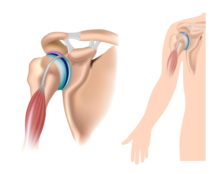
The image provided shows two views of the shoulder joint highlighting the musculature and ligaments involved in its function.
On the left, there is an anterior view of the shoulder joint with the focus on the biceps brachii muscle. This muscle has two heads, as indicated by its name: a long head and a short head. The long head is shown traversing up into the shoulder joint, passing through the bicipital groove of the humerus and attaching to the supraglenoid tubercle of the scapula. This tendon of the long head is kept in place by the transverse humeral ligament. The biceps brachii is a powerful supinator and flexor of the forearm and also assists with shoulder flexion.
The right side of the image shows a lateral view of the shoulder joint, displaying the biceps brachii muscle’s long head, as it extends from the glenoid cavity and transitions into the muscular component along the arm. This view emphasizes the path of the long head of the biceps brachii as it travels through the intertubercular sulcus, also known as the bicipital groove.
These illustrations are valuable in understanding how the biceps tendon contributes to shoulder stability and mobility. This anatomical feature is clinically significant as the long head of the biceps tendon is a common site for injury, which can lead to pain and limited motion in the shoulder and arm.

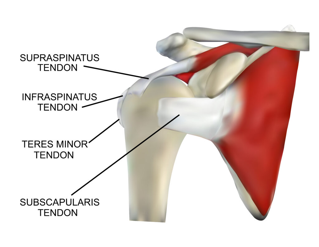
The image displays the tendinous parts of the four rotator cuff muscles of the shoulder, which play a critical role in the function and stability of the shoulder joint.
At the top, we see the supraspinatus tendon, which belongs to the supraspinatus muscle. This muscle is located on the upper part of the scapula, and its tendon passes through the subacromial space to attach to the greater tubercle of the humerus. The supraspinatus is particularly involved in the first 15 degrees of arm abduction and is commonly associated with impingement issues and tears.
Below the supraspinatus tendon is the infraspinatus tendon, associated with the infraspinatus muscle. This muscle occupies the majority of the infraspinatus fossa on the posterior aspect of the scapula and attaches to the greater tubercle of the humerus. It is a powerful external rotator of the shoulder.
Next, we have the teres minor tendon, which comes from the teres minor muscle. This small muscle lies inferior to the infraspinatus and also attaches to the greater tubercle of the humerus. It aids in the external rotation of the shoulder and adds to the overall stability of the joint.
Finally, we see the subscapularis tendon at the bottom. It is the strongest and largest of the rotator cuff muscles and arises from the subscapular fossa on the anterior surface of the scapula. Its tendon inserts into the lesser tubercle of the humerus. The subscapularis muscle is primarily responsible for the internal rotation of the arm.
These tendons are essential components of the rotator cuff, a musculotendinous cuff that encapsulates the glenohumeral joint, providing stability to the inherently unstable shoulder joint. Injuries to these tendons can result in pain, weakness, and reduced range of motion, impacting daily activities and athletic performance.
