7201 Basic Muscles, Tendons, and Ligaments
Muscles
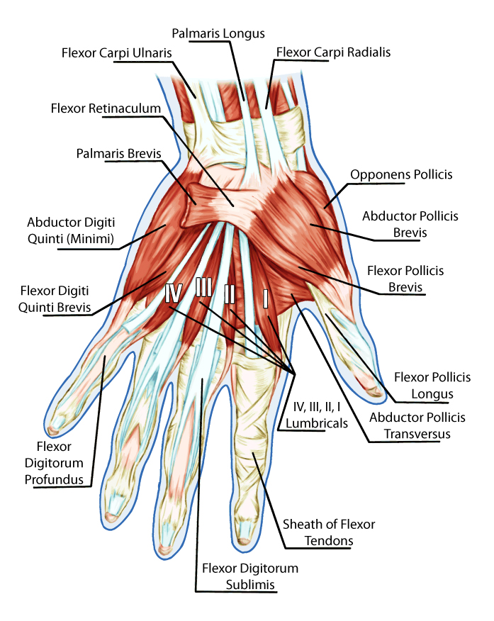
The hand is a complex structure composed of numerous muscles that enable a wide range of movements and functions. These muscles are typically categorized into two groups: extrinsic and intrinsic muscles.

- Extrinsic Muscles: These muscles originate in the forearm and insert into the hand. They are primarily responsible for the gross movements of the hand and fingers. The extrinsic muscles are further divided into two groups:
- Flexor Muscles: Located on the anterior (palm) side of the forearm, these muscles are responsible for flexing the fingers and thumb. Key muscles include the flexor digitorum superficialis and profundus, which flex the fingers, and the flexor pollicis longus, which flexes the thumb.
- Extensor Muscles: Located on the posterior (back) side of the forearm, these muscles extend the fingers and thumb. Important muscles in this group include the extensor digitorum, which extends the fingers, and the extensor pollicis longus and brevis, which extend the thumb.
- Intrinsic Muscles: These muscles are located within the hand and are primarily responsible for the fine motor movements of the fingers and thumb. They are grouped into three categories:
- Thenar Muscles: Located at the base of the thumb, these muscles control thumb movements. Key muscles include the abductor pollicis brevis, flexor pollicis brevis, opponens pollicis, and adductor pollicis.
- Hypothenar Muscles: Situated at the base of the little finger, these muscles control movements of the little finger. They include the abductor digiti minimi, flexor digiti minimi brevis, and opponens digiti minimi.
- Interosseous and Lumbrical Muscles: The interosseous muscles (dorsal and palmar) are located between the bones of the hand and help in finger abduction and adduction. The lumbricals, originating from the tendons of the flexor digitorum profundus, assist in flexing the metacarpophalangeal joints and extending the interphalangeal joints.
The muscles of the hand are innervated primarily by the median, ulnar, and radial nerves, which also provide sensation to different parts of the hand. The muscles work together in a highly coordinated manner, allowing for the complex and precise movements that the hand is capable of performing. This intricate system is essential for everyday tasks, from gripping objects to typing on a keyboard.
Tendons and Ligaments
The hand contains a complex network of tendons and ligaments that work together to provide its remarkable range of motion and dexterity. Here’s a detailed look at the tendons and ligaments in the hand:
Tendons in the Hand
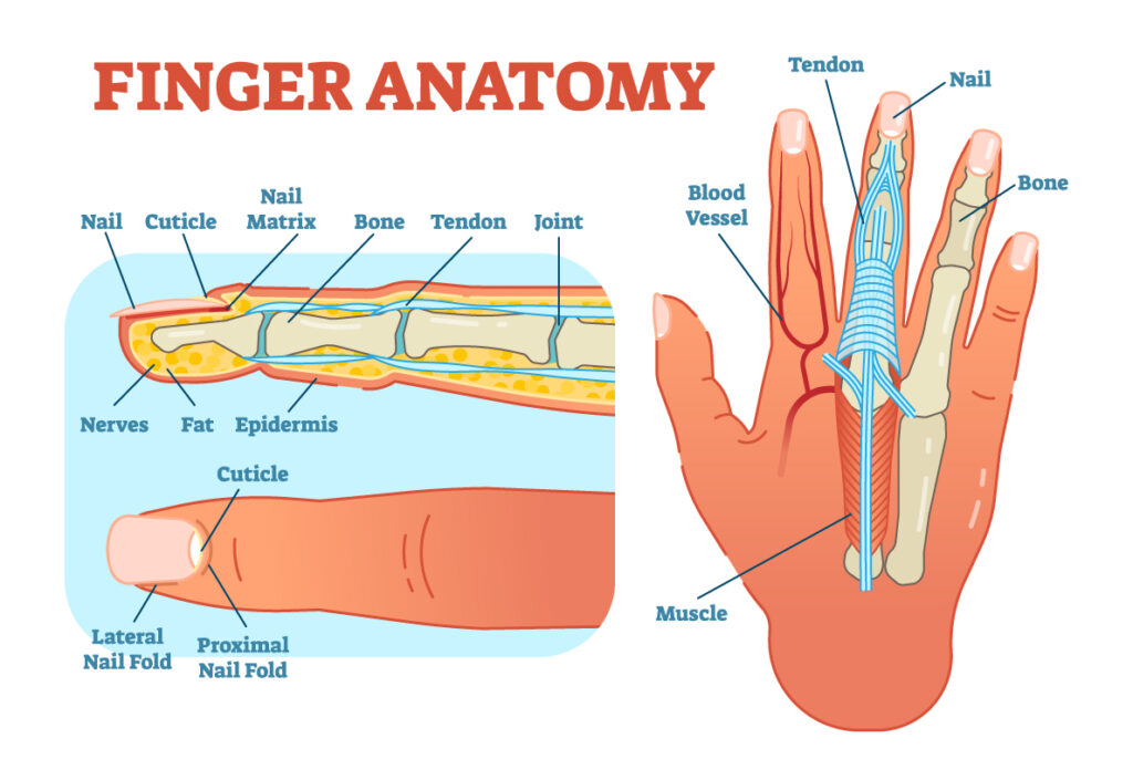
- Flexor Tendons: These tendons run along the palm side of the hand and fingers and are responsible for bending the fingers. They are part of the flexor digitorum superficialis and flexor digitorum profundus muscles.
- Flexor Pollicis Longus Tendon: This tendon is specific to the thumb, aiding in its flexion.
- Extensor Tendons: Located on the back of the hand and fingers, these tendons straighten the fingers. They are connected to muscles in the forearm, such as the extensor digitorum, extensor indicis, and extensor digiti minimi.
- Extensor Pollicis Longus and Brevis Tendons: These tendons extend the thumb.
Ligaments in the Hand
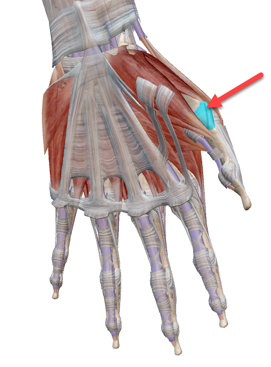
- Volar Plate: A thick ligamentous structure found on the palm side of the finger joints, primarily the proximal interphalangeal (PIP) and metacarpophalangeal (MCP) joints. It prevents hyperextension and contributes to joint stability.
- Dorsal Capsular Ligaments: These are found on the back of the finger joints, providing stability and limiting excessive flexion.

Collateral Ligaments: Located on both sides of each finger and thumb joint, these ligaments provide lateral stability.

Transverse Metacarpal Ligament: Connects the heads of the metacarpal bones, maintaining their alignment and aiding in hand function.

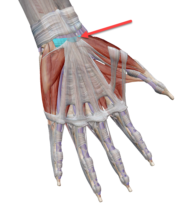
Carpal Ligaments: Including the transverse carpal ligament, which forms the roof of the carpal tunnel, these ligaments are crucial in maintaining the structure of the wrist and the arrangement of the carpal bones.

Tendon Sheaths and Pulleys
- In the hand, tendons run through sheaths which are tube-like structures filled with synovial fluid. This arrangement reduces friction and facilitates smooth tendon movement.
- The flexor tendons are also held in place by a series of pulleys, especially in the fingers, ensuring efficient tendon movement and preventing “bowstringing” during finger flexion.
Common Injuries and Conditions
- Tendon Injuries: Such as cuts or ruptures can impair hand movement. Conditions like tendinitis (inflammation of a tendon) or tenosynovitis (inflammation of the tendon sheath) are common.
- Ligament Injuries: Sprains or tears, particularly in the collateral ligaments, can occur from trauma or overuse.
Understanding the intricate anatomy of tendons and ligaments in the hand is crucial, as it underpins many of the complex motions we perform daily, from grasping objects to intricate finger movements.
