
Table of Contents
Overview of the Thyroid's Function
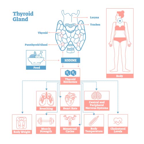
This image provides an informative overview of the thyroid gland’s role in the human body, the importance of iodine for thyroid function, and the systemic effects of thyroid hormones.
At the top, the image features a label “Thyroid Gland” with a detailed anatomical illustration of the thyroid gland in relation to the larynx and trachea. The thyroid gland is shown with its typical butterfly shape, nestled in the lower front of the neck.
Below the anatomical diagram, there is a depiction of the parathyroid glands located at the rear of the thyroid gland. These are essential for regulating calcium levels in the blood, although their function is not elaborated on in this image.
A dotted line connects a bowl labeled “Food” to a molecule labeled “IODINE,” which indicates that iodine, an essential element for thyroid hormone production, is obtained from the diet. The thyroid hormones, T3 and T4, are represented by arrows that suggest their production is dependent on the availability of iodine.
The central part of the image highlights the diverse roles of thyroid hormones in the body. These hormones influence several physiological systems, as shown by the labeled icons:
- Breathing, depicted by a pair of lungs.
- Heart Rate, shown by a heart icon.
- Central and Peripheral Nervous Systems, indicated by a brain illustration.
- Muscle Strength, represented by an arm muscle.
- Menstrual Cycles, symbolized by a calendar.
- Body Temperature, depicted with a thermometer.
- Cholesterol Levels, shown by a test tube with a blood drop.
At the bottom of the image, two additional effects of thyroid hormones are noted: Body Weight, illustrated by a scale, and a representation of a flexing arm for Muscle Strength.
The outline of a human figure to the right connects all these systems, indicating that thyroid hormones act throughout the body.
In summary, this image conveys the critical importance of the thyroid gland and its hormones in regulating various bodily functions. It underscores that iodine from the diet is vital for producing thyroid hormones, which in turn affect breathing, heart rate, nervous system function, muscle strength, menstrual cycles, body temperature, cholesterol levels, and body weight.
Hormonal Feedback Loop
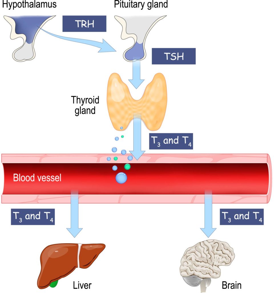
This image illustrates the hormonal feedback loop involving the hypothalamus, pituitary gland, and thyroid gland, as well as the downstream effects on the liver and brain.
At the top left, the hypothalamus, depicted in blue, releases Thyrotropin-releasing hormone (TRH) which signals the pituitary gland, shown in grey. In response, the pituitary gland, which is connected to the hypothalamus by a stalk-like structure, secretes Thyroid-stimulating hormone (TSH), represented by a blue arrow pointing towards the thyroid gland.
The thyroid gland, located centrally in the image and colored in a yellow hue, then produces T3 (triiodothyronine) and T4 (thyroxine) hormones, as indicated by the blue and green dots. These hormones are released into the blood vessel shown as a large red tube running horizontally across the image.
As these thyroid hormones circulate in the bloodstream, they influence various organs. The liver, illustrated at the bottom left, is one target organ, and the brain, depicted at the bottom right, is another significant target. The image signifies the thyroid hormones’ reach to these organs with blue arrows labeled T3 and T4 pointing towards the liver and brain.
In summary, this diagram represents the endocrine control system known as the hypothalamic-pituitary-thyroid axis. The TRH from the hypothalamus stimulates the pituitary gland to release TSH, which in turn stimulates the thyroid gland to produce and secrete T3 and T4. These thyroid hormones then act on various tissues in the body, including the liver and brain, influencing metabolism and other critical functions.
Thyroid and Parathyroid Anatomy
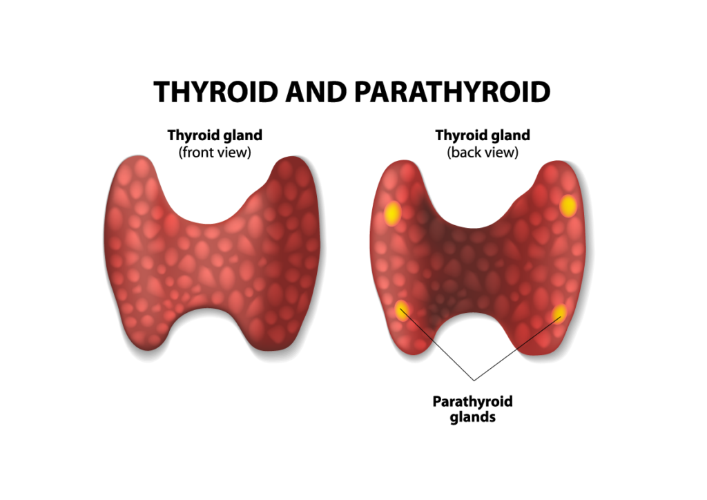
This image provides a detailed depiction of the thyroid gland from both the front and the back view, highlighting the location of the parathyroid glands.
On the left side of the image, we see the front view of the thyroid gland, which has a characteristic reddish color and a butterfly or bow tie shape, wrapping around the trachea. The texture of the gland is represented with a nodular surface suggesting its follicular composition.
On the right side, the image shows the back view of the thyroid gland, with the parathyroid glands indicated in a contrasting color, usually yellow or orange, to distinguish them from the thyroid tissue. These small, round structures are four in number and are positioned on the back of the thyroid gland—two on each lobe.
The parathyroid glands are responsible for regulating the calcium levels in the blood through the secretion of parathyroid hormone (PTH), which is not depicted in this image but is an important function to understand when discussing these glands.
In summary, this visual is aimed at demonstrating the anatomical relationship between the thyroid and parathyroid glands, showcasing the physical distinctiveness and proximity of these critical endocrine glands within the human body.
Thyroid Gland Pathology
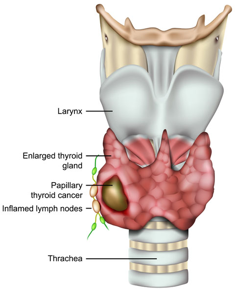
This illustration presents a detailed view of the neck anatomy, focusing on a pathological condition of the thyroid gland. The image shows the larynx at the top, a structure made up of cartilage that houses the vocal cords and is involved in breathing, speech, and protecting the trachea against food aspiration. Below the larynx, the trachea is identified as a tube supported by cartilaginous rings, which conducts air to and from the lungs.
The thyroid gland, depicted in red, is visibly enlarged, a condition known as goiter. This enlargement can be due to various causes, including iodine deficiency, thyroiditis, or the presence of nodules. The illustration points to a specific area on the thyroid labeled “Papillary thyroid cancer,” indicated by a darkened area with an irregular shape. Papillary thyroid cancer is one of the most common types of thyroid cancer and is usually characterized by its slow-growing nature.
Additionally, the image shows several green-colored structures adjacent to the thyroid gland, labeled as “Inflamed lymph nodes.” Lymph nodes are part of the lymphatic system and play a vital role in the body’s immune response. Their inflammation in this context might suggest a reaction to the thyroid pathology, possibly due to the spread of cancer cells or a response to infection or inflammation of the gland.
In summary, this illustration represents an enlarged thyroid gland with a focus on a malignant lesion indicative of papillary thyroid cancer, and it also highlights the associated inflammatory response in the nearby lymph nodes. This visual underlines the interconnectivity between different structures in the neck and the implications of thyroid pathologies on the surrounding tissues.
Anatomical Terms and Definitions
| Term | Definition |
|---|---|
| T3 | A thyroid hormone, also known as triiodothyronine, involved in various bodily functions including metabolism. |
| T4 | Another thyroid hormone, also known as thyroxine, essential for metabolism and growth. |
| Thyroid Gland | An endocrine gland in the neck producing thyroid hormones, crucial for regulating metabolism, growth, and development. |
| Thyroid-stimulating hormone (TSH) | A hormone released by the pituitary gland that stimulates the thyroid gland to produce T3 and T4. |
| Thyrotropin-releasing hormone (TRH) | A hormone released by the hypothalamus that signals the pituitary gland to release TSH. |
| Parathyroid Glands | Small glands located behind the thyroid gland, important for regulating calcium levels in the blood. |
| Iodine | An essential element for the production of thyroid hormones, obtained from the diet. |
| Hypothalamus | A region of the brain that controls the pituitary gland and regulates hormones. |
| Pituitary Gland | An endocrine gland at the base of the brain that controls other glands in the body, including the thyroid gland. |
| Larynx | A structure in the neck containing the vocal cords and involved in breathing, speech, and protecting the trachea. |
| Trachea | A tube that connects the larynx to the lungs, allowing for air passage. |
| Goiter | An enlargement of the thyroid gland, which can be due to various causes including iodine deficiency or thyroiditis. |
| Papillary Thyroid Cancer | A common type of thyroid cancer, characterized by its slow-growing nature. |
| Inflamed Lymph Nodes | Lymph nodes that have become swollen and tender, often in response to infection or inflammation. |
