
Table of Contents
Vascular Spinal Cord Anatomy
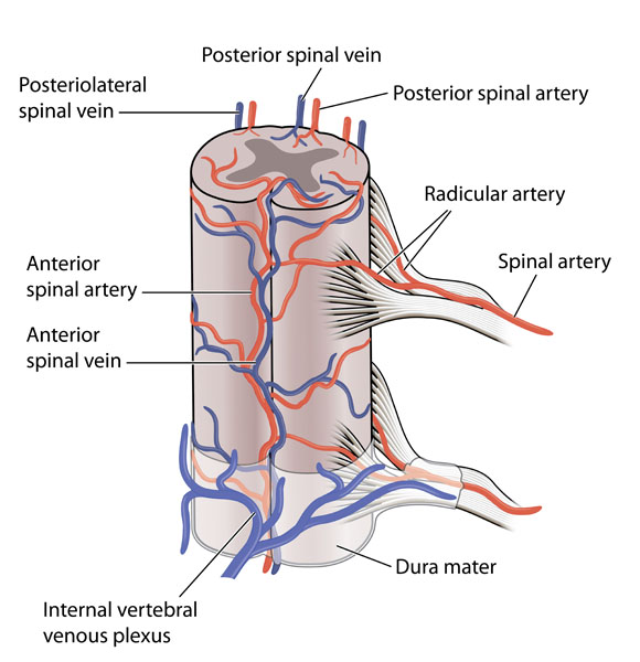
This image depicts the vascular anatomy of the spinal cord, providing a clear illustration of both the arterial and venous blood supply.
At the center, we see the spinal cord itself, encased within the dura mater, a tough and protective membrane. The anterior spinal artery, shown in red, runs along the front of the spinal cord and supplies the anterior two-thirds of the structure. This artery arises from branches of the vertebral arteries and descends along the spinal cord.
The posterior spinal arteries, also in red, are typically paired and run along the dorsal (back) surface of the spinal cord, supplying the remaining one-third of the cord, including the posterior columns. These arteries also originate from the vertebral arteries or sometimes from the posterior inferior cerebellar artery.
Flanking the spinal cord, we observe the radicular arteries, which are small vessels that branch off segmentally from vertebral or deep cervical arteries. These arteries penetrate the dura mater to supply the nerve roots and also contribute to the blood supply of the spinal cord.
The image also shows the venous drainage system of the spinal cord, which includes the anterior spinal vein and the posterolateral spinal veins, both colored in blue. These veins run alongside their corresponding arteries and drain blood away from the spinal cord. The venous blood eventually flows into the internal vertebral venous plexus, a network of veins situated within the layers of the dura mater, noted at the bottom of the illustration.
Understanding this vascular arrangement is crucial because the spinal cord, like the brain, requires a constant, well-regulated blood supply to function properly. Interruptions to this blood flow can result in serious neurological deficits, emphasizing the importance of the spinal cord’s blood supply in maintaining neurological health and function.
Vertebral Anatomy
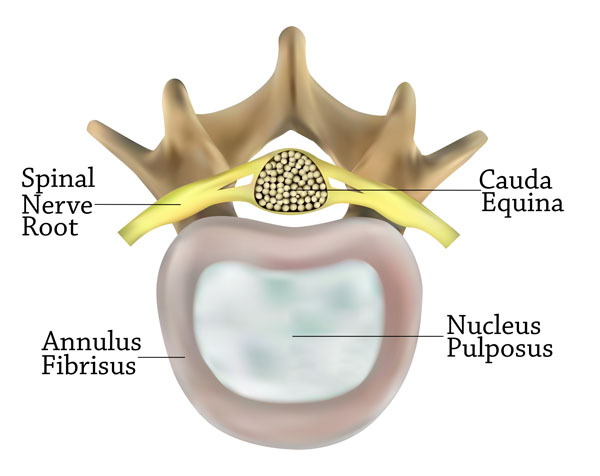
The image displays a cross-sectional view of a vertebral segment focusing on the spinal disc and nearby structures. At the center is the nucleus pulposus, a gel-like substance within the intervertebral disc that gives the disc its shock-absorbing properties. Surrounding the nucleus pulposus is the annulus fibrosus, which is a tough, ring-like structure composed of fibrocartilage. This part provides the disc with structural integrity, allowing it to withstand compressive forces.
Above the disc, we see a spinal nerve root, which is a bundle of nerves exiting the spinal cord through the intervertebral foramen, carrying motor and sensory information from the spinal cord to the rest of the body.
On the upper right side, the cauda equina is indicated, which is a collection of nerve roots at the lower end of the vertebral canal. It resembles a horse’s tail (hence the name ‘cauda equina’, which is Latin for ‘horse’s tail’), and it extends from the lumbar spine down into the sacral region.
The overall structure seen at the top of the image, resembling an inverted “Y,” is part of a vertebra. The specific part is not labeled, but it’s the dorsal aspect of a vertebra where the spinal cord would be located. This type of diagram is typically used in anatomy to teach about the intervertebral discs and their relation to the spinal cord and nerve roots.
Sagittal View of Spinal Column
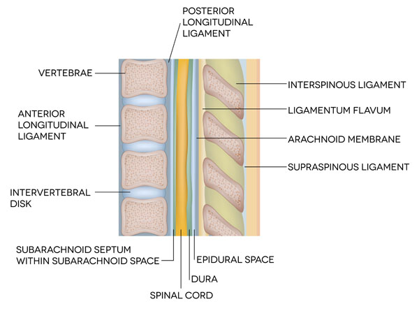
This image provides a sagittal, or side view, of the spinal column, detailing its various anatomical structures and spaces.
The vertebrae are the series of bones that make up the spine. Between each vertebra is an intervertebral disk, which functions as a shock absorber and allows for flexibility of the spine.
On the front (anterior) side of the vertebrae, we see the anterior longitudinal ligament. This ligament runs the entire length of the spine and helps to prevent overextension.
Opposite the anterior longitudinal ligament, on the back (posterior) side of the vertebrae, is the posterior longitudinal ligament. This ligament also runs the full length of the spine and prevents hyperflexion, or excessive forward bending.
The spinal cord, protected within the vertebral column, is a major part of the central nervous system that transmits signals between the brain and the body.
Surrounding the spinal cord is the dura, a tough protective coating. Outside the dura is the epidural space, which contains fat and small blood vessels. This space is clinically important because it is the area into which anesthetics are injected during an epidural block.
The arachnoid membrane is a thin, transparent layer that is part of the meninges, the protective coverings of the brain and spinal cord.
Directly next to the arachnoid membrane, we have the subarachnoid space, which contains cerebrospinal fluid (CSF). This fluid cushions the brain and spinal cord, provides nutrients, and removes waste. Within this space, we see the subarachnoid septum, a partition within the subarachnoid space.
Adjacent to the posterior longitudinal ligament are two important spinal ligaments: the ligamentum flavum, which connects the laminae of adjacent vertebrae and helps to preserve the upright posture, and the interspinous ligament, which connects the spinous processes of adjacent vertebrae.
Above the ligamentum flavum is the supraspinous ligament, which connects the tips of the spinous processes from the seventh cervical vertebra to the sacrum.
These components work in concert to protect the spinal cord, provide structural support to the body, and allow for a range of movements.
Cross-Section of Spinal Cord
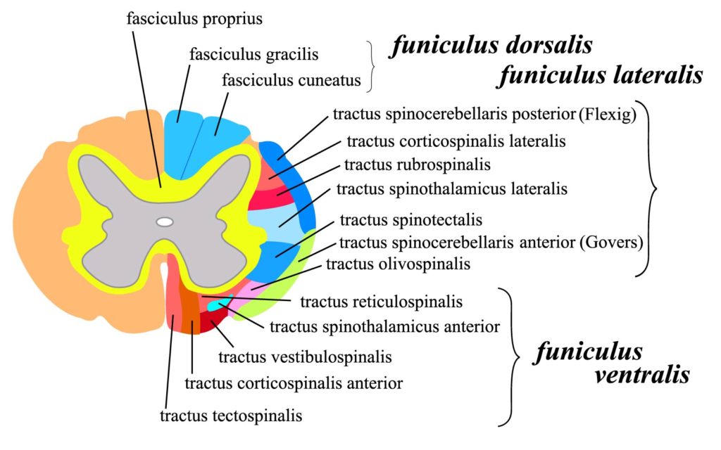
The image is a color-coded schematic of a cross-section of the spinal cord, showing its internal structure and the arrangement of various nerve tracts.
In the center, the grey matter is butterfly-shaped, with anterior and posterior “horns.” The surrounding white matter is divided into three broad regions on each side: the dorsal (posterior), lateral, and ventral (anterior) funiculi. Each funiculus contains multiple ascending and descending nerve tracts responsible for conveying different types of sensory and motor information.
The dorsal funiculus, labeled here, includes the fasciculus gracilis and fasciculus cuneatus, which are ascending tracts involved in carrying fine touch, vibration, and proprioceptive information to the brain.
The lateral funiculus contains several tracts:
- The tractus spinocerebellaris posterior (Flechsig) and tractus spinocerebellaris anterior (Gowers) are involved in proprioception, carrying information about the position of the body to the cerebellum.
- The tractus corticospinalis lateralis is a key pathway for voluntary motor control.
- The tractus rubrospinalis modulates motor input and aids in motor coordination.
- The tractus spinothalamicus lateralis transmits pain and temperature sensations.
- The tractus spinotectalis is involved in reflex movements in response to visual stimuli.
- The tractus olivospinalis is associated with the olivocerebellar system, influencing movement and balance.
- The tractus reticulospinalis is part of the reticular formation’s influence on motor control.
The ventral funiculus, indicated here, contains tracts including:
- The tractus spinothalamicus anterior, which carries information about light touch and pressure.
- The tractus vestibulospinalis, which transmits impulses related to head movement and positional adjustments.
- The tractus corticospinalis anterior, another key motor pathway that influences trunk and proximal limb muscles.
- The tractus tectospinalis, involved in reflex postural movements in response to visual stimuli.
This diagram is a useful educational tool for understanding the complex pathways within the spinal cord that mediate sensory and motor functions.
Sensory and Motor Pathways
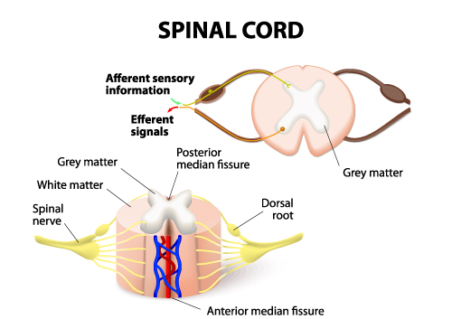
The image provides a simplified view of the spinal cord, highlighting its basic structure and the pathways for sensory and motor information.
The spinal cord is illustrated with a cross-sectional view at the bottom and a longitudinal view at the top. The cross-section is color-coded to distinguish between grey matter, which appears pink and is centrally located, and white matter, which appears as a lighter surrounding area.
The grey matter is known for processing information in the spinal cord, while the white matter contains myelinated axons that act as highways for nerve impulses.
Two distinct features are labeled on the grey matter: the anterior median fissure, a deep groove on the front (ventral) side of the spinal cord, and the posterior median fissure, a shallower groove on the back (dorsal) side.
The dorsal root is shown branching off the spinal cord and is associated with the transmission of afferent sensory information from the body to the spinal cord. This information includes touch, pain, temperature, and proprioceptive signals.
Conversely, efferent signals, which are motor commands from the brain to the muscles, travel from the spinal cord through the ventral root (not labeled in this image, but it would be located opposite the dorsal root).
The spinal nerve is depicted as a bundle that forms from the joining of the dorsal and ventral roots outside the spinal cord, which then transmits both sensory and motor information to and from the body.
This diagram is commonly used to teach the basic anatomy of the spinal cord and the directional flow of neural signals.
Spinal Nerve Pathways
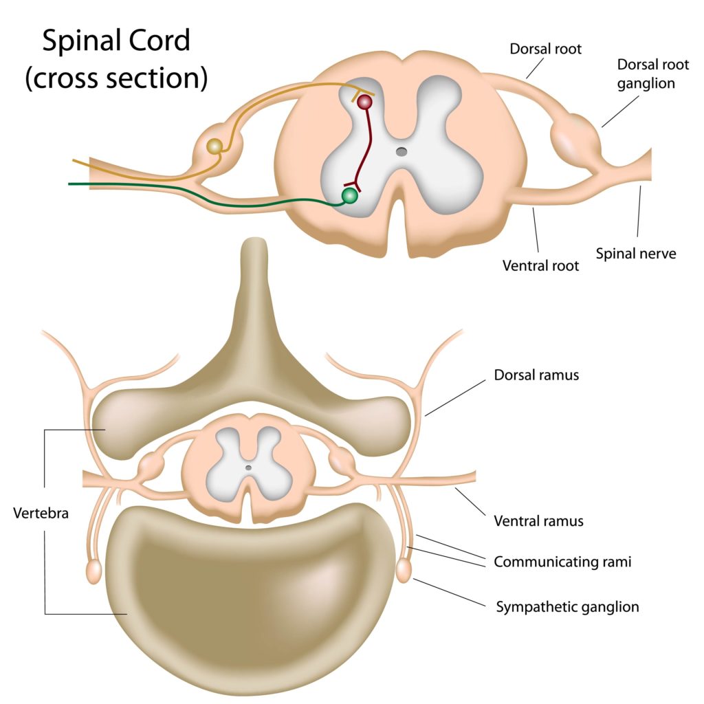
The image presents a detailed view of the spinal cord in cross-section, along with the associated spinal nerves and pathways.
The top part of the illustration shows a lateral cross-sectional view of the spinal cord, where the dorsal root and ventral root come together to form the spinal nerve. The dorsal root contains sensory neurons, which are responsible for transmitting sensory information from the periphery to the spinal cord, and it features a swelling known as the dorsal root ganglion. This ganglion contains the cell bodies of sensory neurons.
The ventral root, on the other hand, contains motor neurons that send signals from the spinal cord to the muscles.
The spinal nerve is the combined structure that arises from the fusion of the dorsal and ventral roots and carries both sensory and motor fibers.
Below, in the larger image, we see the spinal cord ensconced within the vertebra, which is the bony structure that encases and protects the spinal cord. The grey matter in the center of the spinal cord is depicted in a characteristic ‘H’ or butterfly shape and is surrounded by white matter.
Branching off the spinal nerve are the dorsal ramus and ventral ramus. The dorsal ramus carries information to and from the posterior parts of the body, while the ventral ramus does the same for the anterior and lateral parts.
Also shown are the communicating rami, which contain nerve fibers that connect the spinal nerve to a sympathetic ganglion, part of the sympathetic nervous system. The sympathetic ganglion is a cluster of nerve cell bodies that is involved in the fight-or-flight response.
This image is often used in educational contexts to explain the anatomy of the spinal cord, the distinction between sensory and motor pathways, and the organization of the spinal nerves as they relate to the sympathetic nervous system.
Neurological and Biochemical Pathways
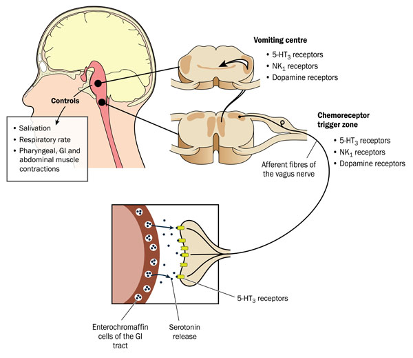
The image illustrates the neurological and biochemical pathways involved in the vomiting reflex. It highlights three key components: the central nervous system control center, the chemoreceptor trigger zone, and the gastrointestinal tract.
At the top of the image, the brain is shown with a section labeled as the vomiting center. This center has receptors for neurotransmitters such as serotonin (5-HT_3 receptors), neurokinin (NK_1 receptors), and dopamine, which, when activated, can initiate the vomiting reflex.
The chemoreceptor trigger zone (CTZ), located in the brain’s area postrema, is sensitive to blood-borne drugs or hormones and is depicted as being in communication with the vomiting center. It also contains receptors for serotonin, neurokinin, and dopamine.
The gastrointestinal tract is represented at the bottom of the image, where enterochromaffin cells release serotonin. This release can activate 5-HT_3 receptors on the afferent fibers of the vagus nerve, which then transmit signals to the vomiting center.
The diagram also shows that the vomiting center controls various physiological responses such as salivation, respiratory rate, and contractions of the pharyngeal, gastrointestinal, and abdominal muscles — all of which are involved in the act of vomiting.
This image serves as an educational tool to explain the complex interactions between the gastrointestinal system and the central nervous system in the control of vomiting.
Anatomical Terms and Definitions
| Term | Definition |
|---|---|
| Anterior Spinal Artery | The artery running along the front of the spinal cord, supplying the anterior two-thirds of the structure with blood. Originates from branches of the vertebral arteries. |
| Anterior Spinal Vein | Vein running alongside the anterior spinal artery, involved in the venous drainage of the spinal cord. |
| Arachnoid Villi | Small protrusions of the arachnoid membrane into the dural venous sinuses, allowing cerebrospinal fluid (CSF) to exit the subarachnoid space and enter the bloodstream. |
| Cauda Equina | A bundle of spinal nerve roots arising from the lower end of the spinal cord and extending downwards within the vertebral canal, resembling a horse's tail. |
| Central Canal | A narrow channel running the length of the spinal cord within the gray matter, filled with cerebrospinal fluid (CSF). |
| Chemoreceptor Trigger Zone (CTZ) | Located in the brain's area postrema, it is sensitive to blood-borne drugs or hormones and can activate the vomiting center. Contains receptors for serotonin, neurokinin, and dopamine. |
| Dorsal Root | A branch of a spinal nerve that carries sensory information from the peripheral nervous system to the spinal cord. |
| Epidural Space | The space outside the dura mater but inside the bony structure of the vertebral column, containing fat and small blood vessels. Clinically important for the administration of epidural anesthesia. |
| Intervertebral Disc | The cartilaginous pad located between the vertebrae of the spine, serving as a shock absorber and allowing flexibility. |
| Nucleus Pulposus | The inner gel-like core of an intervertebral disc, which gives the disc its elastic shock-absorbing properties. |
| Posterior Spinal Arteries | Typically paired arteries that run along the dorsal (back) surface of the spinal cord, supplying the remaining one-third of the cord, including the posterior columns. Originate from the vertebral arteries or the posterior inferior cerebellar artery. |
| Radicular Arteries | Small vessels branching off segmentally from vertebral or deep cervical arteries, penetrating the dura mater to supply the nerve roots and contribute to the blood supply of the spinal cord. |
| Subarachnoid Space | The fluid-filled space surrounding the brain and spinal cord, where cerebrospinal fluid (CSF) circulates, acting as a cushion to protect the central nervous system. |
| Vomiting Center | The brain section with receptors for neurotransmitters such as serotonin, neurokinin, and dopamine, which, when activated, can initiate the vomiting reflex. Located in the medulla oblongata and coordinates the act of vomiting by integrating various bodily responses. |
