
Table of Contents
Lateral Exploded View of the Skull

In this illustration, we have a lateral view of the human skull, with its bones presented in a scattered arrangement to clearly display each individual bone. The frontal bone, forming the forehead, is situated at the top of the image. Just behind it, we find the parietal bone which constitutes a significant portion of the cranial roof and sides.
Below the frontal bone, the sphenoid bone is visible, which is a complex bone that helps form the base of the cranium, the sides of the skull, and the floors and sides of the orbits. The zygomatic bones, commonly referred to as the cheekbones, are shown in symmetry on both sides of the skull.
The nasal bone is found in the central upper part of the image, just below the frontal bone, giving shape to the bridge of the nose. Underneath it, the vomer is shown, which forms the posterior part of the nasal septum, with the perpendicular plate of the ethmoid bone contributing to the upper part of the septum.
The maxilla, or the upper jawbone, is a central feature in the middle of the image. It carries the upper teeth and forms the lower part of the orbits. On each side of the maxilla, the palatine bones are seen, which contribute to the structure of the oral cavity, the nasal cavity, and the eye sockets.
Lower down, the mandible is depicted, which is the lower jaw and the largest, strongest bone of the face.
On the far right of the image, we can see the temporal bone, which houses the structures of the ear, and below it, the occipital bone, which forms the back and base of the skull.
Within the temporal bone area, three smaller bones are displayed: the malleus, incus, and stapes, collectively known as the auditory ossicles. These are the smallest bones in the body and are crucial for the process of hearing, as they transmit sound vibrations from the eardrum to the inner ear.
Lastly, the ethmoid bone is located deep in the skull and is not clearly seen from a lateral perspective, but it’s a key part of the cranial floor, and the nasal cavity walls and septum.
The disarticulated presentation of these bones serves to show the relationships and connections between the various components of the cranial and facial structures.
Anterior Exploded View of the Skull
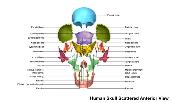
This image provides an anterior view of the human skull with the bones displayed in a scattered format to enhance the understanding of their positions and relationships. At the top, the frontal bone is prominently displayed, forming the forehead and the upper part of the orbital cavities.
Flanking on either side of the frontal bone are the parietal bones, which together form the greater portion of the sides and roof of the cranial cavity. Towards the base of the skull, the occipital bone is situated, which forms the back and base of the skull’s cranium.
The sphenoid bone is located beneath the frontal bone, slightly central in the image, and contributes to the floor of the cranial cavity. It’s not fully visible from this view but plays a critical role in the structure of the skull.
On the lateral aspects, the zygomatic bones, also known as the cheekbones, are visible, and they articulate with the maxilla, the sphenoid, and the temporal bones. The nasal bones are small and rectangular, situated at the center top of the image, forming the bridge of the nose.
Below the nasal bones, the vomer is seen, a thin, flat bone forming the lower part of the nasal septum. The nasal conchae, which are not clearly visible in this view, are small scroll-like bones that contribute to the nasal cavity’s structure.
The maxilla or upper jawbone is central in the image, showing the alveolar process where the teeth are anchored. The mandible, or lower jawbone, forms the lower facial structure and houses the lower teeth.
Adjacent to the maxilla, the palatine bones are visible, which form the back part of the nasal cavity and a portion of the hard palate.
The temporal bones are lateral to the maxilla and form part of the sides and base of the skull, including the cranial cavity and the ear canals. Within the temporal bones, the malleus, incus, and stapes (the auditory ossicles) are represented. These tiny bones are essential for hearing as they transmit sound from the eardrum to the inner ear.
At the center, just below the nasal bone and the sphenoid, the ethmoid bone is indicated, which is a complex structure contributing to the cranial floor, the nasal cavity, and the nasal septum.
This scattered view elucidates how the bones of the skull are interconnected, providing a comprehensive understanding of the craniofacial anatomy.
Spatial Relationship of Skull Bones
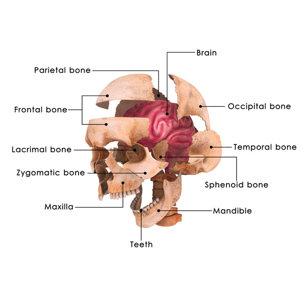
This illustration is an exploded view of the human skull, providing a spatial relationship between the various bones and the brain within the cranial cavity.
At the top of the image is the parietal bone, which, along with its contralateral partner, forms the sides and roof of the cranial cavity. The frontal bone is shown below the parietal bone; it creates the forehead and the upper orbits where the eyes are situated.
Below the frontal bone, to the left of the image, is the lacrimal bone, the smallest and most fragile bone of the face, sitting inside the orbit, part of the structure that supports the lacrimal or tear ducts.
The zygomatic bone, commonly known as the cheekbone, is adjacent to the lacrimal bone. Below the zygomatic bone is the maxilla, which forms the upper jaw and carries the upper teeth, as can be seen in the image.
The mandible, the lower jawbone, is depicted below the maxilla, and it holds the lower teeth. It is the only movable bone of the skull not forming a joint with the temporal bone.
In the center of the image, a section of the brain is visible, showcasing the convoluted surface of the cerebral cortex, which is responsible for many higher-order brain functions.
Towards the back of the image is the occipital bone, which forms the back and base of the skull and contains the foramen magnum, where the spinal cord exits the skull.
On the right side of the image, the temporal bone is indicated. It forms part of the side and base of the cranium, houses the structures of the ear, and forms the temporomandibular joint with the mandible.
Lastly, the sphenoid bone can be seen in front of the temporal bone. This bone is located at the base of the skull in front of the temporals and basilar part of the occipital bone. It is considered the keystone of the cranial floor because it is in contact with all other cranial bones, helping to stabilize their position.
This perspective allows for an understanding of how the bones of the skull protect the brain and form the structure of the face.
Internal Structures of the Skull
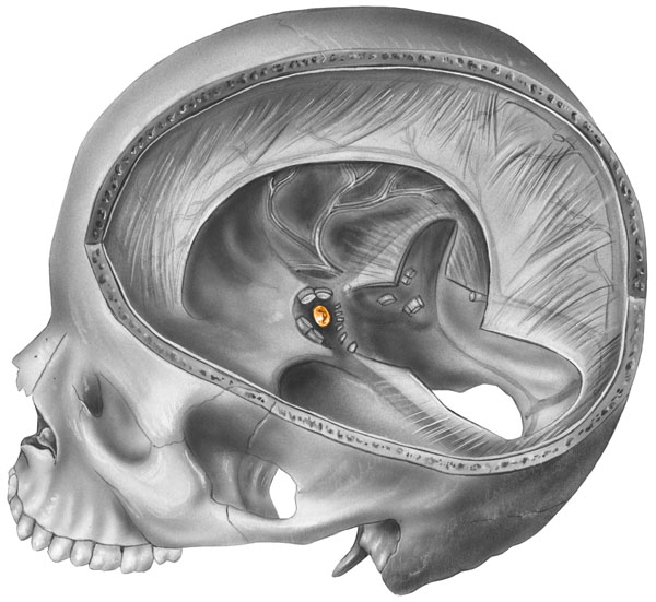
This illustration provides a sagittal section of the human skull, allowing us to observe the internal structure and features that are not visible from the exterior. The sagittal plane divides the skull into left and right halves, and this particular view shows the left side.
Starting from the top, we see the interior aspect of the cranial vault, which is the inner surface of the parietal bone. This bone, along with the frontal, temporal, and occipital bones, forms the cranium, protecting the brain.
Moving downward, the intricate bony structure within the cranial base is visible. The sphenoid bone, which shapes part of the floor of the cranial cavity, can be seen here with the sella turcica in the center. The sella turcica is a saddle-shaped depression that houses the pituitary gland, an important part of the endocrine system.
In the lower part of the image, the upper jaw or the maxilla is present, with the teeth in situ showing the alveolar process where the teeth are anchored. The mandible or lower jaw is not in view in this section as it would be located further forward in a complete skull.
The large opening at the base of the skull is the foramen magnum, through which the spinal cord connects to the brain. The occipital bone, which forms the back and base of the skull, surrounds this opening.
On the interior surface of the cranium, we can observe the grooves for the meningeal vessels, which supply blood to the meninges, the protective coverings of the brain.
The image also shows the intricate detail of the temporal bone, where we find the external acoustic meatus, leading to the middle and inner ear structures. These are essential for hearing and balance.
Notably, the zygomatic arch, which is part of the temporal bone and forms the prominence of the cheek, is visible on the far left of the image.
This cross-sectional view provides insight into the complex anatomy of the skull, revealing how the bones are constructed to protect the brain and support the facial structures.
Skull Suture Anatomy

The image illustrates two aspects of cranial anatomy: the intact skull and a detailed view of a suture.
On the left, we see a lateral view of the human skull. The skull is composed of several bones that are joined together by sutures, which are fibrous joints. These sutures are visible as the slightly jagged lines where the bones meet.
The right side of the image zooms in to show a close-up view of a suture. A suture is a type of fibrous joint that connects two bones in the skull. The bones are held together by a suture ligament, which allows for slight movement at the joint. This slight movement is essential during birth when the flexible sutures allow the bones of the skull to overlap, making it easier for the baby’s head to pass through the birth canal. In adults, this mobility provides a minor accommodation for brain movement.
The section of bone also demonstrates the inner structure, including the trabeculae of spongy bone that provide structural support while minimizing weight. The spongy bone is located between two layers of a denser type of bone known as compact bone. This arrangement contributes to the strength and durability of the skull while allowing for a degree of flexibility at the sutures.
The anatomy showcased here is fundamental to understanding how the skull protects the brain and accommodates growth throughout human development.
Composition of the Cranial Bones
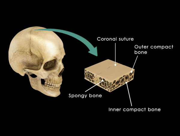
This illustration presents a lateral view of the human skull and an illustrative detail showing the composition of a cranial bone.
In the full view of the skull, we see the coronal suture, which is a fibrous joint that runs across the top of the skull, nearly ear to ear, dividing the frontal bone from the parietal bones. Sutures like this one are found only in the skull and are essential for brain growth during infancy and childhood.
Next to the full skull is a cutaway section of a cranial bone. This section illustrates the typical structure of a cranial bone, with an outer layer of compact bone, a middle layer of spongy bone, and an inner layer of compact bone. The compact bone provides strength and protection, while the spongy bone, which contains bone marrow and blood vessels, lightens the weight of the skull without compromising its structural integrity.
The compact bone, also known as cortical bone, is dense and forms the outer shell of all bones. It provides strength for weight-bearing and protection. The spongy bone, also known as cancellous or trabecular bone, is much lighter and less dense than compact bone. It is found in the interior of bones and is characterized by a honeycomb-like arrangement of trabeculae (the small, needle-like pieces of bone).
This specific structure of bone is vital to the function of the skull, as it must be strong enough to protect the brain but light enough to be supported by the neck and muscles. The coronal suture and bone composition play a crucial role in the skull’s ability to protect the brain while accommodating movement and growth.
Lateral View the the Cranial Layers
The image presents a lateral view of the human skull and an illustrative detail showing the composition of a cranial bone.
In the full view of the skull, we see the coronal suture, which is a fibrous joint that runs across the top of the skull, nearly ear to ear, dividing the frontal bone from the parietal bones. Sutures like this one are found only in the skull and are essential for brain growth during infancy and childhood.
Next to the full skull is a cutaway section of a cranial bone. This section illustrates the typical structure of a cranial bone, with an outer layer of compact bone, a middle layer of spongy bone, and an inner layer of compact bone. The compact bone provides strength and protection, while the spongy bone, which contains bone marrow and blood vessels, lightens the weight of the skull without compromising its structural integrity.
The compact bone, also known as cortical bone, is dense and forms the outer shell of all bones. It provides strength for weight-bearing and protection. The spongy bone, also known as cancellous or trabecular bone, is much lighter and less dense than compact bone. It is found in the interior of bones and is characterized by a honeycomb-like arrangement of trabeculae (the small, needle-like pieces of bone).
This specific structure of bone is vital to the function of the skull, as it must be strong enough to protect the brain but light enough to be supported by the neck and muscles. The coronal suture and bone composition play a crucial role in the skull’s ability to protect the brain while accommodating movement and growth.
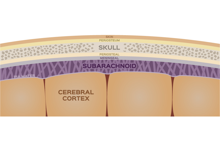
Meninges Anatomy
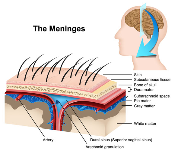
This image is a detailed representation of the meninges, the protective coverings of the brain, and the upper layers of the scalp.
On the right, we see a profile of a human head, highlighting the area of the scalp and skull where the meninges are located underneath. The blue arrow indicates a section that has been magnified on the left side of the image to show the different layers in detail.
Starting from the outermost layer, we see the skin, which is the external covering of the scalp. Below the skin is the subcutaneous tissue, which contains fat and connective tissue that cushions and insulates the skull.
Beneath the subcutaneous tissue is the bone of the skull, which forms the rigid structure that protects the brain. Below the bone, we encounter the dura mater, the outermost layer of the meninges. It is a thick, durable membrane that provides additional protection to the brain.
Under the dura mater is the subarachnoid space, which contains cerebrospinal fluid (CSF). This fluid-filled space helps cushion the brain, acting as a shock absorber.
The pia mater is the innermost layer of the meninges. It is a delicate membrane that closely adheres to the surface of the brain, following its contours and blood vessels.
Within the subarachnoid space, we see an artery and the dural sinus (labeled as the superior sagittal sinus in this diagram), which is a channel through which venous blood and CSF are returned from the brain to the heart. The arachnoid granulations are small protrusions of the arachnoid (the middle layer of the meninges, not labeled in this image) through the dura mater; they allow CSF to pass into the dural sinus.
The image also shows a cross-section of the brain’s outer layer, the gray matter, which contains the cell bodies of neurons, and the inner layer, the white matter, which is composed of myelinated nerve fibers that connect different areas of the brain.
This layered anatomical structure is critical for the protection and function of the brain, allowing it to be well-protected within the skull while also ensuring that it is nourished and can communicate effectively with the rest of the body.
Anatomical Terms and Definitions
| Term | Definition |
|---|---|
| Arachnoid Granulations | Small protrusions of the arachnoid membrane through the dura mater, allowing cerebrospinal fluid (CSF) to pass into the dural sinus for drainage into the venous system. |
| Cerebrospinal Fluid (CSF) | A clear, colorless body fluid found in the brain and spinal cord, acting as a cushion or buffer for the brain's cortex, providing basic mechanical and immunological protection to the brain inside the skull, and serving a vital function in the cerebral autoregulation of cerebral blood flow. |
| Dura Mater | The outermost, toughest, and most fibrous of the three membranes (meninges) covering the brain and spinal cord, providing significant protection to the brain. |
| Gray Matter | The darker-colored regions of the brain and spinal cord, consisting mainly of neuronal cell bodies and branching dendrites, responsible for processing and integrating information. |
| Pia Mater | The delicate innermost layer of the meninges that closely adheres to the surface of the brain and spinal cord, following all its contours and blood vessels. |
| Skin | The outer covering of the body, consisting of a layer of tissues that protect the underlying muscles and organs. |
| Skull | The bone structure that forms the head in vertebrates, supporting the structures of the face and protecting the brain. |
| Subarachnoid Space | The interval between the arachnoid membrane and the pia mater, filled with cerebrospinal fluid and containing large blood vessels. |
| Subcutaneous Tissue | The layer of tissue lying immediately below the dermis of the skin, consisting of connective tissue and fat, providing insulation and cushioning to the body. |
| Superior Sagittal Sinus | A dural sinus (large vein) located within the folds of the dura mater along the midline on top of the brain, responsible for draining blood from the brain back to the heart. |
| White Matter | The paler tissue of the brain and spinal cord, consisting mainly of nerve fibers with their myelin sheaths, responsible for transmitting signals within the nervous system. |
