
Table of Contents
Basic Lung Anatomy
Depicted here is the anatomy of the human lungs, featuring a detailed and labeled illustration of both the right and left lung structures.
Starting at the top, the trachea is shown as a vertical tube that bifurcates into the right and left bronchus. These bronchi act as the main passageways through which air travels into each lung.
The right lung is divided into three lobes by two fissures: the horizontal and the oblique fissure. The right upper lobe is situated at the top, the right middle lobe below it, and the right lower lobe at the bottom. Each lobe is supplied by its own bronchus, which branches off from the right main bronchus.
The left lung, on the other hand, consists of two lobes separated by the oblique fissure. The left upper lobe includes a unique indentation, known as the cardiac notch, which accommodates the heart. Below the upper lobe is the left lower lobe. Similar to the right lung, each lobe is supplied by its own bronchus, stemming from the left main bronchus.
Both lungs feature an apex at the topmost portion, which extends slightly above the collarbone, and a base at the bottom, which rests on the diaphragm. The hilum is a region on the medial aspect of each lung through which bronchi, blood vessels, lymphatics, and nerves enter and exit the lungs.
Lastly, the bronchioles are depicted as smaller branches stemming from the bronchi, indicating the further division of air passageways within the lungs, which eventually lead to the alveoli where gas exchange occurs.
Pleural Cavities
This illustration provides a simplified view of the pleural cavities of the lungs and their relation to the pleurae.
On the left side of the image, we see the right lung, and on the right side, the left lung, both enveloped by a two-layered membrane known as the pleura. The pleura is divided into two distinct layers with a space in between: the inner layer is the visceral pleura, which closely covers the lung tissue, and the outer layer is the parietal pleura, which lines the inside of the chest wall.
The space between these two layers is the pleural cavity. This cavity is typically filled with a small amount of pleural fluid, which allows the lungs to move smoothly within the chest cavity during respiration.
An inset at the bottom of the image provides a magnified cross-sectional view of the pleural layers. It shows the chest wall, the parietal pleura, the pleural cavity with pleural fluid, and the visceral pleura closely adhering to the lung tissue.
Additionally, the image indicates where the visceral and parietal pleurae join at the root of the lung. This is a critical junction where the structures such as bronchi, blood vessels, and nerves enter and exit the lungs.
The pleural fluid collecting within the pleural cavity is a crucial component, as it lubricates the surfaces of the pleura to prevent friction during breathing. The pleurae play an essential role in respiratory mechanics and in maintaining the negative pressure within the thoracic cavity that is necessary for lung expansion.
Pleura Anatomy
Here is a depiction of a frontal section of the thoracic cavity, showcasing the pleura and pleural cavity in relation to the lungs and diaphragm.
The pleura is a serous membrane that folds back onto itself to form a two-layered membranous structure. The outer layer, known as the parietal pleura, lines the inner surface of the thoracic cavity, covering the thoracic wall and superior surface of the diaphragm. The inner layer, called the visceral pleura, directly covers the lung tissue. The space between these two layers is the pleural cavity, which contains a thin layer of pleural fluid. This fluid serves as a lubricant, allowing the lungs to expand and contract smoothly within the thoracic cavity during breathing.
In this depiction, the left lung is shown in blue, and the right lung in purple, each occupying its respective side of the thoracic cavity. The heart is visible in the center, draped by the lungs, with the aorta and other vessels shown in red and blue, representing the arterial and venous systems, respectively.
The diaphragm is illustrated at the bottom of the image in brown, separating the thoracic cavity from the abdominal cavity. The diaphragm is the primary muscle of respiration, contracting and flattening to enlarge the thoracic cavity and allow air to be drawn into the lungs.
The trachea is represented at the top as a white tube bifurcating into the left and right bronchi, which then branch into the lungs.
Overall, this image serves to emphasize the relationship between the pleurae, the pleural cavity, and the lungs, as well as their position within the thoracic cavity above the diaphragm.
Respiratory Process
Here are two views of the human ribcage, crucial for the respiratory process as it offers structural support and facilitates the expansion and contraction of the chest cavity.
On the left side, we have an anterior view of the ribcage with the sternum, or breastbone, at the center. This flat bone forms the front of the ribcage and connects to the ribs via costal cartilages. The ribs are curved bones that form the cage-like structure which protects vital thoracic organs such as the heart and lungs. Below the ribcage, the diaphragm is shown, a dome-shaped muscle critical for breathing. During inhalation, the diaphragm contracts and moves downward, expanding the chest cavity and allowing the lungs to fill with air.
On the right side, the image provides a lateral view of the ribcage, emphasizing the intercostal muscles. These muscles are located between the ribs and come in two layers: the internal and external intercostal muscles. They assist in the mechanical aspect of breathing; during inhalation, the external intercostal muscles contract to elevate the ribs and sternum, increasing the volume of the thoracic cavity. During exhalation, the internal intercostal muscles can contract to depress the ribs and sternum, which helps to expel air from the lungs.
This diagram effectively highlights the bones and muscles involved in the respiratory mechanism, underscoring their significance in protecting respiratory organs and facilitating the movement of air in and out of the lungs. The coordinated action of the diaphragm and intercostal muscles is essential for the pulmonary ventilation process.
Process of Respiration
Now let’s take a closure look at the process of respiration, focusing on the mechanics of breathing in (inhalation) and breathing out (exhalation).
On the left side of the image, we see a profile of a human torso during inhalation. The diaphragm is shown contracting and moving downward, indicated by the red arrow. This movement increases the volume of the thoracic cavity, causing a reduction in pressure within the lungs compared to the outside air, resulting in air being drawn into the lungs (as shown by the blue arrows).
On the right side, the image shows exhalation. The diaphragm is relaxing and moving upwards, as denoted by the red dashed arrow, which decreases the volume of the thoracic cavity and increases the pressure in the lungs, pushing air out (as shown by the red arrows).
At the bottom of the image, a pair of ribs is depicted during inspiration and expiration to demonstrate their movement. During inspiration, the rib cage moves upwards and outwards to further increase the thoracic volume. Conversely, during expiration, the rib cage moves downwards and inwards as the thoracic volume decreases.
This visual explanation helps to understand the dynamic changes that occur within the chest cavity during the respiratory cycle. It emphasizes the role of the diaphragm as the primary muscle of respiration, along with the contribution of the rib cage movement in the process of ventilation.
Gas Exchange Process
This comprehensive visual illustrates the human gas exchange process, tracing the path of oxygen from the air into the bloodstream and the transport of carbon dioxide from the blood back into the air for exhalation.
The top of the image shows a profile of a human figure, illustrating the air pathway into the body through the trachea, leading to the lungs where the air is directed into increasingly smaller airways until reaching the alveoli, the tiny air sacs where gas exchange occurs.
A detailed inset highlights the alveoli, depicting oxygen (O2) molecules entering the blood through the thin walls of the alveoli, while carbon dioxide (CO2) molecules transfer from the blood into the alveolar space to be expelled from the body. This close-up view shows the intimate contact between the air in the alveoli and the blood in the capillaries, enabling this exchange.
The heart is central in the image, emphasizing its role in pumping oxygenated blood to the organs and tissues of the body and receiving deoxygenated blood back. The blood’s path is indicated by red arrows for oxygen-rich blood and blue arrows for carbon dioxide-rich blood, denoting the systemic and pulmonary circuits, respectively.
An additional illustration on the right details capillary gas exchange at the cellular level. Here, oxygen is shown leaving the capillaries and entering the body cells, where it is used for metabolic processes, while carbon dioxide produced as a metabolic waste product moves from the cells into the blood.
The lower portion of the image showcases major organs, such as the brain, kidney, and muscle, receiving oxygenated blood through arteries, and veins depicted in red carry the deoxygenated blood back to the heart, completing the circuit.
The entire image underscores the interconnectivity of the respiratory and circulatory systems, highlighting the significance of the heart, lungs, and blood vessels in maintaining the vital process of gas exchange essential for life.
Oxygen and Hemoglobin
The image illustrates the process of gas exchange involving oxygen and hemoglobin within the blood, highlighting the role of red blood cells in transporting oxygen from the lungs to the body tissues and returning carbon dioxide to the lungs for exhalation.
At the upper left, we see an alveolus, a small air sac in the lungs where the exchange of gases takes place. Oxygen, depicted as blue dots, diffuses from the alveolus into a red blood cell, which is rich in hemoglobin, a protein that binds oxygen. This process is detailed in the first step, where oxygen molecules bind to the hemoglobin within the red blood cell.
As the red blood cell moves through the bloodstream, shown in the second step, the oxygenated hemoglobin (now indicated by the red blood cell containing blue dots) transports the oxygen to the body’s tissues.
At the tissues, illustrated at the bottom of the image, oxygen is released from hemoglobin and diffuses into the cells, where it will be used for various metabolic processes.
Simultaneously, carbon dioxide, shown as white dots, is a waste product produced by the cells during metabolism. It diffuses out of the cells and into the red blood cells, as seen in the third step. Here, the red blood cells carry the carbon dioxide, now represented by white dots within the red blood cell, from the tissues back to the lungs.
The green arrows indicate the direction of oxygen moving from the alveoli to the red blood cells and from the red blood cells to the body’s tissues, while the carbon dioxide moves in the opposite direction, from the cells back to the red blood cells and ultimately to the alveoli for exhalation.
This exchange is essential for maintaining the oxygen supply for cellular functions and removing the carbon dioxide produced as a byproduct.
Hemoglobin Physiology
Displayed here is a detailed visualization of the hemoglobin molecule, essential for the transportation of oxygen in the bloodstream. Hemoglobin is contained within red blood cells and is composed of subunits that contribute to its oxygen-carrying capacity.
The graphic shows hemoglobin as a complex structure with multiple components:
Polypeptide chains (globin): These are the protein strands shown in pink, intertwined and folded into a specific three-dimensional structure. Hemoglobin is made up of four polypeptide chains.
Heme: Each globin chain is associated with a heme group, depicted in darker red with a central iron atom. The heme groups are responsible for binding oxygen molecules.
Iron: Highlighted within the heme group, the iron atom is the critical binding site for oxygen. The iron’s ability to alternate between different oxidation states allows it to bind and release oxygen molecules effectively.
Oxygen molecule: The small, paired figures in white and blue represent oxygen molecules. Each hemoglobin molecule can bind up to four oxygen molecules, one on each iron atom in the heme groups.
This molecular arrangement allows hemoglobin to pick up oxygen in the lungs, where oxygen concentration is high, and release it in the tissues, where oxygen concentration is lower. The red backdrop symbolizes the red blood cell, emphasizing that hemoglobin operates within these cells to facilitate oxygen transport throughout the body.
Alveloli Gas Exchange Process
Here is a more detailed illustration showing the process of gas exchange in the alveoli of the lungs, which are the microscopic air sacs where oxygen is absorbed into the blood and carbon dioxide is released from the blood into the lungs.
On the left side of the image, we see an overview of the branching airways in the lung, leading to the alveoli. The alveoli are represented as small, round structures at the end of the airways.
The main feature of the image is a detailed cross-section of an individual alveolus. The alveolar wall is a thin barrier that allows gases to pass through. Surrounding the alveolus is a network of capillaries, the smallest blood vessels, which are shown in blue and red indicating deoxygenated and oxygenated blood, respectively.
Inside the alveolus, the movement of gases is shown by arrows: Oxygen (O2) enters the alveolus from the air and moves into the blood through the alveolar wall, while carbon dioxide (CO2) moves from the blood into the alveolar space to be exhaled. This exchange is driven by the concentration gradients of the gases; oxygen moves from a high concentration in the alveolus to a lower concentration in the blood, and carbon dioxide moves from a higher concentration in the blood to a lower concentration in the alveolus.
Red blood cells within the capillaries are depicted carrying oxygen away from the alveoli once it has diffused through the alveolar wall. This image encapsulates the crucial step in respiration that ensures oxygen is delivered to the tissues of the body and carbon dioxide is expelled from the bloodstream.
Hemoglobin Physiology
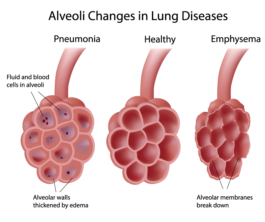
The image illustrates a comparison of alveolar structures in the lungs under different conditions: pneumonia, healthy, and emphysema. Alveoli are small air sacs within the lungs where the exchange of oxygen and carbon dioxide takes place.
On the left, the depiction of pneumonia shows alveoli filled with fluid and blood cells, which is indicative of infection and inflammation. The alveolar walls are visibly thickened by edema (accumulation of fluid), which can impair gas exchange.
The middle image represents healthy alveoli. They are clean, open, and unobstructed, allowing for efficient gas exchange. The walls between the alveoli (interalveolar septa) are thin and well-maintained, which is essential for proper lung function.
On the right, the alveoli affected by emphysema are shown. In this condition, the alveolar membranes are breaking down, which leads to larger but fewer alveoli. This reduces the surface area available for gas exchange and can lead to breathing difficulties.
The image captures the structural changes that these lung diseases cause, highlighting the importance of the integrity of the alveolar walls and space for the lung’s function of gas exchange. The healthy alveoli serve as a reference point, showing the stark contrast in structure and highlighting the pathological changes seen in disease states.
COPD Pathology
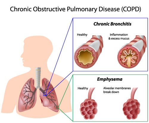
This image provides an educational overview of Chronic Obstructive Pulmonary Disease (COPD), emphasizing its two main conditions: chronic bronchitis and emphysema.
In the upper portion of the image, chronic bronchitis is depicted. It shows a comparison between a healthy bronchial tube and one affected by chronic bronchitis. The healthy bronchus has a clear lumen allowing free airflow. In contrast, the bronchus with chronic bronchitis is narrowed due to inflammation and is filled with excess mucus, which can obstruct airflow and cause difficulty in breathing.
Below, the focus is on emphysema, a condition where the alveoli, the small air sacs in the lungs, are damaged. The comparison here is between healthy alveoli and those affected by emphysema. Healthy alveoli are numerous and tightly packed, providing a large surface area for gas exchange. In emphysema, the alveolar membranes break down, leading to fewer and larger air spaces and a reduced surface area, which impairs the lungs’ ability to oxygenate blood and remove carbon dioxide.
The image also includes an illustration of a human silhouette with the lungs visible, and blue and green boxes linking the conditions to the respective parts of the respiratory system they affect.
The image is designed to educate viewers about the structural changes in the lungs and airways that characterize COPD, how these changes disrupt normal respiratory function, and the relationship between the anatomy and the disease process.
Pneumothorax Pathology
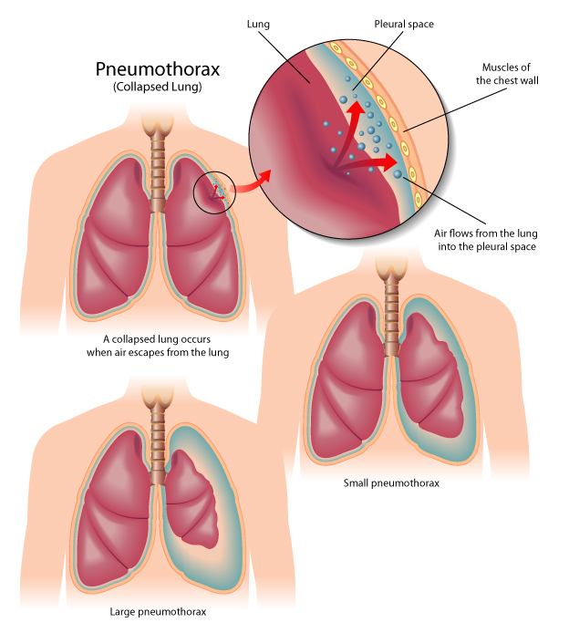
The image provides a visual explanation of pneumothorax, commonly known as a collapsed lung. It consists of three panels, each depicting a different aspect or severity of the condition within the human torso.
In the first panel, we see an overview of the lungs within the chest cavity with a red circle highlighting the area of interest. A detailed inset gives a closer look at the pleural space—the thin gap between the lung and the chest wall. Normally, this space is filled with a small amount of lubricating fluid, allowing the lung to move smoothly during breathing. In the case of pneumothorax, air has entered the pleural space, represented by red dots, which disrupts the pressure balance and causes the lung to collapse.
The second panel illustrates a large pneumothorax. It shows a significant portion of the lung collapsed, with a notable empty space where the lung has retracted away from the chest wall. This condition can lead to severe respiratory distress and requires immediate medical attention.
In the third panel, a small pneumothorax is shown, where there is a minor collapse of the lung with a small amount of air in the pleural space. This may cause less severe symptoms and, in some cases, can resolve on its own without invasive treatment.
The image caption explains that a collapsed lung occurs when air escapes from the lung into the pleural space. The mechanics of breathing are also indicated; the muscles of the chest wall expand and contract to facilitate lung inflation and deflation. However, when air is present in the pleural space, it prevents the lung from expanding fully, which can result in difficulty breathing and reduced oxygenation of the blood.
The educational graphic is designed to help viewers understand the physical changes of pneumothorax and how it affects lung function. It’s a condition often caused by chest injury, lung disease, or can spontaneously occur in otherwise healthy individuals. Treatment may involve procedures to remove the air from the pleural space and allow the lung to re-expand.
Pulmonary Embolism
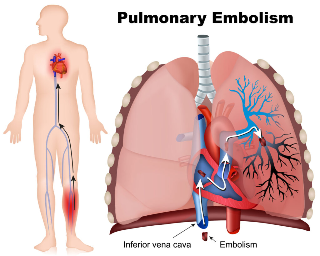
This image depicts a pulmonary embolism, which is a blockage in one of the pulmonary arteries in the lungs, typically caused by blood clots that travel to the lungs from the legs or other parts of the body (deep vein thrombosis).
On the left side of the image, a silhouette of a human figure is shown with a network of blood vessels highlighted. It indicates the possible path (shown with arrows) that a blood clot might take from the lower extremities, through the veins, and up into the heart and lungs. The red and blue colors represent oxygenated and deoxygenated blood vessels, respectively.
On the right side, the focus is on the heart and lungs, providing a detailed look at where the embolism occurs. The heart is shown pumping blood into the lungs, but a clot (labeled as “Embolism”) is lodged in one of the pulmonary arteries, obstructing the blood flow.
The image also points out the inferior vena cava, the large vein that carries deoxygenated blood from the lower body to the heart, which is a common route for a clot on its way to the lungs.
Pulmonary embolism is a serious condition that can be life-threatening and requires immediate medical attention. Symptoms may include shortness of breath, chest pain that may become worse when breathing in, cough, and leg pain or swelling, typically in the calf. Treatments often involve anticoagulants to prevent further clotting, thrombolytics to dissolve clots, and sometimes surgical intervention.
Anatomy Terms and Definitions
| Term | Definition |
|---|---|
| Alveoli | Small air sacs in the lungs where the exchange of oxygen and carbon dioxide takes place. |
| Bronchi | The main passageways through which air travels into each lung, branching from the trachea. |
| Bronchioles | Smaller branches stemming from the bronchi, leading to the alveoli where gas exchange occurs. |
| Cardiac Notch | A unique indentation in the left lung that accommodates the heart. |
| Diaphragm | The primary muscle of respiration, separating the thoracic cavity from the abdominal cavity and facilitating lung expansion. |
| Fissures | Lines that divide the lungs into lobes; the right lung has two (horizontal and oblique), and the left lung has one (oblique). |
| Hemoglobin | A protein within red blood cells that binds oxygen and transports it from the lungs to the body's tissues. |
| Hilum | A region on the medial aspect of each lung through which bronchi, blood vessels, lymphatics, and nerves enter and exit the lungs. |
| Intercostal Muscles | Muscles located between the ribs that assist in breathing by elevating or depressing the ribcage. |
| Lobes | Divisions within the lungs, with three in the right lung (upper, middle, lower) and two in the left lung (upper, lower). |
| Oxygenation | The process of oxygen molecules binding to hemoglobin in the red blood cells within the capillaries. |
| Pleura | A two-layered membrane enveloping each lung, consisting of the visceral pleura (inner layer) and the parietal pleura (outer layer). |
| Pleural Cavity | The space between the visceral and parietal pleurae, containing pleural fluid that lubricates the lungs during respiration. |
| Trachea | A vertical tube that bifurcates into the right and left bronchus, serving as the main airway to the lungs. |
