
Table of Contents
Basic Bronchi Anatomy
Presented here is an anterior view of the human respiratory system, focusing on the trachea and the lungs, along with their individual lobes and segments.
At the top, we have the trachea, also known as the windpipe, which is a tubular structure that facilitates the passage of air to the lungs. The trachea is supported by tracheal cartilages, which are C-shaped rings that prevent the trachea from collapsing and maintain an open airway. Above the trachea, the thyroid cartilage and cricoid cartilage are parts of the larynx, which plays a critical role in voice production and also serves as a passageway for air.
The trachea bifurcates into the right and left bronchi, which lead to the right and left lungs, respectively. Each lung is divided into lobes by fissures. The right lung consists of three lobes: the right upper lobe, right middle lobe, and right lower lobe, separated by the horizontal and oblique fissures. The left lung, on the other hand, consists of two lobes: the left upper lobe and left lower lobe, separated by the oblique fissure.
The apex of the lung is the uppermost part of the lung, located just below the collarbone. The base of the lung rests on the diaphragm, which is the main muscle involved in breathing.
The hilum is a medial region on each lung where the bronchi, blood vessels, and nerves enter and exit the lungs. This area is crucial for the organ’s function and connectivity to the rest of the body.
A unique feature of the left lung is the cardiac notch, which is an indentation on the surface of the lung where the heart lies against it, reflecting the close relationship between the heart and lungs within the thoracic cavity.
The branching patterns within the lungs represent the bronchial tree, with the bronchi dividing into smaller bronchioles and eventually leading to the tiny air sacs known as alveoli, where gas exchange occurs.
This diagram is instrumental in understanding the basic anatomy and division of the respiratory system, providing a clear visualization of the airways and the structure of the lungs.
Bronchial Tree Anatomy
Depicted here is a detailed illustration showcasing the structure of the bronchial tree within the human respiratory system.
At the top, the lumen of the primary bronchus is visible, which is the central airway passage within the bronchus. Surrounding the lumen, we can see longitudinal muscle fibers that help control the diameter of the airway.
Just beneath the lumen, mucous glands are depicted, which produce mucus to trap debris and microorganisms, keeping the airways clean. Moving outward, we have a layer of connective tissue which supports the bronchus structure.
The primary bronchus is seen branching into secondary bronchi, indicated on the left and right sides of the image. These secondary bronchi further subdivide into smaller passages, eventually leading to the bronchioles, shown at the bottom of the image. Bronchioles are the smallest airways that lead directly to the alveoli where gas exchange occurs.
The bronchus is also associated with the circulatory system, with the pulmonary artery running adjacent to it, carrying deoxygenated blood to the lungs for oxygenation. Additionally, a branch pulmonary venous is indicated, which will carry oxygenated blood away from the lungs back to the heart.
An essential feature of the bronchi are the cartilaginous rings, which provide structural support and prevent the airway from collapsing during the breathing process. These rings are visible throughout the bronchus’ length and are a defining characteristic of larger airways.
Overall, this image is a comprehensive representation of the bronchial anatomy, highlighting the airway structure and its relationship with the circulatory system within the lungs.
Cross-section of the Bronchus
This illustration provides a cross-sectional view of the bronchus, a major air passage of the lungs that branches from the trachea.
At the center, we observe the airway, which is the hollow part through which air is conducted. Lining the interior of the bronchus is the ciliated lining, comprised of ciliated epithelial cells. These cilia move rhythmically to propel mucus, which traps dust and pathogens, upwards towards the throat to be swallowed or coughed out, thereby protecting the respiratory system.
Surrounding the ciliated lining, gland ducts are shown. These ducts are connected to mucous glands that secrete mucus onto the surface of the epithelial lining. This mucus plays a vital role in humidifying the air and trapping particulates.
The section through cartilage hoop indicates a slice of the cartilaginous rings that provide structural support to the bronchus. These cartilaginous structures are crucial as they keep the airway open and prevent it from collapsing, particularly during exhalation when the pressure within the thorax increases.
The entire structure is indicative of a typical bronchus, which bifurcates into smaller bronchi and subsequently into bronchioles, finally leading to the alveolar ducts and alveoli where gas exchange occurs. This anatomical illustration is key for understanding the pathways of air conduction as well as the protective mechanisms in place within the respiratory system.
Respiratory Tract Infection
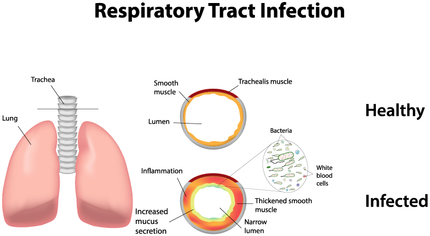
The image is an educational diagram contrasting a healthy respiratory tract with one that is suffering from a respiratory tract infection.
On the left, we see an illustration of the lungs and trachea, which make up parts of the lower and upper respiratory tract, respectively. This sets the anatomical context for the diagram.
In the upper right, there is a cross-section of a healthy trachea. The key features labeled here are the lumen (the open space within the trachea through which air flows), surrounded by a layer of smooth muscle, and the trachealis muscle, which helps adjust the diameter of the tracheal lumen.
Below this is a cross-section showing an infected trachea. The inflammation is visible as a thickened red layer, indicative of the body’s response to infection. The diagram highlights the results of this inflammation: increased mucus secretion, which is the body’s way of trapping and expelling pathogens, and a narrowed lumen, which can restrict airflow and cause symptoms like coughing and difficulty breathing. The presence of bacteria and white blood cells within the lumen demonstrates the infection and the immune response, respectively.
Overall, the image serves to educate on how a respiratory infection can physically alter the structure of the respiratory tract, leading to symptoms, and how the body’s immune response is actively engaged in fighting the infection. It also subtly emphasizes the importance of an open airway for proper breathing and how infections can compromise this.
Chronic Bronchitis
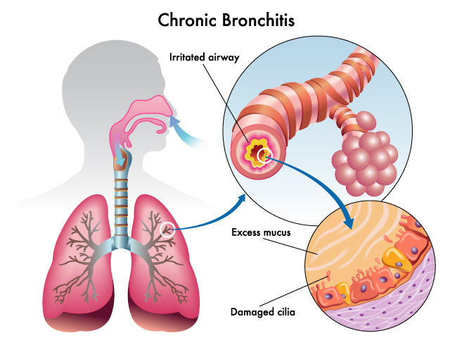
The image presented here illustrates the condition known as chronic bronchitis, which is characterized by prolonged inflammation of the airways within the respiratory system.
Starting on the left side of the image, we see a silhouette of a human head and torso, with the respiratory system highlighted. The trachea, or windpipe, is shown leading down from the back of the mouth and splitting into two main bronchi, each leading to a lung. The lungs are depicted in pink, and the branching structure of the smaller airways, or bronchioles, is visible within them.
Moving to the right, there is a detailed view of an irritated airway. The airway is depicted as a tube with a swollen and inflamed inner lining, indicating irritation. This is a hallmark of bronchitis, where the lining of the bronchial tubes becomes inflamed.
Further to the right, there is a close-up of the wall of the airway. Two key features are noted here: excess mucus and damaged cilia. The excess mucus is represented by a cluster of light pink blobs, accumulating within the airway and partially obstructing it. This mucus production is the body’s response to the irritation and serves to trap pathogens and particles, but in chronic bronchitis, it is produced in excess and can lead to coughing and difficulty breathing.
Below the mucus, the wall of the airway is shown with tiny, hair-like structures called cilia, which appear damaged and flattened. These cilia normally beat in a coordinated fashion to move mucus and trapped particles out of the airways, but when damaged, their function is compromised. This can lead to an accumulation of mucus and debris in the airways, further exacerbating the condition.
Overall, the image serves to highlight the key pathological features of chronic bronchitis, which include the persistent inflammation of the airway lining, excessive mucus production, and damage to the cilia that are essential for clearing mucus and particles from the respiratory tract.
Asthma Pathology
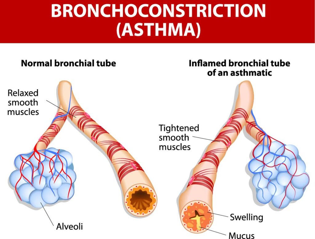
This image depicts a comparison between a normal bronchial tube and an inflamed bronchial tube of an individual with asthma, illustrating the concept of bronchoconstriction.
On the left, the normal bronchial tube is shown with relaxed smooth muscles surrounding it. These muscles are not constricted, allowing the airway to remain open and making breathing easy. The alveoli, which are the tiny air sacs located at the end of the bronchial tubes, are depicted in blue. They are where gas exchange takes place—oxygen enters the blood and carbon dioxide is expelled.
In contrast, on the right, the bronchial tube of an asthmatic individual is shown with tightened smooth muscles. This tightening, or bronchoconstriction, narrows the airway, making it more difficult for air to flow in and out. Additionally, there is visible swelling of the airway walls and the presence of mucus within the bronchial tube, both of which contribute to the narrowing of the airway and the characteristic difficulty in breathing experienced during an asthma attack.
The image illustrates the key differences in the airway dynamics between a normal functioning respiratory system and one affected by asthma, a chronic respiratory condition that can trigger episodes of wheezing, coughing, and shortness of breath.
Anatomical Terms and Definitions
| Term | Definition |
|---|---|
| Airway | The hollow part of the bronchus through which air is conducted. |
| Alveoli | Tiny air sacs where gas exchange occurs, connected by bronchioles. |
| Apex | The uppermost part of the lung, located just below the collarbone. |
| Base | The bottom part of the lung that rests on the diaphragm. |
| Bronchioles | The smallest airways leading directly to the alveoli. |
| Bronchus | A major air passage of the lungs that branches from the trachea. |
| Cartilaginous Rings | Structural supports that prevent the airway from collapsing. |
| Ciliated Lining | Lining of the bronchus made of ciliated epithelial cells that propel mucus. |
| Gland Ducts | Ducts connected to mucous glands, secreting mucus onto the bronchus lining. |
| Hilum | A medial region on each lung where the bronchi, blood vessels, and nerves enter and exit. |
| Lumen | The central airway passage within the bronchus. |
| Mucous Glands | Glands that produce mucus to trap debris and microorganisms. |
| Pulmonary Artery | The artery carrying deoxygenated blood to the lungs for oxygenation. |
| Pulmonary Vein | The vein carrying oxygenated blood away from the lungs back to the heart. |
| Secondary Bronchi | Branches of the primary bronchus that further subdivide into smaller passages. |
| Trachea | The windpipe, a tubular structure that facilitates the passage of air to the lungs. |
| Trabeculae | Fibrous bands providing internal support within the spleen. |
| Vascular Sinuses | Spaces in the spleen where blood is exposed to macrophages for phagocytosis. |
