
Table of Contents
Muscles and Tendons of the Ankle
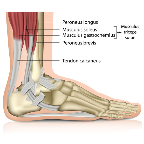
This image depicts the posterior aspect of the lower leg and foot, highlighting the musculature and tendinous structures that play pivotal roles in movement and stability.
At the top, we have the peroneus longus, also known as the fibularis longus, which is a superficial muscle on the lateral side of the leg. It plays a key role in eversion of the foot and stabilization of the ankle.
Beneath it lies the musculus soleus, a powerful muscle of the calf located underneath the gastrocnemius. It is important for plantar flexion of the foot at the ankle, crucial for actions like standing on tiptoes.
The musculus gastrocnemius is a large, superficial muscle on the back of the leg. It has two heads that create its distinctive shape and is the primary muscle for plantar flexion of the ankle, as well as flexing the knee.
Below the gastrocnemius, the peroneus brevis, or fibularis brevis, is situated. It is a shorter muscle than the peroneus longus and lies deep to it. It also assists in eversion of the foot.
Lastly, we see the tendon calcaneus, commonly known as the Achilles tendon, which is the thickest and strongest tendon in the body. It connects the calf muscles, including the gastrocnemius and soleus, to the heel bone (calcaneus). This tendon transmits the force from these powerful muscles to the foot, allowing for plantar flexion.
These muscles and tendons work in concert to control movements of the foot and ankle, providing the power for locomotion and balance.
Plantar Surface of the Foot
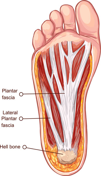
The image displays the anatomical structures on the underside of a human foot, providing a detailed view of the plantar fascia and related components.
In the center, we see the plantar fascia, a thick band of fibrous tissue that extends from the heel bone to the base of the toes. It supports the arch of the foot and plays a critical role in walking and absorbing shock. Its integrity is essential for the mechanical functioning of the foot during the stance and propulsive phases of gait.
Adjacent to the plantar fascia is the lateral plantar fascia, a portion of the overall plantar fascia structure, which specifically refers to the fascia located on the lateral, or outer, side of the foot. It helps to maintain the foot’s arch and manage the stresses placed upon the foot.
At the bottom of the image is the heel bone, known anatomically as the calcaneus. It is the largest bone in the foot and serves as the foundation for the posterior part of the foot structure. The calcaneus also serves as the attachment point for the plantar fascia and the Achilles tendon, making it integral to the function and stability of the foot.
The illustration shows these tissues in a healthy state, with the plantar fascia appearing as a strong and intact white band. This view is essential for understanding conditions such as plantar fasciitis, where the plantar fascia becomes inflamed, typically resulting in pain in the heel or bottom of the foot.
Posterior View of the Ankle
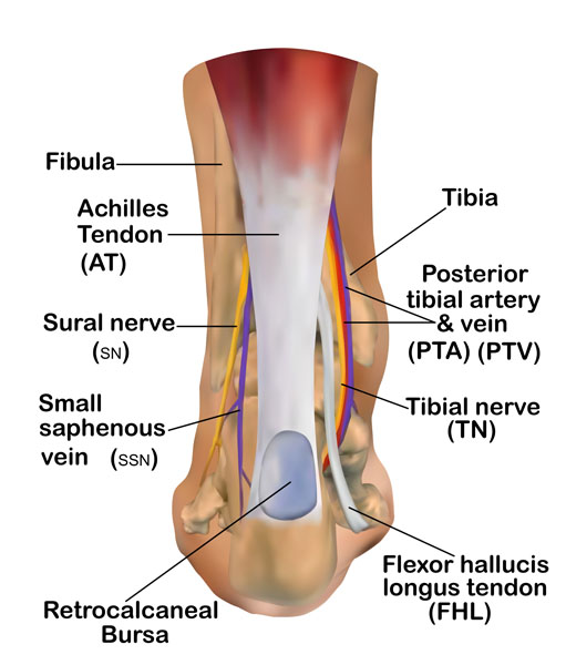
The image depicts the anatomical structures located at the back of the lower leg and ankle, detailing both the osseous (bony) and soft tissue components, as well as the neurovascular elements.
The two bones of the lower leg are labeled: the fibula and the tibia. The fibula is the smaller of the two and is located on the lateral side of the leg, while the tibia, also known as the shinbone, is larger and medially located.
The Achilles Tendon (AT), also known as the calcaneal tendon, is prominently displayed. It is the thickest and strongest tendon in the body, attaching the calf muscles to the heel bone and enabling plantar flexion of the foot and flexion of the knee.
Just anterior to the Achilles tendon, we can see the sural nerve (sN), which provides sensory innervation to the lower leg and foot.
The small saphenous vein (ssn) is a superficial vein that runs along the back of the leg and drains blood from the lateral side of the foot.
In the midline, the posterior tibial artery (PTA) and vein (PTV) are seen, which supply blood to the tissues of the lower leg and foot. Alongside these vessels runs the tibial nerve (TN), a branch of the sciatic nerve that innervates the lower leg and foot.
Located medially is the flexor hallucis longus tendon (FHL), which facilitates flexion of the big toe and supports the arch of the foot.
Lastly, the retrocalcaneal bursa is shown, which is a small sac of fluid that cushions and facilitates smooth movement between the Achilles tendon and the calcaneus (heel bone). This bursa can become inflamed, a condition known as retrocalcaneal bursitis, often associated with pain and swelling in the heel.
The illustration effectively captures the complexity of the lower leg and ankle, indicating how these structures interact to enable movement and support the body’s weight.
Ligaments of the Ankle: Lateral View
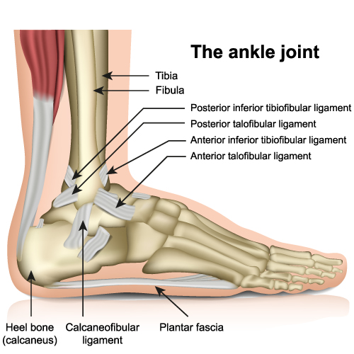
The image offers a detailed view of the ankle joint, highlighting its complex structure and the different components that allow for its function.
The tibia and fibula, the bones of the lower leg, frame the ankle joint at the top of the image. The tibia is the thicker bone located medially (toward the center of the body), while the fibula is thinner and found laterally (toward the outer side of the body).
Several ligaments are illustrated, which are crucial for the stability of the ankle joint. The posterior inferior tibiofibular ligament and the anterior inferior tibiofibular ligament connect the tibia and fibula, reinforcing the joint where these bones meet. These ligaments are part of the syndesmosis joint that allows for slight movement between the tibia and fibula and provides stability especially during the dorsiflexion of the foot.
The anterior talofibular ligament is shown on the front (anterior) side of the ankle, connecting the fibula to the talus bone of the foot. It is the most commonly injured ligament during an ankle sprain. The posterior talofibular ligament is located at the back (posterior) of the ankle, also connecting the talus to the fibula, and provides posterior stability to the ankle joint.
The calcaneofibular ligament, which stretches between the fibula and the calcaneus (heel bone), is visible on the lateral side of the ankle. This ligament provides lateral stability to the ankle and is also susceptible to injury when the ankle is inverted.
The plantar fascia is shown at the bottom of the foot. It is a thick band of connective tissue that extends from the calcaneus to the toes, supporting the arch of the foot.
Lastly, the calcaneus, or heel bone, is labeled at the bottom of the image. It forms the prominence of the heel and serves as the attachment point for the Achilles tendon and various ligaments of the foot.
This anatomical representation aids in understanding how these various bones, ligaments, and connective tissues contribute to the movement and stability of the ankle joint.
Anatomical Structure of the Foot
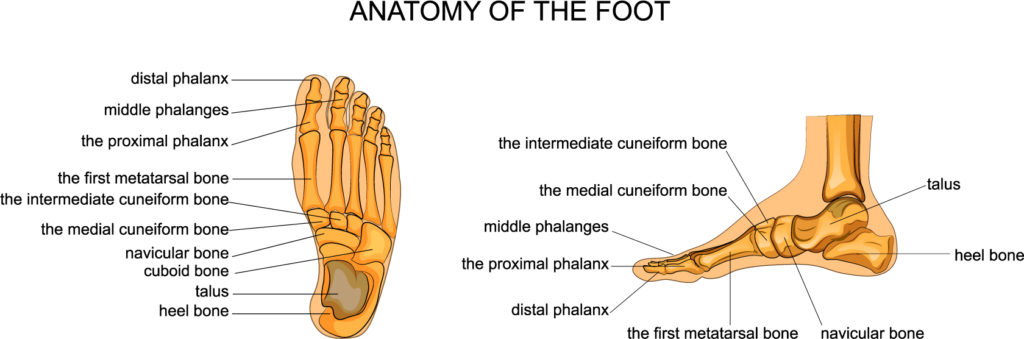
This diagram provides two perspectives of the anatomical structure of the human foot, delineating the various bones that compose its complex architecture.
On the left, we see a dorsal view, essentially a top-down look at the foot. It begins with the phalanges, the small bones of the toes. Each toe has three phalanges – distal, middle, and proximal, except for the big toe, which typically has only the distal and proximal. Moving back toward the heel, the metatarsals are shown, which are the long bones that connect the phalanges to the mid-foot. The first metatarsal bone is identified, which is associated with the big toe.
In the mid-foot, we have the tarsal bones, including the navicular bone, which sits medially in the arch of the foot, and the three cuneiform bones – medial, intermediate, and lateral, which are stacked between the navicular and the first three metatarsals. Additionally, the diagram indicates the cuboid bone, which is not labeled but would be situated laterally next to the cuneiforms. The talus bone is located just above the navicular and is a pivotal bone for ankle movement as it articulates with the tibia above.
At the rear of the foot lies the heel bone, known as the calcaneus, which is the largest bone in the foot and serves as the foundation for the external projection of the heel.
On the right, the image shifts to a lateral view of the foot, offering a side profile. Here, we can see the relationship between the talus and the calcaneus more clearly, along with the alignment of the first metatarsal bone and the phalanges of the big toe. The navicular bone is visible in front of the talus, and the medial cuneiform is directly in front of the navicular.
Understanding the anatomy of the foot is crucial for a wide range of disciplines, from medicine to sports science, as it plays an integral role in balance, movement, and absorbing the impact associated with activities like walking and running.
Plantar Fasciitis Pathology
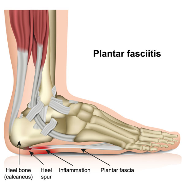
This image illustrates a condition known as plantar fasciitis, which affects the foot. At the bottom of the foot, we see the plantar fascia, a thick band of tissue that connects the heel bone (calcaneus) to the toes. It supports the arch of the foot and absorbs shocks when we walk.
Plantar fasciitis is characterized by inflammation of the plantar fascia, particularly where it attaches to the heel bone. This inflammation is indicated on the diagram with a red highlight, suggesting pain or irritation. The cause of this inflammation can be due to overuse, such as from running or standing for long periods, or from having a foot structure that puts extra stress on the plantar fascia.
Additionally, the image shows a heel spur, a bone growth on the heel bone. Heel spurs are often associated with plantar fasciitis, though they are not always the cause of the pain. They can be the result of long-term strain on the muscles and ligaments of the foot, and they are often found in athletes who perform a lot of jumping.
Treatment for plantar fasciitis typically involves rest, ice, exercises to stretch the plantar fascia and Achilles tendon, and anti-inflammatory medications. In persistent cases, physical therapy or orthotics may be recommended.
Tendonitis Pathology
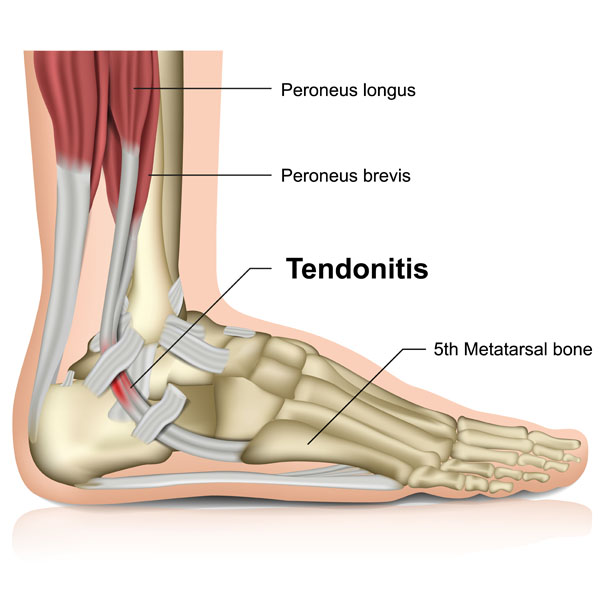
This image focuses on the lateral aspect of the lower leg and foot, showing both the muscular and skeletal structures, as well as a condition affecting the tendons.
Labeled at the top, the peroneus longus, known anatomically as the fibularis longus, is a superficial muscle that extends down the lateral side of the leg. It functions primarily to evert and plantarflex the foot. It passes behind the lateral malleolus and under the foot to attach at the base of the first metatarsal and the medial cuneiform bone.
Beneath the peroneus longus is the peroneus brevis, a shorter muscle that also lies on the lateral side of the leg and assists in the eversion and plantarflexion of the foot. The peroneus brevis tendon attaches to the base of the fifth metatarsal bone.
The area highlighted as “Tendonitis” indicates inflammation of one or both of the tendons of the peroneal muscles. Tendonitis here can result from overuse, acute injury, or chronic irritation, and it commonly presents with pain and swelling around the lateral malleolus, potentially affecting mobility.
Lastly, the 5th metatarsal bone is shown, which is the bone at the outer side of the midfoot. Fractures of this bone are common sports injuries, often occurring near the base of the bone, which can be compromised by the pull of the peroneus brevis during forceful activities or an ankle sprain.
In managing tendonitis, treatment typically involves rest, ice, compression, and elevation (RICE protocol), along with anti-inflammatory medications and physical therapy to strengthen and stretch the affected tendons and muscles.
Ankle Sprain Pathology
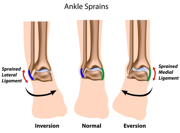
The image displays three foot positions related to ankle sprains, a common injury where the ligaments that support the ankle stretch beyond their limits and, in severe cases, tear.
The first on the left shows an ‘Inversion’ injury, where the sole of the foot is turned inwards. This movement is the most common mechanism of injury for ankle sprains and typically affects the lateral ligaments located on the outside of the ankle. The illustration marks the sprained lateral ligament in blue with a red area indicating the site of injury or pain.
The middle image represents a ‘Normal’ ankle alignment where the ligaments are not under abnormal stress.
The third on the right shows an ‘Eversion’ injury, where the sole of the foot is turned outwards. This type of sprain is less common and usually results in injury to the medial ligament, located on the inside of the ankle. The sprained medial ligament is highlighted in green with a red area indicating the injury site.
Ankle sprains are graded based on their severity, from mild stretching to complete tears, and the treatment varies accordingly, from rest and ice application to physical therapy and, in severe cases, surgery. It is important to properly rehabilitate ankle sprains to prevent chronic instability.
Achilles Tendonitis Pathology
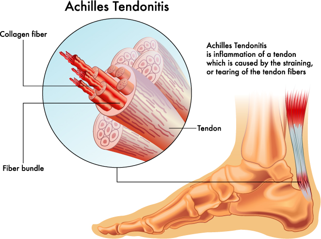
This illustration presents an overview of Achilles tendinitis, which is a condition characterized by inflammation of the Achilles tendon. The Achilles tendon is a robust and fibrous cord that connects the muscles in the back of your calf to your heel bone.
The image shows the human foot from the side, with the Achilles tendon highlighted in red to signify inflammation. Inflammation of a tendon can result from straining the tendon, which happens when the tendon is overloaded or overstretched, or from micro-tears in the tendon fibers due to acute injury or chronic overuse.
A magnified view of the affected area is provided to give a closer look at the tendon’s structure. The tendon is composed of collagen fibers, which are bundled together to form fiber bundles. These bundles collectively make up the tendon itself. The magnified section reveals fraying of these collagen fibers, which is indicative of the damage that occurs with tendonitis.
Achilles tendinitis can lead to pain and swelling in the heel area and can make walking or running difficult. Treatment typically involves rest, ice, anti-inflammatory medications, and in some cases, physical therapy to strengthen the surrounding muscles and support the healing process. If left untreated, Achilles tendinitis can lead to a greater risk of tendon rupture, which may require surgical intervention.
Bone Spur Pathology
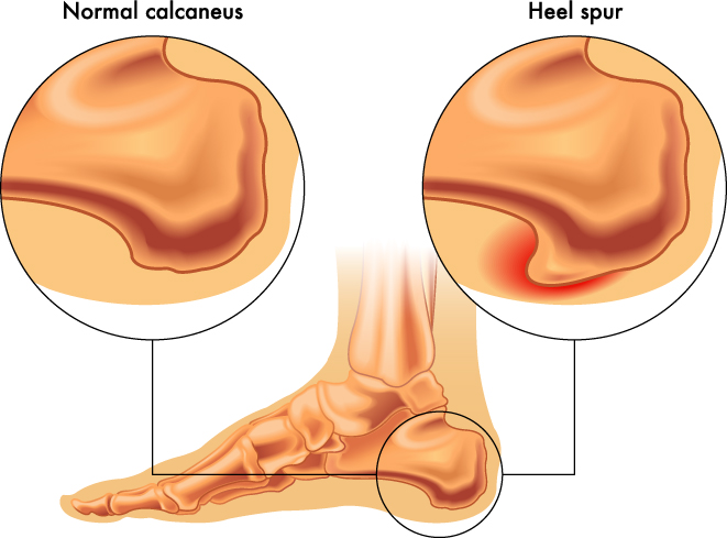
The image displays a comparison between a normal calcaneus, or heel bone, and one affected by a heel spur. The calcaneus is a critical bone in the foot that forms the heel and serves as a point of attachment for the Achilles tendon and the plantar fascia.
On the left, a magnified view of a normal calcaneus reveals a smooth, rounded surface without any protrusions. This is the typical shape of a healthy heel bone, which supports the weight of the body and aids in walking.
On the right, the magnified view shows a calcaneus with a heel spur. A heel spur is a bony growth or calcified deposit that forms on the underside of the heel bone. It is represented in the illustration by a hook-like protrusion extending from the bone’s surface. The red shading near the spur indicates inflammation, which often accompanies the presence of a spur and can contribute to pain.
A heel spur is frequently associated with plantar fasciitis, as the chronic pull of a tight plantar fascia on the heel bone can lead to the formation of a spur. Heel spurs can cause discomfort or pain when standing or walking, though they may not always be symptomatic.
The central part of the image shows the side profile of the foot with the location of the heel bone circled, linking it to the magnified views for clear identification. Treatment for a heel spur may include rest, ice, anti-inflammatory medications, orthotic inserts, and in some cases, surgery to remove the spur if conservative treatments fail to relieve pain.
Anatomical Terms and Definitions
| Term | Definition |
|---|---|
| Achilles Tendon | A robust and fibrous cord that connects the muscles in the back of the calf to the heel bone, critical for walking, running, and jumping. |
| Calcaneus | The heel bone, the largest bone in the foot that forms the heel's contour and serves as an attachment point for the Achilles tendon and plantar fascia. |
| Flat Foot (Pes Planus) | A condition where the arch on the inner side of the foot is flattened, allowing the entire sole of the foot to touch the ground when standing. |
| Heel Spur | A bony growth or calcified deposit on the underside of the heel bone, often associated with plantar fasciitis. |
| Inversion Injury | A common mechanism of ankle sprain where the sole of the foot is turned inwards, typically affecting the lateral ligaments on the outside of the ankle. |
| Lateral Ankle Sprain | A type of injury affecting the ligaments on the outer side of the ankle, classified into grades based on severity from stretching to complete tears. |
| Plantar Fascia | A thick band of fibrous tissue that runs across the bottom of the foot, connecting the heel bone to the toes, supporting the arch of the foot. |
| Plantar Fasciitis | A condition characterized by inflammation of the plantar fascia, causing heel pain, especially with the first steps after waking up or after long periods of rest. |
| Tendonitis | Inflammation of a tendon, often resulting from overuse, acute injury, or chronic irritation, and typically presents with pain and swelling. |
| Tendinosis | Degenerative change in the tendon fibers, characterized by tiny tears within and around the tendon tissue, leading to persistent pain and an increased risk of tendon rupture. |
| Tendon Rupture | A complete tear through the tendon, leading to sudden and severe pain, inability to bend the foot downward, and difficulty walking, often requiring surgical repair. |
