
Table of Contents
Thoracic and Cervical Lymphatic System
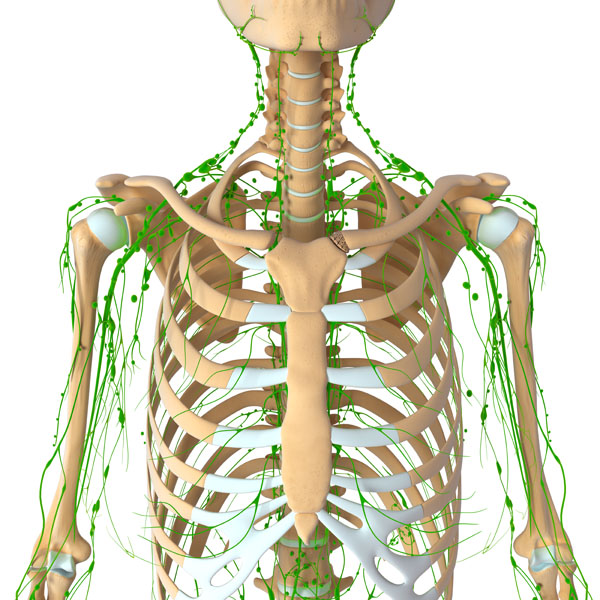
The image depicts the thoracic region of the human body with a focus on the lymphatic system. At the center, we see the sternum, a flat bone that forms the front part of the rib cage. Flanking the sternum on either side are the ribs, arching bones that protect the thoracic cavity.
What stands out most prominently in this representation is the network of green lines, which indicate the pathways of the lymphatic vessels. These vessels are part of the lymphatic system, which plays a crucial role in maintaining fluid balance in the body and is a key component of the immune system. The lymphatic vessels transport lymph, a clear fluid that contains white blood cells, especially lymphocytes that fight infection.
Above the sternum, the green lines converge towards the neck, indicating the location of major lymph nodes. Lymph nodes are small, bean-shaped structures that act as filters for harmful substances. They contain immune cells that can help fight infection by attacking and destroying germs that are carried in through the lymph fluid.
The arrangement of the lymphatic vessels around the shoulder region, extending towards the arms, suggests the presence of axillary lymph nodes, which are responsible for draining lymph from the upper limbs, breast, and upper wall of the abdomen.
In summary, the image provides a visual overview of the superficial lymphatic drainage of the thoracic region, highlighting the network of lymphatic vessels and their connections to lymph nodes, which are instrumental in immune function and fluid regulation.
Pelvic Region Lymphatic System
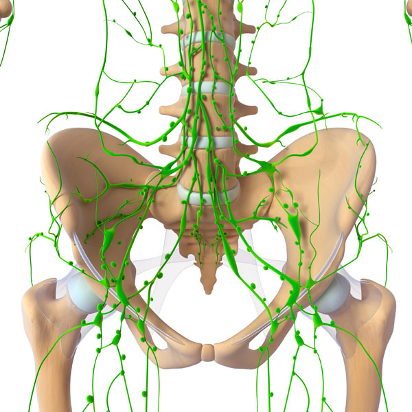
This image shows a detailed view of the pelvic region, with an emphasis on the lymphatic system. The pelvis is formed by the hip bones, sacrum, and coccyx. The hip bones are depicted in a lighter shade, highlighting the iliac crests at the top, which form the upper border of the pelvis.
The intricate network of green lines represents the lymphatic vessels, which are crucial for transporting lymph fluid throughout the body. This fluid contains lymphocytes, which are pivotal for the immune response. In the pelvic region, these vessels are responsible for draining lymph from the lower limbs, the pelvis, and the genital region.
The convergence of these green lines toward the center of the pelvic cavity indicates the presence of numerous lymph nodes, which are key elements in the lymphatic system. These nodes filter the lymph fluid, trapping bacteria, viruses, and other foreign substances, which are then destroyed by specialized white blood cells known as lymphocytes.
In the center, above the convergence of the lymph vessels, we can observe the lumbar spine, which includes the vertebrae and intervertebral discs. The central placement of the lumbar spine within the network of lymphatic vessels underscores its anatomical importance, serving as a structural foundation for the upper body while also being centrally located within the lymphatic drainage system of the lower body.
Overall, the image provides an anatomical overview of the lymphatic drainage in the pelvic area, demonstrating the complexity and vital role of the lymphatic system in this region.
Upper Limb Lymphatic System
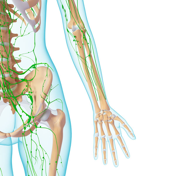
The provided image illustrates the lymphatic system of the upper limb, specifically the arm. The bones of the upper limb are shown in a lighter color, with the humerus visible in the upper arm, the radius, and ulna in the forearm, and the intricate arrangement of carpals, metacarpals, and phalanges in the hand.
Overlaying the skeletal structure, the green lines represent the lymphatic vessels, which form a complex network designed to transport lymph—a fluid containing white blood cells that are vital for immune function—throughout the limb. These vessels are particularly dense around the elbow and the axillary region (armpit), where several lymph nodes are located.
Lymph nodes are small, kidney-shaped structures that serve as filters for the lymphatic system. They are strategically placed in clusters where the lymphatic vessels converge, such as the axillary lymph nodes in the armpit, which are responsible for draining lymph from the upper limb, breast, and upper chest area.
The lymphatic vessels extend down the arm, running parallel to the major blood vessels. They continue across the elbow, where they follow the brachial artery and then diverge into two paths along the radius and ulna bones in the forearm. These vessels extend to the wrist and into the hand, ending in the fingertips.
This network of lymphatic vessels and nodes plays a critical role in the body’s defense mechanisms by transporting lymphocytes to areas of infection or inflammation and by removing cellular debris and pathogens from the tissues. The lymphatic system also contributes to fluid balance by returning excess tissue fluid back into the circulatory system.
The image effectively demonstrates the extensive reach of the lymphatic system in the arm, highlighting its importance in both the immune response and the maintenance of homeostasis.
Lymphatic System Anatomy
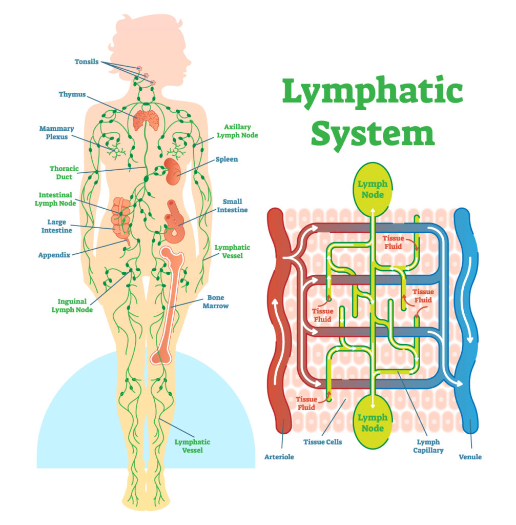
The image presents a comprehensive view of the human lymphatic system, which is a part of the circulatory system and a vital component of the immune system. It features two main components: a schematic representation of the lymphatic system within the human body on the left, and a close-up diagrammatic illustration of the interstitial fluid exchange within the lymphatic and blood circulatory systems on the right.
On the left side, the outline of a human body is shown with a network of green lines indicating the lymphatic vessels that transport lymph—a fluid containing white blood cells that help fight infection. Key lymphatic organs and collections of lymph nodes are identified and named. Starting from the head, the tonsils are marked as part of the lymphatic system, which contributes to defending the body against inhaled or ingested pathogens. The thymus, situated in the chest, is noted for its role in the maturation of T-cells, which are critical for adaptive immunity.
The diagram also identifies clusters of lymph nodes, such as the axillary lymph nodes in the armpits, which filter lymph from the upper limbs, breasts, and upper chest. The spleen, on the left side of the body under the ribs, is marked as an organ that filters blood, recycles old red blood cells, and helps mount an immune response. The inguinal lymph nodes are indicated in the groin area, where they filter lymph from the legs and the lower torso.
Other highlighted areas include the mammary plexus, thoracic duct, and various nodes associated with the gastrointestinal tract, such as the intestinal lymph nodes. The thoracic duct is the largest lymphatic vessel and plays a crucial role in transporting lymph from the majority of the body back into the bloodstream.
On the right side, the image zooms in to show how the lymphatic system interacts with the blood circulatory system. Here, we see arterioles and venules, small branches of arteries and veins, respectively. The tissue fluid, which bathes and surrounds the cells of the tissues, is depicted in blue. Excess tissue fluid is drained by lymph capillaries, which are part of the lymphatic system. The lymph nodes act as filters for the lymph before it is returned to the venous blood circulation.
This detailed diagram serves to explain the structure and function of the lymphatic system, highlighting its role in fluid balance, nutrient absorption, and immune defense throughout the body.
Cardiovascular and Lymphatic System Interraction
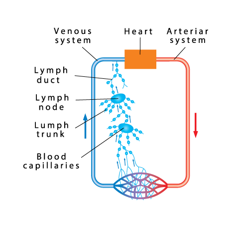
The image illustrates the relationship between the cardiovascular and lymphatic systems. On the right, we have the arterial system shown in red, which carries oxygenated blood away from the heart to the rest of the body. On the left, the venous system in blue returns deoxygenated blood back to the heart.
Connecting these two systems are the blood capillaries, represented by the intertwined blue and red lines at the bottom of the image. These capillaries are the sites of nutrient and gas exchange between the blood and the tissues. The blue capillaries are where oxygen and nutrients diffuse from the blood to the tissues, while the red capillaries represent the return path of deoxygenated blood.
The lymphatic system is depicted in light blue, featuring a network that includes lymph capillaries, lymph nodes, lymph trunks, and a lymph duct. Lymph capillaries collect excess tissue fluid—now called lymph—from the interstitial spaces, which is a byproduct of the exchange processes in the blood capillaries.
Lymph then travels through the lymph trunks and is filtered by lymph nodes, where immune responses can be initiated. The lymph nodes are depicted as enlarged blue beads along the path of the lymphatic vessels. After passing through the lymph nodes, the lymph is directed into the lymph duct, which ultimately drains into the venous system, thus maintaining fluid balance and playing a role in the body’s defense mechanisms.
The diagram effectively communicates the circulation of fluids in the body, showing how the lymphatic system complements the cardiovascular system by returning interstitial fluid to the bloodstream and participating in immune surveillance.
Capillary Fluid Exchange
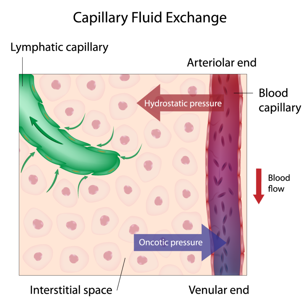
The diagram you’re looking at provides a visual explanation of capillary fluid exchange, a process that occurs at the microscopic level in the body’s tissues.
In this image, we see a blood capillary, which is part of the body’s circulatory system. The blood capillary is depicted in red with an arrow indicating the direction of blood flow. This flow is from the arteriolar end (where blood enters the capillary from an arteriole) to the venular end (where blood exits the capillary to a venule).
At the arteriolar end, blood pressure within the capillary is high, creating what is known as hydrostatic pressure. This pressure is depicted with outward-facing arrows, showing that it forces fluid out of the blood capillary and into the surrounding interstitial space, which is the area between cells.
The interstitial space is shown containing numerous pink circles, representing tissue cells. Around these cells, we have what is called the interstitial fluid, which is the fluid that surrounds and bathes the cells.
Also illustrated is the concept of oncotic pressure, which is indicated by the blue arrow pointing into the blood capillary. Oncotic pressure is primarily due to plasma proteins in the blood, which draw water back into the capillary from the interstitial space. This inward draw of fluid helps to balance the outward force of the hydrostatic pressure.
Lymphatic capillaries are shown in green, with small arrows depicting the flow of interstitial fluid into them. These capillaries are part of the lymphatic system and serve to collect excess fluid from the interstitial space and return it to the bloodstream, thus maintaining fluid balance in the tissues.
The diagram simplifies a complex physiological process, demonstrating how the balance of fluid between blood and tissue cells is maintained through the forces of hydrostatic and oncotic pressures and how the lymphatic system plays a role in this balance by collecting excess fluid. This exchange is vital for delivering nutrients to cells and removing waste products.
Diagram of Lymphatic Capillary
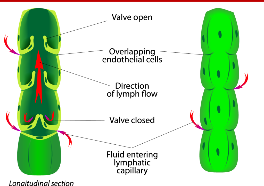
The image provides a diagrammatic representation of a section of a lymphatic capillary, demonstrating how the structure of the capillary facilitates the flow of lymph.
On the left, we see a longitudinal section of a lymphatic capillary with several key features labeled. The main feature shown is a series of valves that ensure unidirectional flow of lymph. These valves are depicted in two states: ‘valve open’ and ‘valve closed’. The open valve shows the flaps apart, allowing the flow of lymph through the vessel, indicated by the red arrows pointing upwards. This movement of lymph is essential for transporting immune cells throughout the body and for returning excess interstitial fluid to the bloodstream.
The ‘valve closed’ state shows the flaps of the valve meeting together, preventing the backflow of lymph, ensuring that lymph moves towards the larger lymphatic vessels and ultimately towards the venous circulation. The closed valve action is crucial for maintaining the flow of lymph even when there is a change in pressure inside the lymphatic capillary or surrounding tissues.
Surrounding the vessel are overlapping endothelial cells. These cells form the inner lining of the lymphatic capillary and their overlapping nature allows fluid from the surrounding tissue to enter the lymphatic system while preventing the lymph from leaking out.
The diagram also indicates fluid entering the lymphatic capillary. This fluid is collected from the interstitial space, which contains proteins, waste products, and other cells, including immune cells.
To the right, the image presents a three-dimensional view of the lymphatic capillary, showing the bulging sections where the valves are located, which correspond to the areas marked as ‘valve open’ and ‘valve closed’ in the left image.
Overall, the diagram serves to illustrate the mechanism by which lymphatic capillaries collect and transport lymph fluid, playing a crucial role in the immune system and in maintaining fluid homeostasis within the body.
Lymph Node Structure
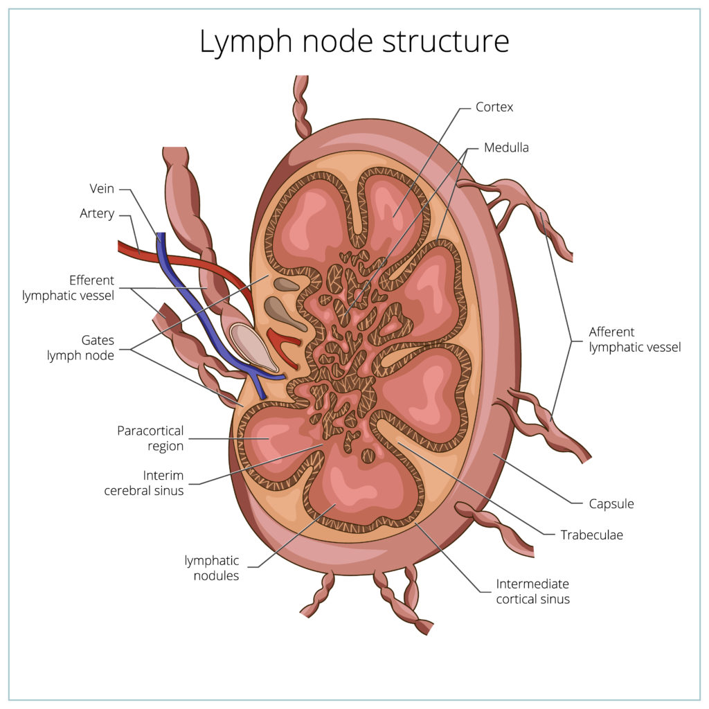
The image provides an anatomical illustration of a lymph node structure, which is an integral component of the lymphatic system.
At the outermost layer, we see the capsule, which is the tough outer covering of the lymph node. Extending inward from the capsule are trabeculae, which are supportive structures that divide the node into compartments.
Lymph nodes have a characteristic internal structure that includes the cortex and medulla. The cortex is the outer part of the lymph node and contains densely packed immune cells, such as lymphocytes, which are crucial for the immune response. Within the cortex, we can identify the paracortical region, which is rich in T-cells, and lymphatic nodules that are often the sites of B-cell activation and germinal center formation. These germinal centers are where B-cells proliferate and differentiate into antibody-producing cells.
The medulla is the innermost part and contains medullary cords, which are strands of lymphatic tissue, and medullary sinuses, which are channels through which lymph flows.
Lymph enters the lymph node through multiple afferent lymphatic vessels, which are indicated by arrows showing the direction of lymph flow towards the node. Once inside, lymph percolates through the sinuses of the cortex and medulla, allowing immune cells to detect and respond to pathogens or foreign particles.
The lymph then exits the lymph node via the efferent lymphatic vessel, which is singular and usually found at the hilum of the lymph node, where blood vessels also enter and exit. The efferent vessel carries the filtered lymph away from the lymph node and towards the venous circulation.
Arteries and veins are shown adjacent to the efferent lymphatic vessel, indicating the close association of the lymphatic system with the circulatory system in terms of nutrient supply and waste removal.
This diagram is a valuable tool for understanding the structure and function of lymph nodes, showcasing how they act as filters for the lymphatic fluid and play a vital role in the body’s immune defense.
Overview of the Lymphatic System
The lymphatic system serves as a critical subsystem within our body, playing an instrumental role in our immune defense and fluid balance. Unlike the heart’s driving force behind the cardiovascular system, the lymphatic system lacks an inherent pump. Consequently, it heavily relies on surrounding muscle contractions and general body movements to propel the lymph fluid.
The rhythmic contraction and relaxation of adjacent skeletal muscles predominantly govern the lymph fluid’s circulation. When these muscles contract, typically during physical activity, they exert pressure on lymphatic vessels, which facilitates the lymph fluid’s forward propulsion. Thus, every movement, every flex of a muscle, contributes to the circulation of this crucial fluid. It underscores the importance of regular exercise and physical activity in maintaining a healthy lymphatic system.
However, inactivity poses a threat to this system’s fluid flow. Extended periods of sitting or lying down, typical of sedentary lifestyles, mean fewer muscle contractions and thus less pressure on lymphatic vessels. Consequently, this reduces lymph fluid flow, hampering the efficient removal of waste products and excess interstitial fluid, potentially impacting tissue health adversely.
Aided by specialized valves within their walls, lymphatic vessels overcome the challenge posed by gravity. Functioning as one-way gates, these valves allow fluid to flow in a single direction—toward the lymph nodes and eventually back into the bloodstream—while preventing backward movement. This unidirectional flow is critical to the system’s effective operation, maintaining the proper balance of interstitial fluid, avoiding edema (swelling), and supporting immune function by facilitating the removal of toxins, waste products, and pathogens from the tissues.
Lymph nodes, as small, bean-shaped structures scattered throughout our body along the lymphatic vessels, play a pivotal role in the lymphatic system’s defense mechanism and waste disposal. As the lymph fluid journeys back toward the heart, it passes through sinuses within these nodes, where it undergoes a thorough cleansing process. Specialized immune cells housed within the nodes, including lymphocytes and macrophages, recognize and eradicate pathogens, foreign substances, and abnormal cells.
Apart from this recognition and elimination function, lymph nodes also break down toxins and large particles into smaller ones for further processing. This crucial role aids in neutralizing harmful substances and ensuring their efficient removal from the body. Strategically located at lymphatic vessels’ convergence points or where the lymph flow is concentrated, these nodes efficiently process and purify lymph fluid from different body parts before it reenters the circulatory system.
Finally, after purification, the lymphatic fluid continues its journey back into the cardiovascular system. Merging into larger collecting vessels, it drains into the subclavian vein situated behind the left collarbone. Here, it reenters the bloodstream, becoming part of the plasma and resuming its circulation throughout the body.
In conclusion, the lymphatic system, intricately intertwined with our daily movements, serves as an unsung hero in our body’s defense mechanisms and fluid balance. Its reliance on muscle contractions for lymph fluid flow underscores the importance of regular physical activity. Furthermore, the specialized lymph nodes ensure the efficient processing and purification of the lymphatic fluid, contributing to the overall health of our body’s tissues.
Lymph Nodes and Processing of Fluids
Lymph nodes play a vital role in the lymphatic system’s function as a defense mechanism and waste disposal system. Let’s explore the significance of lymph nodes in processing fluids and maintaining overall health:
Structure and Cleansing: Lymph nodes are small, bean-shaped structures distributed throughout the body along the lymphatic vessels. They consist of a network of sinuses, which are interconnected spaces where lymphatic fluid flows. As lymphatic fluid travels back toward the heart, it passes through these sinuses within the lymph nodes.
Fluid Cleansing: The sinuses within the lymph nodes act as filtration sites, where lymphatic fluid is cleansed and processed. Lymph nodes contain specialized immune cells, such as lymphocytes and macrophages, which play a crucial role in recognizing and eliminating pathogens, foreign substances, and abnormal cells.
Breakdown of Toxins: One of the primary functions of lymph nodes is to break down toxins and large particles into smaller, more manageable particles for further processing. This breakdown process aids in neutralizing harmful substances and facilitating their removal from the body. The immune cells within the lymph nodes work to dismantle and eliminate these toxins, ensuring the overall purification of the lymphatic fluid.
Location and Distribution: Lymph nodes are strategically located throughout the body, primarily in regions where lymphatic vessels converge or where lymph flow is concentrated. The largest clusters of lymph nodes can be found in specific areas, including behind the knees, at groin leg creases, in the armpits, around the face and neck, in the upper chest, and deep inside the abdomen. These locations reflect regions where lymphatic fluid from different body parts converges and undergoes further processing and purification.
Drainage and Return to Circulation: After passing through the lymph nodes, the cleansed lymphatic fluid continues its journey back into the circulatory system. The lymphatic vessels ultimately merge into larger collecting vessels, which drain into the subclavian vein located behind the left collarbone. At this point, the lymphatic fluid reenters the bloodstream and becomes part of the plasma, continuing its circulation throughout the body.
Understanding the role of lymph nodes in fluid processing highlights their essential function in maintaining a healthy immune response and removing waste materials from the body. By filtering lymphatic fluid and breaking down toxins, lymph nodes contribute to the overall purification of the fluids and support the body’s defense against pathogens and harmful substances. The strategic distribution of lymph nodes ensures effective processing and purification of the lymphatic fluid before its return to the cardiovascular system.
Anatomical Terms and Definitions
| Term | Definition |
|---|---|
| Afferent | Referring to vessels or nerves that carry fluid or signals towards a particular organ or structure, such as lymphatic vessels bringing lymph into a lymph node. |
| Arteries | Blood vessels that carry oxygenated blood away from the heart to the rest of the body. |
| Axillary Lymph Nodes | Lymph nodes located in the armpits, responsible for draining lymph from the upper limbs, breast, and upper wall of the abdomen. |
| Capsule | The tough outer covering of a lymph node. |
| Cortex | The outer part of the lymph node, containing densely packed immune cells. |
| Efferent | Referring to vessels or nerves that carry fluid or signals away from an organ or structure, such as lymphatic vessels taking filtered lymph away from a lymph node. |
| Endothelial Cells | Cells that form the inner lining of lymphatic capillaries, blood vessels, and the heart, allowing fluid from the surrounding tissue to enter the lymphatic system while preventing the lymph from leaking out. |
| Germinal Center | Areas within the lymphatic nodules of the cortex of a lymph node where B-cells proliferate and differentiate into antibody-producing cells. |
| Hip Bones | The bones forming the pelvis, including the iliac crests, which are the upper borders of the pelvis. |
| Iliac Crests | The upper borders of the pelvis, part of the hip bones. |
| Intercostal Spaces | The spaces between the ribs, which are supplied blood by the intercostal arteries. |
| Intestinal Lymph Nodes | Lymph nodes associated with the gastrointestinal tract, involved in filtering lymph from this region. |
| Lymph | A clear fluid that circulates in the lymphatic system, containing white blood cells, especially lymphocytes, which fight infection. |
| Lymph Capillaries | Small lymphatic vessels that collect excess tissue fluid, now called lymph, from the interstitial spaces. |
| Lymph Nodes | Small, bean-shaped structures that act as filters for harmful substances, containing immune cells that help fight infection by attacking and destroying germs carried in through the lymph fluid. |
| Lymphatic System | A part of the circulatory system, comprising a network of lymphatic vessels that transport lymph—a fluid containing white blood cells—to fight infection. |
| Lymphatic Vessels | Vessels that form a complex network designed to transport lymph throughout the body. |
| Lumbar Spine | The part of the spine located in the lower back, consisting of vertebrae and intervertebral discs. |
| Medulla | The innermost part of a lymph node, containing medullary cords and sinuses through which lymph flows. |
| Pelvis | The lower part of the torso, formed by the hip bones, sacrum, and coccyx. |
| Rib Cage | The part of the skeleton that encloses the thoracic cavity, consisting of the ribs and sternum. |
| Sacrum | A large, triangular bone at the base of the spine, forming the upper part of the pelvis. |
| Spleen | An organ under the ribs on the left side of the body, filtering blood, recycling old red blood cells, and helping mount an immune response. |
| Sternum | A flat bone that forms the front part of the rib cage, providing attachment for the ribs. |
| Thymus | An organ situated in the chest, involved in the maturation of T-cells, which are critical for adaptive immunity. |
| Trabeculae | Supportive structures within a lymph node that divide it into compartments. |
| Veins | Blood vessels that return deoxygenated blood back to the heart from the rest of the body. |
