
Table of Contents
Adrenal Gland Anatomy
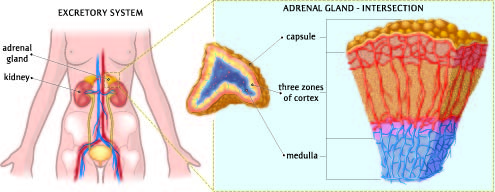
This image illustrates two aspects of human anatomy, focusing on the excretory system and a detailed view of the adrenal gland.
On the left side of the image, we have a depiction of the excretory system. This system is responsible for the removal of wastes and excess substances from the bloodstream. The kidneys play a crucial role here, filtering blood to produce urine, which is then passed to the bladder through structures called ureters. The bladder stores urine until it is expelled from the body. The image shows the kidneys located at the back of the abdominal cavity, one on each side of the spine, and the ureters descending from the kidneys to the bladder. Above each kidney, there is an adrenal gland.
Zooming in on the right side of the image, we have an intersection view of the adrenal gland, which sits atop the kidney. The adrenal gland is divided into two main parts: the cortex and the medulla. The outer region, called the cortex, is shown in three distinct zones, each responsible for producing different hormones. These hormones are vital for regulating metabolism, immune system suppression, and the body’s salt and water balance. The inner region, the medulla, produces adrenaline and noradrenaline, which are crucial in the body’s fight-or-flight response.
The adrenal gland is encapsulated by a protective tissue layer called the capsule. This capsule helps maintain the structure of the gland and protects it from direct injury.
Overall, this image provides a clear overview of the location and structure of the kidneys and adrenal glands within the human excretory system and a detailed view of the internal anatomy of the adrenal gland itself.
Hormones of the Adrenal Glands
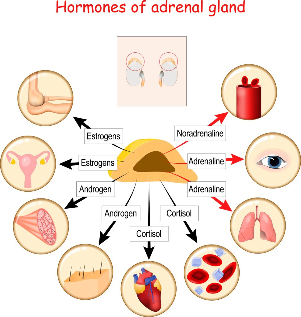
This diagram is an informative display of the hormones produced by the adrenal gland and their respective functions within the human body.
At the center of the illustration is the adrenal gland, depicted in yellow, and arrows point from the gland to various organs, indicating the target effects of its hormones.
Starting at the top and moving clockwise:
- Noradrenaline, also known as norepinephrine, affects blood vessels as shown by the red symbol indicating a blood pressure cuff. It causes vasoconstriction which increases blood pressure.
- Adrenaline, also known as epinephrine, is represented by the symbol of an eye and lungs. In the eye, it causes dilation of the pupil, and in the lungs, it relaxes the muscles of the bronchial tubes to improve breathing.
- Cortisol is depicted targeting muscle, the heart, and blood cells. In muscles, it stimulates gluconeogenesis, which is the production of glucose to provide energy. For the heart, cortisol has a supporting role in maintaining cardiovascular function. In blood cells, it influences both the stress response and the immune system.
- Androgen targets the skin and hair follicles, where it can stimulate hair growth and increase oil production.
- Estrogens, typically known as female sex hormones, have a systemic influence on the female reproductive system as indicated by the symbol of the uterus and also affect bone health, as shown by the representation of a bone.
In the inset at the top, the adrenal glands are positioned above the kidneys, highlighting their anatomical location.
Each hormone has multiple effects on the body, and this diagram simplifies these to show one or two key target organs or systems for each hormone. It’s important to note that the balance of these hormones is crucial for health and can affect a wide range of physiological processes.
Adrenal Gland Cross-Section
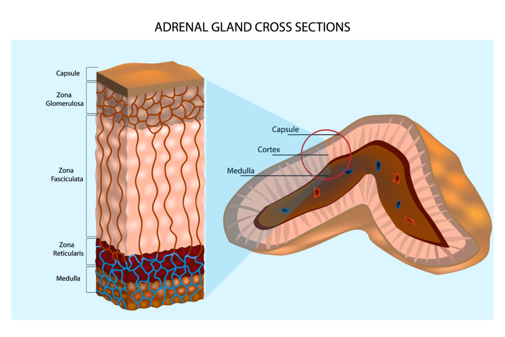
Presented in this illustration are two cross-sectional views of the adrenal gland, offering a visual breakdown of its internal structure.
On the left, we have a detailed, elongated cross-section of the adrenal gland, showing its various layers from the outermost to the innermost regions. The outermost layer is the capsule, a thin layer of connective tissue that encases and protects the gland. Beneath the capsule is the zona glomerulosa, the first and most superficial layer of the adrenal cortex. This layer is responsible for the production of mineralocorticoids, such as aldosterone, which regulate sodium and potassium balance in the body.
Below the zona glomerulosa is the zona fasciculata, which is the thickest section of the adrenal cortex. The cells in this layer appear arranged in straight columns and it is primarily responsible for producing glucocorticoids, including cortisol, which are critical for stress response, metabolism, and immune response.
Next is the zona reticularis, the innermost layer of the adrenal cortex, where cells are clustered in a network-like arrangement. This zone produces sex hormones, particularly androgens, which are precursors to sex steroids.
Finally, at the core of the gland lies the medulla. The medulla produces catecholamines, such as adrenaline and noradrenaline, which are vital for the body’s fight-or-flight response.
On the right, a different angle of cross-section shows the adrenal gland with its medulla in the center, surrounded by the cortical zones. This view provides a more anatomically correct representation of the gland’s shape and the distribution of the cortex and medulla within it.
Together, these cross-sections illustrate the complex structure and organization of the adrenal gland, emphasizing the distinct functions of its different regions.
Anatomy Terms and Definitions
| Term | Definition |
|---|---|
| Adrenal gland | An organ located above each kidney, responsible for producing hormones like adrenaline, noradrenaline, cortisol, androgens, and estrogens. |
| Adrenaline | A hormone and neurotransmitter produced by the adrenal medulla, involved in the body's fight-or-flight response. |
| Aldosterone | A mineralocorticoid hormone produced by the zona glomerulosa of the adrenal cortex, regulating sodium and potassium balance. |
| Androgen | A sex hormone produced by the zona reticularis of the adrenal cortex, involved in the development of male characteristics and influencing skin and hair follicles. |
| Benzene ring | A stable hexagonal arrangement of atoms found in the adrenaline molecule and many organic compounds. |
| Bladder | A hollow organ in the lower abdomen that stores urine until it is expelled from the body. |
| Capsule | A thin layer of connective tissue encasing the adrenal gland, providing protection and structural maintenance. |
| Cortex | The outer region of the adrenal gland, responsible for producing various hormones including cortisol, androgens, and mineralocorticoids. |
| Cortisol | A glucocorticoid hormone produced by the zona fasciculata of the adrenal cortex, involved in stress response, metabolism, and immune function. |
| Estrogen | A group of hormones produced by the adrenal cortex, influencing the female reproductive system and bone health. |
| Glucocorticoids | A class of corticosteroids involved in metabolism and stress response, produced by the adrenal cortex. |
| Gluconeogenesis | The metabolic process of producing glucose from non-carbohydrate sources, stimulated by cortisol. |
| Kidneys | A pair of organs in the abdominal cavity involved in filtering blood, removing waste, and producing urine. |
| Medulla | The inner region of the adrenal gland, producing catecholamines such as adrenaline and noradrenaline. |
| Mineralocorticoids | Hormones produced by the adrenal cortex, involved in regulating the body's salt and water balance. |
| Noradrenaline | A hormone and neurotransmitter produced by the adrenal medulla, involved in increasing blood pressure and preparing the body for action. |
| Ureters | Tubes that transport urine from the kidneys to the bladder. |
| Zona fasciculata | The thickest layer of the adrenal cortex, producing glucocorticoids like cortisol. |
| Zona glomerulosa | The outermost layer of the adrenal cortex, producing mineralocorticoids such as aldosterone. |
| Zona reticularis | The innermost layer of the adrenal cortex, producing sex hormones like androgens. |
