1916 Forearm and Elbow Basic Anatomy
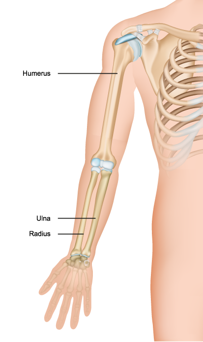
The image showcases a simplified representation of the upper limb skeletal anatomy, specifically focusing on the arm and forearm bones. At the top, the humerus is identified as the long bone of the upper arm, extending from the shoulder to the elbow. This bone is central to the function of the arm, providing structural support and serving as an attachment for several muscles that enable movement at the shoulder and elbow joints.
Below the humerus, the two bones of the forearm are labeled: the ulna and the radius. The ulna is situated on the medial side (the side closest to the body when in the standard anatomical position), and it is primarily involved in forming the elbow joint with the humerus. Its proximal end articulates with the humerus at the elbow, while its distal end forms part of the wrist joint.
The radius is the lateral bone of the forearm (the side furthest from the body in the anatomical position). It is thinner at the elbow and widens as it approaches the wrist. The radius is crucial for the movement of the forearm, allowing for the supination and pronation, which are the actions of turning the palm up and down, respectively.
These bones are connected to each other at the proximal and distal radioulnar joints, which facilitate the rotational movements of the forearm. The joints at either end of these bones, the shoulder joint (glenohumeral joint) for the humerus and the wrist joint (radiocarpal joint) for the radius and ulna, allow for a wide range of motion and dexterity of the upper limb.
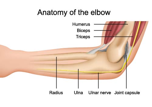
This illustration provides a simplified view of the anatomy of the elbow, highlighting the primary structures associated with this complex joint.
The humerus, the long bone of the upper arm, terminates at the elbow joint where it articulates with the two bones of the forearm: the radius and the ulna. This hinge joint allows for the flexion and extension of the forearm relative to the upper arm.
The biceps muscle, positioned anteriorly (or in front) of the humerus, is partially visible. This
muscle is responsible for flexing the elbow when contracted. Its tendon attaches to the radius, enabling the forearm to bend and also to supinate, which is the action of turning the palm upwards.
On the posterior side (or back) of the humerus is the triceps muscle, which serves as the antagonist to the biceps. The triceps is the main extensor of the elbow, straightening the arm when contracted. Its tendon attaches to the ulna at the olecranon, which is the pointed bone that forms the tip of the elbow.
The radius and ulna are the two bones that make up the forearm. The radius is located on the thumb side of the forearm and is involved in the supination and pronation of the forearm. The ulna, on the other hand, is on the little finger side and forms a hinge joint with the humerus, allowing for bending and straightening of the elbow.
Lying within the groove between the humerus and ulna, we can see the ulnar nerve. This nerve is responsible for the “funny bone” sensation when hit, due to its superficial position at the elbow. It provides sensation to the little finger and part of the ring finger and controls most of the fine movements of the hand.
Encasing the entire elbow joint is the joint capsule, a tough, fibrous sheath that provides stability and contains the synovial fluid that lubricates the joint, allowing for smooth movement.
Understanding the anatomy of the elbow is crucial not only for grasping how the joint operates but also for diagnosing and treating injuries commonly associated with this region, such as fractures, dislocations, and tendonitis.
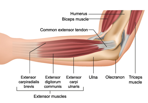
This image illustrates the posterior compartment of the forearm, focusing on the group of muscles that primarily extend the wrist and fingers. These extensor muscles are located on the backside of the forearm and are contrasted with the flexor muscles, which reside on the front side.
At the upper part of the image, the humerus is the long bone of the upper arm, connecting the shoulder to the elbow. Overlaid on the humerus is the biceps muscle, a powerful flexor of the elbow.
The common extensor tendon is a convergence point for several muscles in the forearm that extend the wrist and fingers. This tendon anchors these muscles to the lateral epicondyle of the humerus, which is not visible in this image but is located on the outer aspect of the elbow.
The extensor carpi radialis brevis is one of the muscles that extends and abducts the wrist, moving it away from the midline of the body. Adjacent to it is the extensor digitorum communis, which extends the fingers and the wrist. Next is the extensor carpi ulnaris, which extends and adducts the wrist, moving it towards the body’s midline.
The ulna is one of the two long bones in the forearm, positioned on the side opposite the thumb. At the elbow, the pointed part of the ulna, known as the olecranon, is what forms the tip of the elbow and serves as an attachment point for the triceps muscle, which extends the elbow.
The triceps muscle, visible at the upper right corner of the image, is the large muscle responsible for extending the elbow, counteracting the action of the biceps muscle.
The coordination of these muscles allows for complex movements of the wrist and fingers, enabling various activities such as typing, playing a musical instrument, or performing a backhand in tennis. The health and integrity of the common extensor tendon are particularly important, as conditions like lateral epicondylitis, also known as tennis elbow, can arise from overuse or strain of this area.
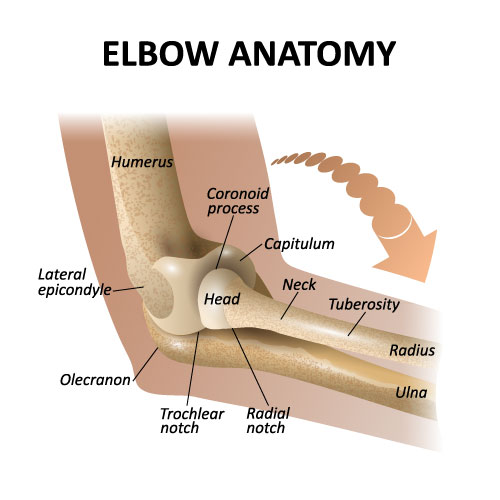
This image provides a detailed look at the elbow anatomy, highlighting the bone structures and the articulations that make up the elbow joint.
The humerus is the long bone of the upper arm, and at its lower end, it features two important landmarks: the lateral epicondyle and the capitulum. The lateral epicondyle is a small, tuberous bump on the outer side of the humerus to which the forearm muscles attach. The capitulum is a rounded knob on the humerus that articulates with the head of the radius, allowing for the rotation of the forearm.
The radius is one of the two bones of the forearm, shown with its head, neck, and tuberosity. The head of the radius is disc-shaped and interacts with the capitulum of the humerus, contributing to the hinge movement of the elbow and the rotational movement of the forearm. The neck is the narrowed portion just below the head, and the tuberosity is a bony prominence below the neck where the biceps tendon attaches.
The ulna, the other bone of the forearm, is shown with two important features: the olecranon and the coronoid process. The olecranon is the pointed bone at the elbow’s tip, which fits into the olecranon fossa of the humerus when the arm is extended. The coronoid process is a triangular eminence that projects forward from the upper part of the ulna and fits into the trochlear notch during elbow flexion.
The trochlear notch is a deep groove in the ulna that hugs the trochlea of the humerus, forming the hinge portion of the elbow joint. The radial notch is a small, smooth area on the ulna where the head of the radius sits, allowing for the pivot-like movement of the radius around the ulna.
These components work together to allow the elbow joint to perform its functions: the bending and straightening of the arm (flexion and extension) and the rotation of the forearm (pronation and supination). The elbow is a complex hinge joint that is crucial for many daily activities, from lifting objects to throwing a ball.
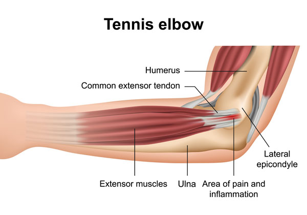
The diagram illustrates the condition known as tennis elbow, or lateral epicondylitis, which is a form of tendinopathy that occurs due to overuse or strain of the extensor muscles in the forearm.
The humerus, the upper arm bone, has at its lower end a bony prominence on the outside called the lateral epicondyle. The common extensor tendon is attached to this point and anchors the extensor muscles of the forearm, which are responsible for extending the wrist and fingers.
These extensor muscles run along the forearm and connect to the lateral epicondyle via the common extensor tendon. When these muscles are overused, particularly through repetitive wrist and arm motions, the common extensor tendon can develop small tears or become inflamed, leading to pain and tenderness around the lateral epicondyle.
The area of pain and inflammation is typically located on the outside of the elbow, over or near the lateral epicondyle, which is why this condition is referred to as “tennis elbow.” Despite its name, tennis elbow doesn’t only affect tennis players. It can occur in anyone who engages in activities that put a repetitive strain on the wrist extensors.
The ulna, one of the two bones of the forearm, is shown parallel to these extensor muscles. The ulna and the radius (not labeled here) work together to facilitate movement at the wrist and elbow.
Tennis elbow is a common musculoskeletal disorder, and management typically includes rest, ice, nonsteroidal anti-inflammatory drugs (NSAIDs), physical therapy, and in some cases, more invasive treatments like steroid injections or surgery if conservative measures fail to provide relief.
